Flaccidity of the Vocal Cords

Vocal cord flaccidity correlates to some degree with atrophy of the muscle comprising them. Bowing also accompanies flaccidity most of the time. It is possible to have bowed/slender vocal cords that are not particularly flaccid—they still vibrate with good firmness and resilience.

Similarly, vocal cords that appear to have good bulk (and are not atrophied) can nevertheless have a flaccid vibratory pattern. Photos below show the visual findings of flaccidity as distinct from bowing and atrophy. Voice manifestations of flaccid vocal cords are similar to bowing in cases such as:

  • Loss of “edge”
  • Reduced ability to be heard in noisy places
  • Reduced vocal endurance (The voice becomes fuzzier or raspier and more air-wasting as the day progresses and the atrophied muscles tire).

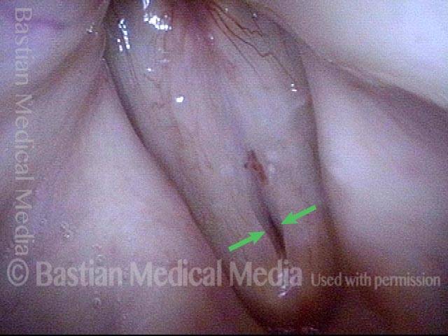
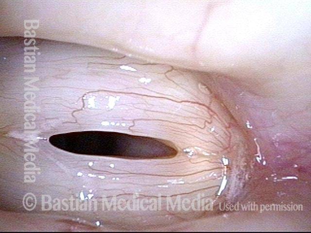
A key visual finding of vocal cord flaccidity

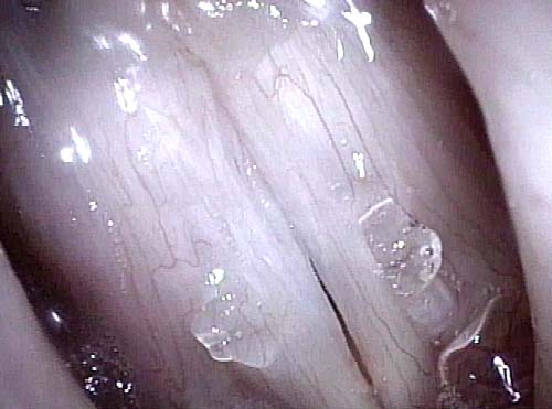
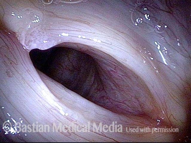
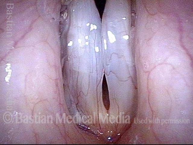
This seventy-something woman has noted a weak voice for at least a decade. She came for evaluation due to what seemed like further reduction of strength following a bout of laryngitis.

At her evaluation, her faintly fuzzy speaking voice can “pass for normal for age” but when asked to project, her voice manifests a lack of edge and power. At low pitch there is one moment of “luffing.” From her history and vocal phenomenology, vocal cord bowing is suspected.

Visual Portfolio, Posts & Image Gallery for WordPress

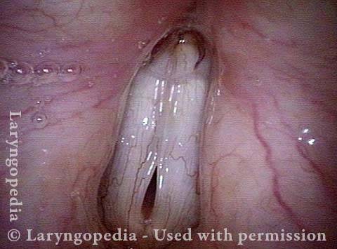
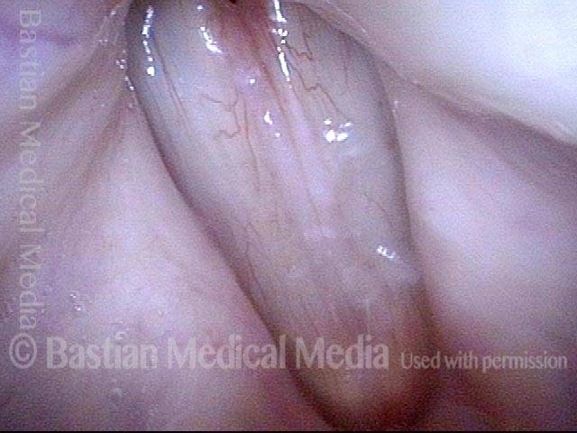
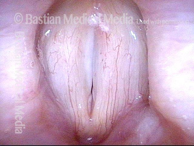
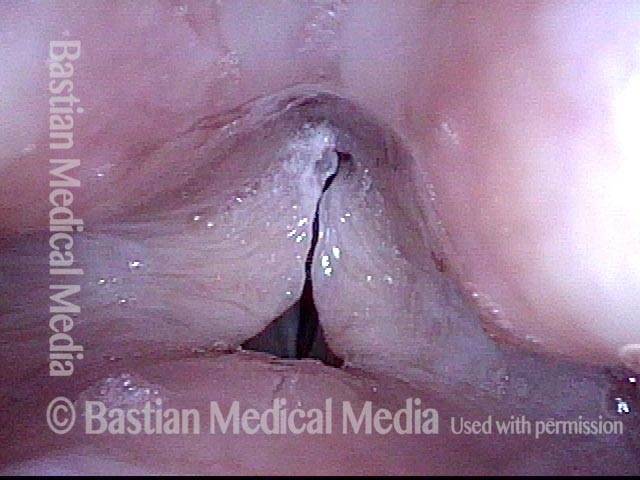
Vocal cords (1 of 4)

Distant view, while breathing. No abnormality is seen.

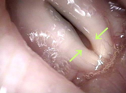
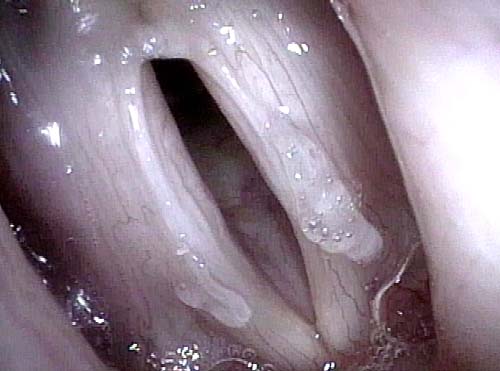
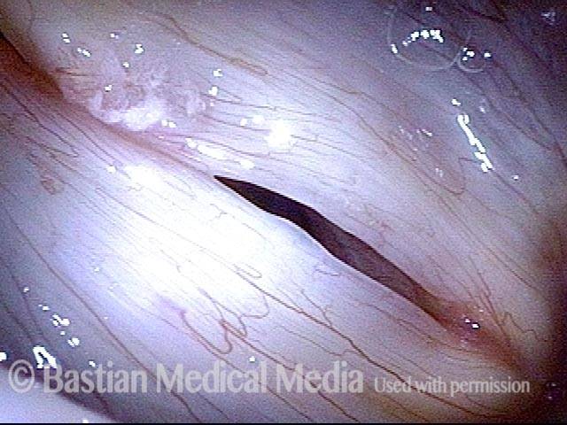
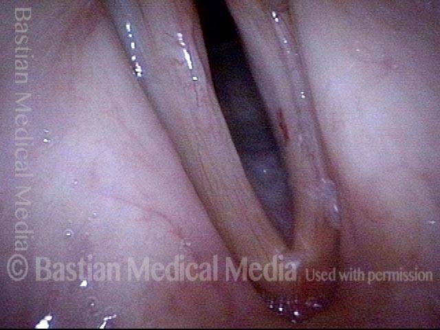
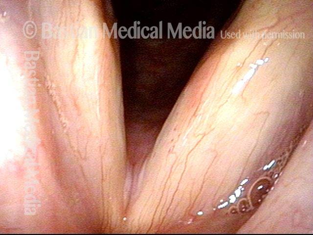
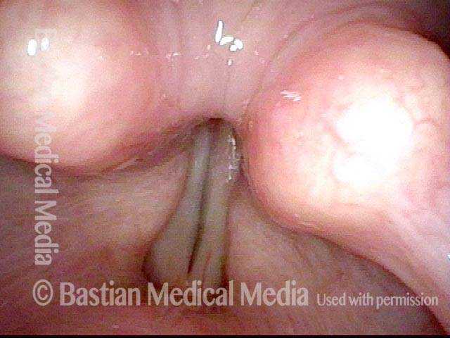
Vocal cords phonating (2 of 4)

Distant view, phonating. Again, nothing unusual is seen.

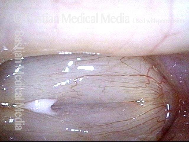
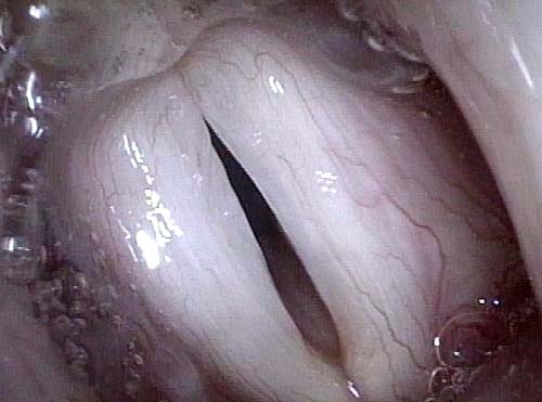
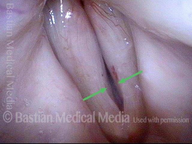
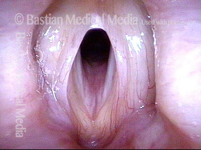
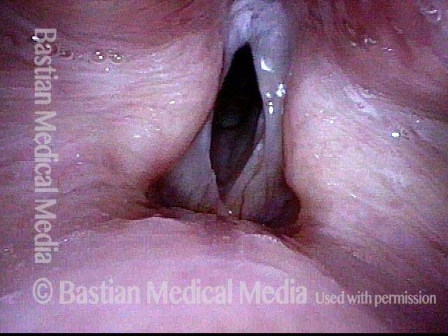
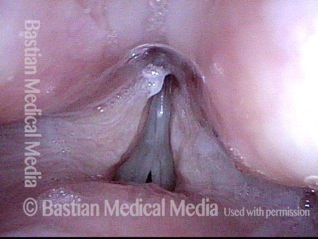
Large amplitude of vibration (3 of 4)

At close range and under strobe light: At estimated A3 (220 Hz), open phase of vibration. An unusually large amplitude of vibration (lateral excursion of the folds) is seen. This is a typical finding suggesting flaccidity.

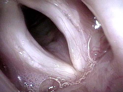
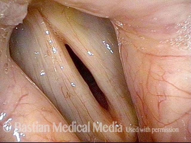
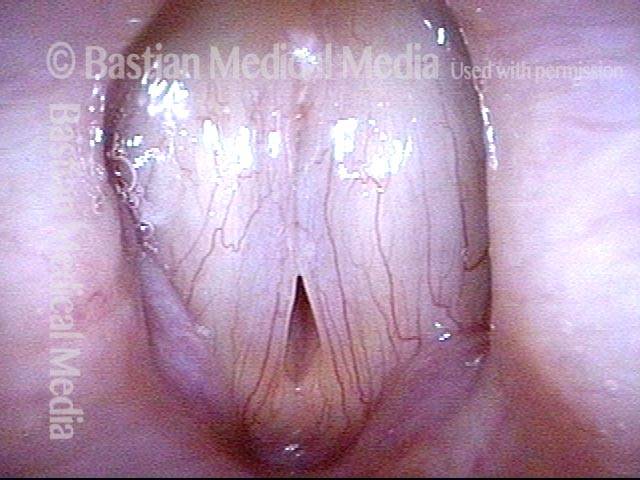
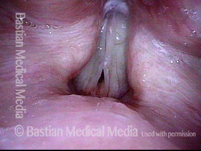
Vocal cord flaccidity (4 of 4)

“Closed phase” of vibration is not fully closed and a highly specific finding of flaccidity is the anterior vocal cords’ failure to close seen here.

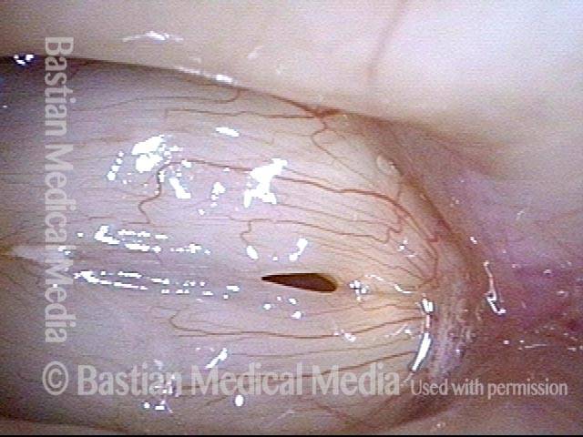
Vocal Cord Flaccidity at Two Pitches

Visual Portfolio, Posts & Image Gallery for WordPress

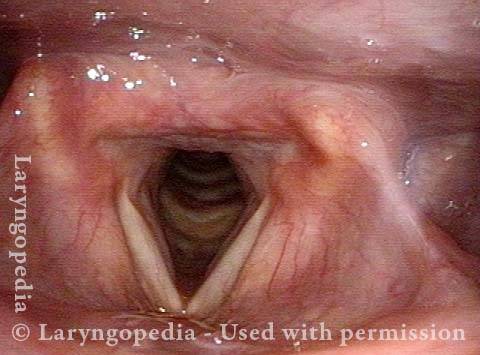
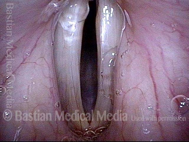
Anterior vocal cord gap (1 of 5)

This woman in her sixties is experiencing loss of vocal strength. Her voice becomes raspier as the day progresses. Under standard light during voicing at her average pitch for speech, F3 (175 Hz), vibrating cords have blurred margins. Notice that the gap at the anterior vocal cords is wider (arrow) than between the rest of the cords. This anterior gap is a typical finding of flaccidity.

Wide anterior gap (2 of 5)

Still at her habitual pitch for speech, F3 (175 Hz), the closed phase is not closed at all! And again, the gap is wider anteriorly. This incomplete closure explains her air wasting, fuzzy voice quality.

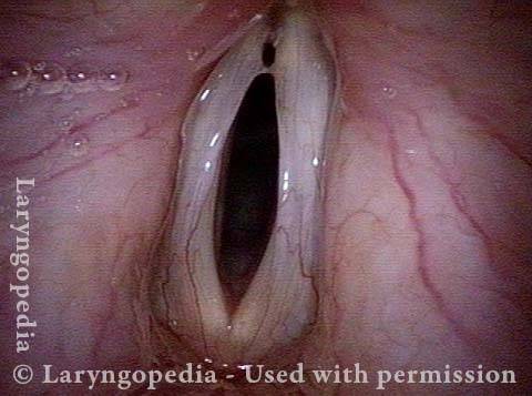
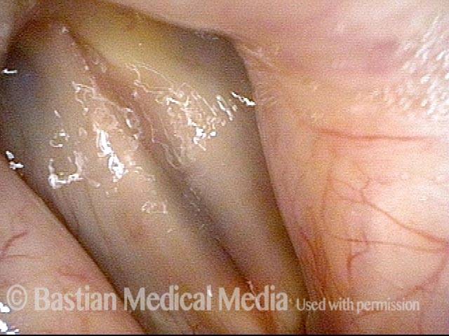
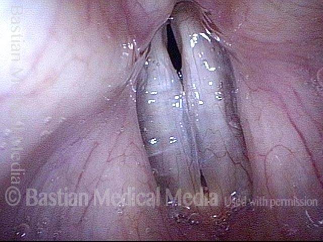
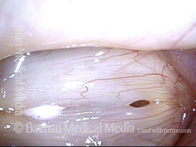
Closed phase! (3 of 5)

Still at her habitual pitch for speech, F3 (175 Hz), the closed phase is not closed at all! And again, the gap is wider anteriorly. This incomplete closure explains her air wasting, fuzzy voice quality.

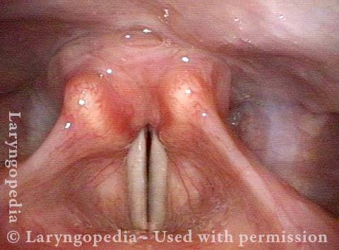
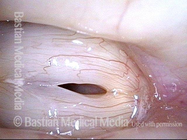
Telltale small gap (4 of 5)

Higher pitch lengthens the vocal cords (stretches them longitudinally, making them less flaccid: think “rubber band”) Yet under strobe light at C4 (252 Hz), that telltale small gap is still seen anteriorly during closed phase of vibration.

Vocal Cords (5 of 5)

At the same pitch, the vocal folds fly less far laterally, but definitely farther than would normally be seen at moderate loudness at this pitch.

Flaccid Vocal Cords

Visual Portfolio, Posts & Image Gallery for WordPress

Flaccid vocal cords (1 of 4)

Pre-phonatory instant, standard light.

Flaccid vocal cords (2 of 4)

Vibratory blur with standard light. Note relatively wide “gray” zone of blur.

Flaccid and bowing (3 of 4)

Dramatic lateral excursions due not only to bowing, but also flaccidity.

Flaccid vocal cords (4 of 4)

Eventual recoil towards midline, but cords never come into full contact before they are again thrown to their maximum lateral vibratory excursion, as in photo 3.

Example 2

Visual Portfolio, Posts & Image Gallery for WordPress

Flaccid vocal cords (1 of 3)

Open phase of vibration showing large amplitude in middle voice.

Flaccid vocal cords (2 of 3)

Maximum closed phase, but with persistent open area anteriorly (at arrow) If this area of exaggerated flaccidity oscillates independently, a rough quality is added to the voice.

Flaccid vocal cords (3 of 3)

Between open and closed phase, showing vibratory contact is aberrant.

Vocal Cord Bowing

Visual Portfolio, Posts & Image Gallery for WordPress

Vocal cord bowing (1 of 4)

Open phase vibration, strobe light. Notice the large amplitude of vibration. The wide lateral excursions suggest flaccidity, especially when this is seen in middle voice.

Cause of a gravely voice (2 of 4)

Partially closed phase, strobe light. Notice that the anterior cords are more flaccid, with delayed return to midline contact. When this is seen, that anterior segment may vibrate independently and cause a rough, gravelly voice quality. The capillary ectasia, left vocal cord is an incidental finding an not related to the patient’s rough voice quality.

Vocal cords do not close completely (3 of 4)

Coming to closed phase, but with persistent anterior open segment.

Vocal cord bowing (4 of 4)

Closed phase of vibration, strobe light.

Flaccidity Without Bowing

Visual Portfolio, Posts & Image Gallery for WordPress

Flaccidity without bowing (1 of 4)

The patient exhibits typical symptoms of bowing/atrophy/flaccidity, but in this case there is little bowing or atrophy—primarily flaccidity is seen. In this view, the abducted vocal cords appear full, with no exaggeration of the ventricles. (The apparent asymmetry between the vocal cords is due to the viewing angle; both cords are the the same.)

Flaccidity without bowing (2 of 4)

Now seen under the strobe light, the amplitude of vibration of the vocal cords is excessive. These flaccid vocal cords lack the firmness to “recoil” back to the mid-line, until maximal separation of the cords is reached.

Flaccidity without bowing (3 of 4)

As the patient is reaching the closed phase of the vibratory cycle, the anterior cords are arriving late to closure, a typical finding with flaccid vocal cords.

Flaccidity without bowing (4 of 4)

Now at the maximum closed phase of vibration, a pinhole of incomplete closure is seen. In this case, there is no independent oscillatory vibration of the anterior segment of the vocal cord.

False Cord Phonation

Visual Portfolio, Posts & Image Gallery for WordPress

False cord phonation due to flaccid true cords (1 of 5): before false cords begin to vibrate

An elderly man, quiet by nature who uses the voice little, complains of weak, gravelly voice quality. This view of phonation, standard light, shows a slightly wider dark line of phonatory blurring. Also, the false vocal cords are overly approximated, but not yet participating in vibration (for that, see images 4 and 5).

False cord phonation due to flaccid true cords (2 of 5): before false cords begin to vibrate

Strobe light reveals an unusually wide amplitude of vibration, denoting flaccidity of the true vocal cords.

False cord phonation due to flaccid true cords (3 of 5): before false cords begin to vibrate

Maximum closed phase shows the telltale residual opening at the anterior commissure (from this perspective, the lowermost end of the true cords), also a potent indicator of flaccidity.

False cord phonation due to flaccid true cords (4 of 5): after false cords begin to vibrate

When asked to produce louder voice, the false cords begin to participate in vibration, and a rough, gravelly superimposed “ godfather” quality arrives. Notice that the true cords are in at least partial open phase of vibration.

False cord phonation due to flaccid true cords (5 of 5): after false cords begin to vibrate

Now the false cords are in open phase of vibration, and the true cords are in maximum closed phase of vibration, with the same residual opening anteriorly seen in photo 3. False cord phonation is not to be seen most accurately as primary here, but as a secondary result of effort necessitated by flaccid true cords.

Flaccidity

Visual Portfolio, Posts & Image Gallery for WordPress

Flaccidity (1 of 5)

Under strobe light, the vocal cords begin opening at the area of flaccidity anteriorly. The patient has a husky and gravelly voice quality (view here rotated 90 degrees counterclockwise).

Flaccidity (2 of 5)

Vibratory separation continues and is still greatest anteriorly.

Flaccidity (3 of 5)

Open phase of vibration now complete. Distance of lateral excursions is large, indicating the flaccidity of cords.

Flaccidity (4 of 5)

The cords are returning to closed vibratory position, but anterior cords close late due to flaccidity. Sometimes this anterior segment vibrates independently and this causes a rough voice quality.

Flaccidity (5 of 5)

At maximum closed phase, there is still a tiny incomplete closure anteriorly.