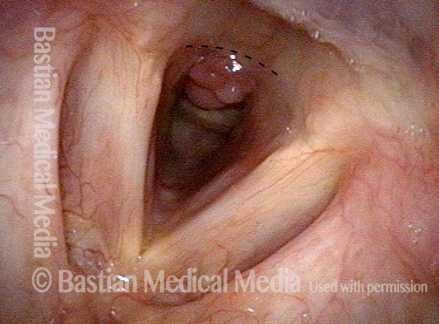
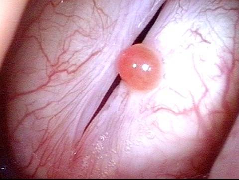
Granulation tissue is tissue that develops as an exuberant “over-healing” response to irritation or injury. This irritation or injury could be due to an endotracheal tube, or a superficial cordectomy wound from surgery, or a number of other causes. Granulation tissue that forms on the posterior vocal cord is called a contact granuloma.


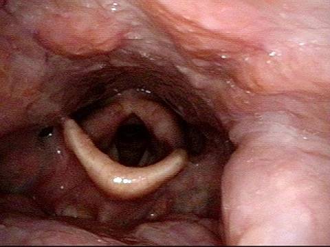
Subglottic granulation and curving airstream

Visual Portfolio, Posts & Image Gallery for WordPress

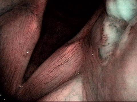
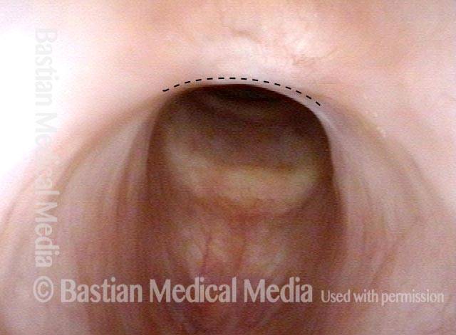
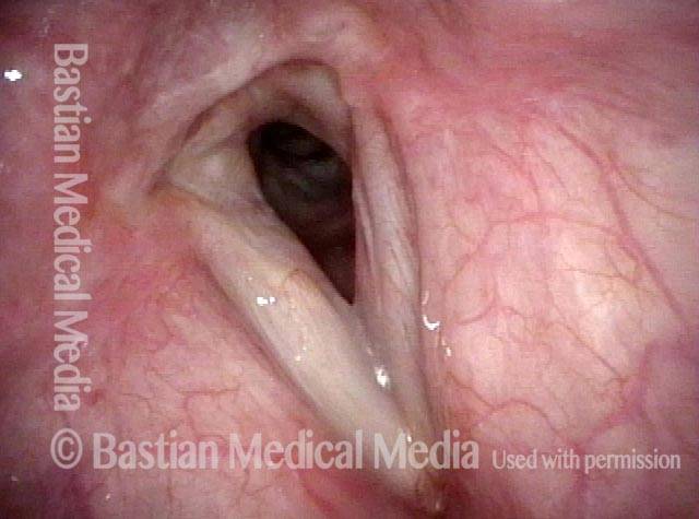
Intubation injury (1 of 4)
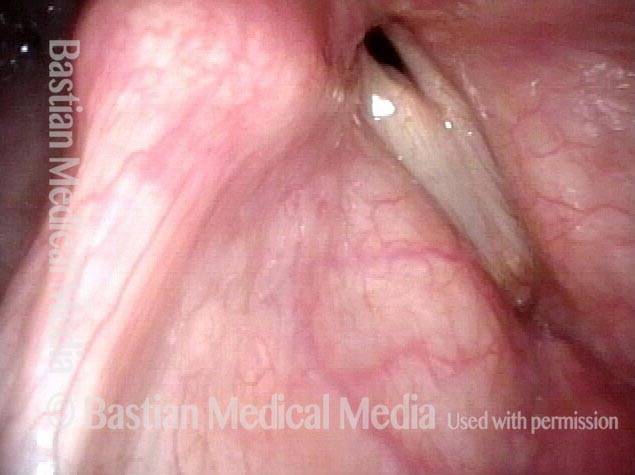
After a 9-day intubation for serious illness, the patient has difficulty breathing due to this “proud flesh” response to injury within the cricoid ring, posteriorly. Breathing has improved in the past week, leading to a decision to await further maturation, rather than proceeding to microlaryngoscopic removal.

Lobules (2 of 4)

Close-up view of the lobules of granulation tissue. Air can easily pass around the obstruction as indicated by the arrows.

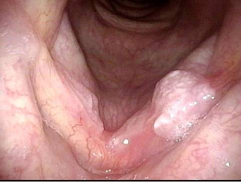
2 months later (3 of 4)
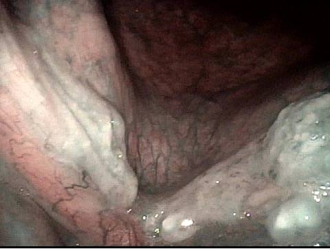
As predicted, breathing continued to improve to the point of seeming normal to the patient, and 2 months later, the granulation tissue has matured and detached, leaving behind a subglottic scar band (parallel lines).

Scar band (4 of 4)

Here is a close-up of the scar band. A solid line denotes the anterior border of the scar band for reference in all 4 photos, but compare especially to photo 2.

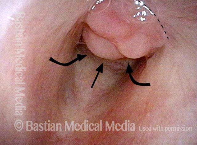
Sometimes you DO remove granulation to avoid tracheotomy

Visual Portfolio, Posts & Image Gallery for WordPress

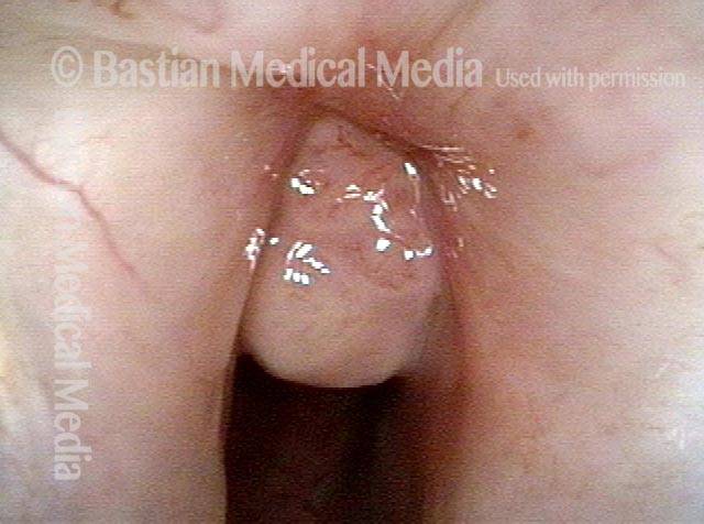
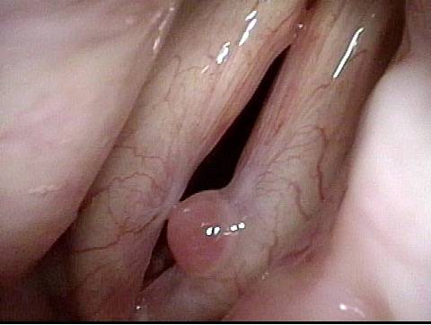
Granulation (1 of 8)

Prior to this first visit, this person suffered extensive burns, was intubated for 10 days, and then underwent tracheotomy, and then was decannulated (tracheotomy removed). She has scarring of the posterior commissure outlined by the dotted line. The granulation extends well down into the subglottis. She is uncomfortable with a marginal airway and noisy breathing. Laser and microdebrider are planned to try to avoid having to reinsert the tracheotomy.

Closer view (2 of 8)

Tip of the iceberg view of granulation and scarred area.

Post microlaryngoscopies (3 of 8)
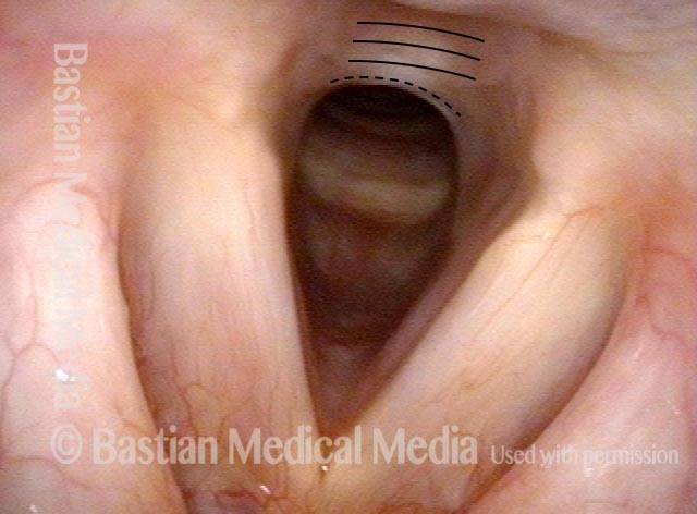
After a series of microlaryngoscopies purely to improve airway and avoid tracheotomy, the granulation has finally matured. Airway is no longer marginal, but is still very limited for significant activity.

Scarring (4 of 8)

At close range, the area of posterior scarring is again indicated by dotted line; the dark area of the actual airway is narrow and slit-like.

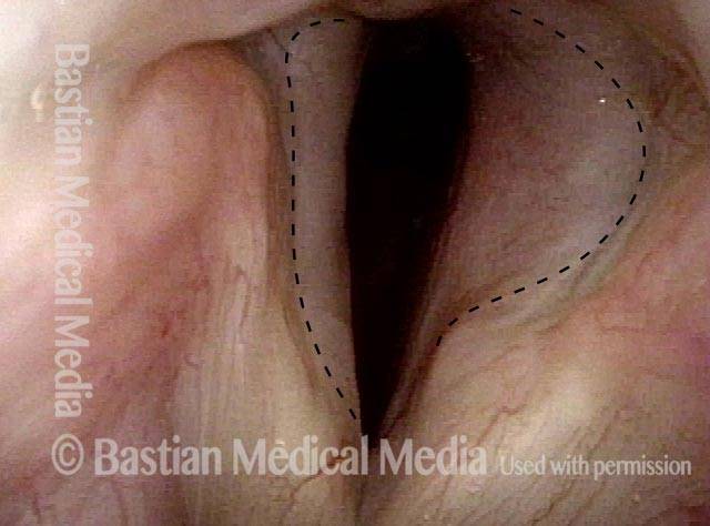
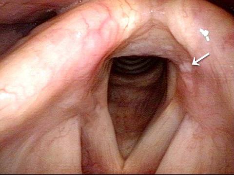
Post posterior commissuroplasty (5 of 8)

A month after posterior commissuroplasty, breathing is improved due to the widened space posteriorly. Compare the dark area for breathing with photo 3.

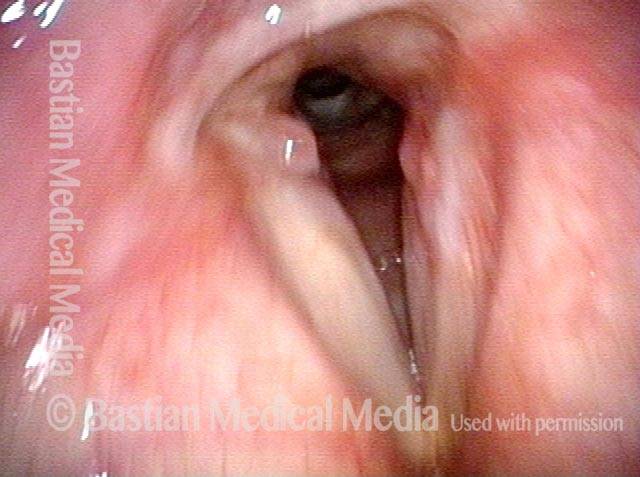
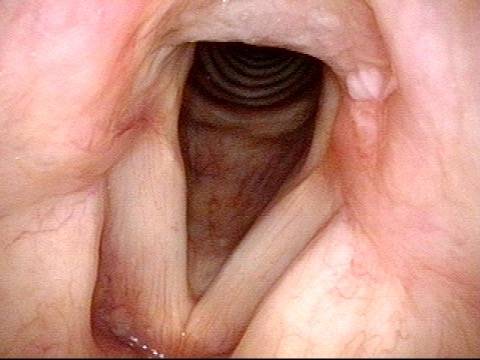
Breathing improved (6 of 8)

Six months after posterior commissuroplasty, breathing remains much improved. Compare dark airway contour again with photo 3 above.

Closer view (7 of 8)

A closer view of the airway, which is much wider posteriorly than preoperatively (photo 4).

Phonatory view (8 of 8)

When patient makes voice, there is a persistent space posteriorly, where the airway was surgically widened, but again, this has not significantly affected the voice.

Laser surgery for Bilateral Vocal Cord Cancer

Visual Portfolio, Posts & Image Gallery for WordPress

Squamous cell carcinoma (1 of 6)

This man developed hoarseness across a few months. Biopsy elsewhere revealed squamous cell carcinoma, seen here on both vocal cords. Based upon a friend’s experience, he rejected radiotherapy, opting for laser resection, knowing it could be very hard on voice.

Tumor on the vocal cords (2 of 6)

At closer range and under narrow band (blue-green) light to accentuate the vascular abnormalities associated with this tumor.

Granuloma delays voice recovery (3 of 6)

Six weeks after superficial laser cordectomy, the larynx is almost healed with the exception of a small granuloma, left vocal cord (right of photo). When healing includes granulation, voice recovery is delayed as the granuloma resolves.

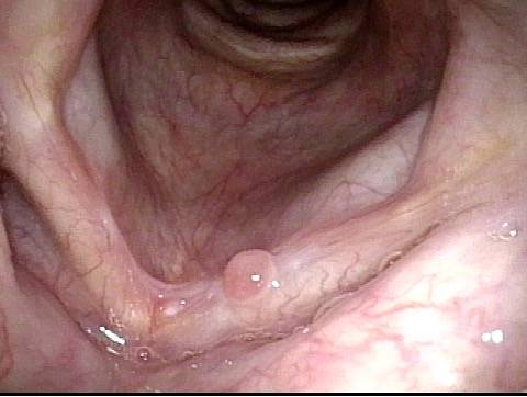
Closer view of granuloma (4 of 6)

He has hoarse but functional voice, but under strobe light, the granuloma prevents vibratory closure. Note the medial-to-lateral capillary reorientation so typical after laser cordectomy.

Granuloma is smaller (5 of 6)

Now 3 ½ months from surgery, voice has improved further and he considers it “75%” of original...One can see that the granuloma is smaller.

Granuloma doesn't impede voice (6 of 6)

Note that the granuloma no longer prevents vibratory closure and this explains further improvement of voice. Compare with photo 4.

Arytenoid Perichondritis—an Issue of “Festering”

This middle-aged man has had a chronically sore throat on the left for at least 6 months.  When asked to indicate where, he doesn’t gesture towards the throat as a whole, but points with one finger to the upper part of the thyroid cartilage fairly far laterally.  He doesn’t remember being ill, or any other explanation. After seeing the lesion below, read the introductory article of this post for treatment of this condition.

Visual Portfolio, Posts & Image Gallery for WordPress

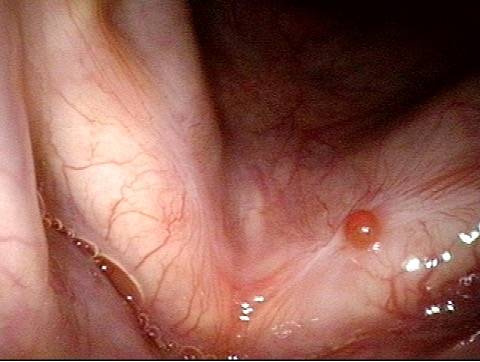
Arytenoid perichondritis (1 of 4)

A distant, panoramic view shows some cobblestoning but nothing (yet) focal to the left.

Lesion (2 of 4)

At a mid-range view, a small lesion is seen on the anterior face of his left arytenoid (arrow). There is mild surrounding redness.

Closer look at lesion (3 of 4)

At closer range, the lesion is more easily seen to be significant and quite different from the right. It is not a typical contact granuloma, which would be found lower, on the vocal process.

Vascular atypia (4 of 4)

At very close range, under narrow band light. One can see that there is a “pitted” center, and some vascular atypia of chronic inflammation/healing.