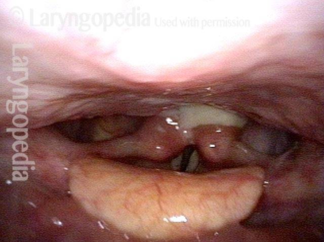
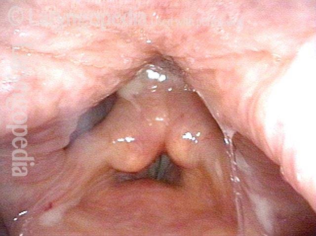
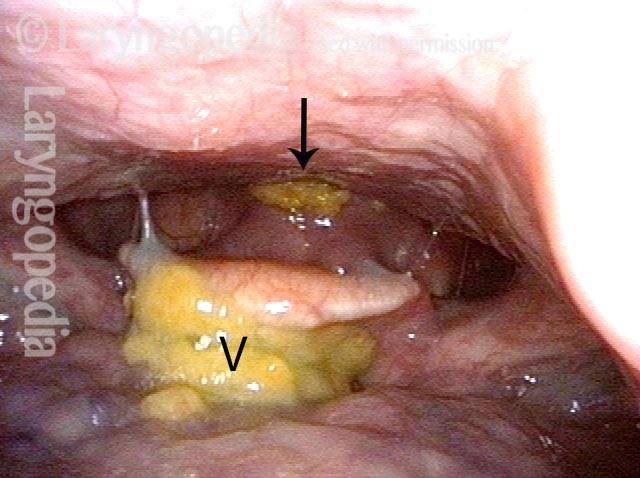
Antegrade cricopharyngeal dysfunction (A-CPD) is the failure of the tonically contracted upper esophageal sphincter to relax and open when one swallows. It is also known as cricopharyngeal achalasia. The cause is usually unknown.
The upper esophageal sphincter is also known as the cricopharyngeus muscle and is located at the lower level of the voicebox or larynx. This muscle is always contracted except at the moment of swallowing, when it relaxes briefly to let food or liquid pass through.
Symptoms and treatment for A-CPD
Typically, individuals with A-CPD first notice that pills or solid food begin to lodge at the level of the lower part of the larynx. The problem tends to progress inexorably, though often slowly, as the years pass, until the individual must limit himself or herself to liquid and soft foods.
Cricopharyngeal dysfunction is fully resolved through a straightforward surgical procedure (cricopharyngeal myotomy), performed through the mouth with the laser or, only occasionally, through a neck incision.
See also: Zenker’s diverticulum.
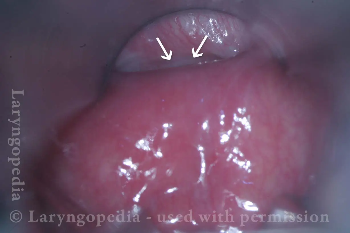
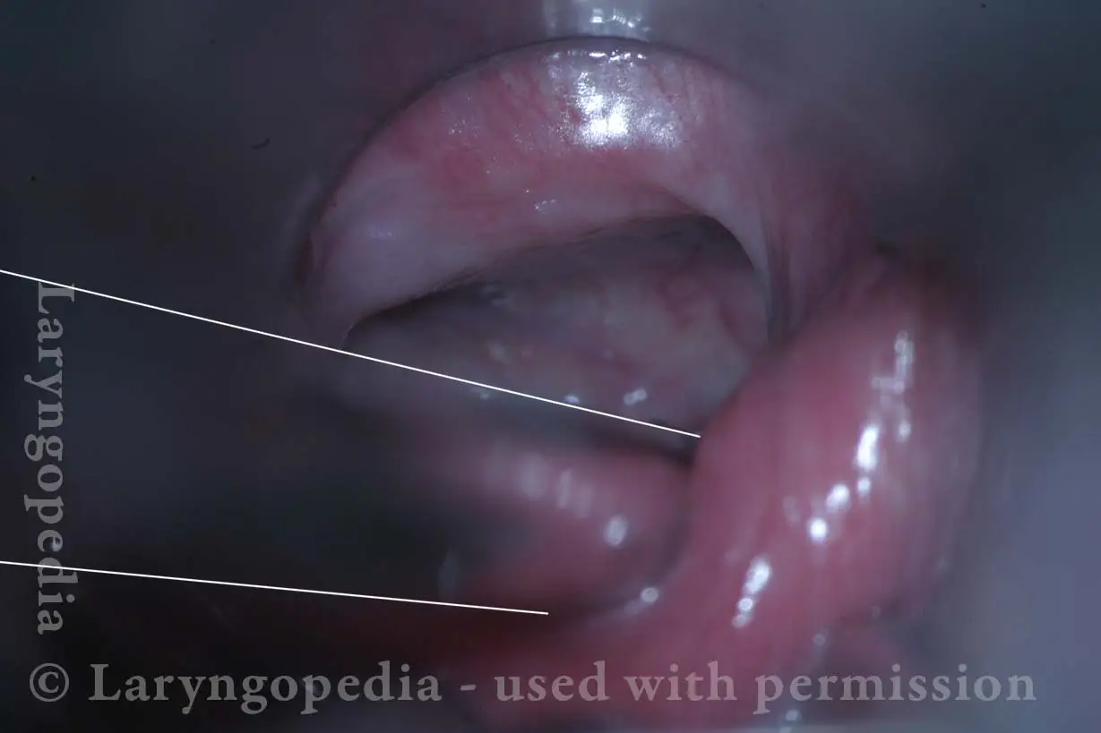
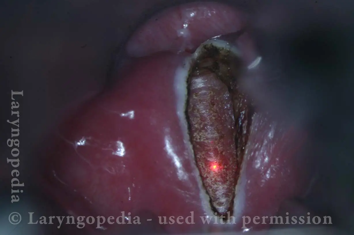
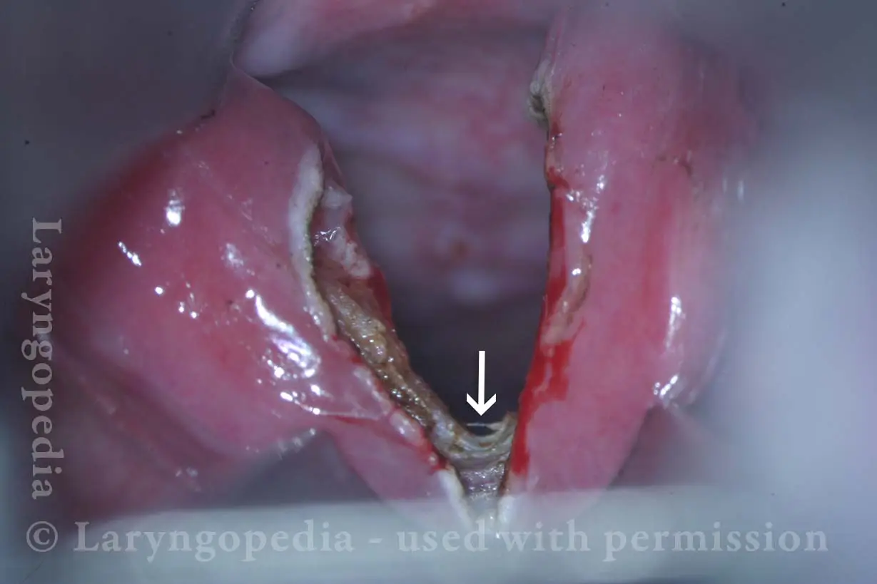
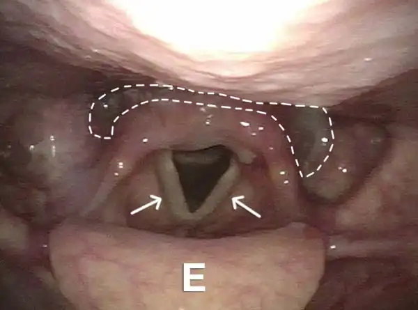
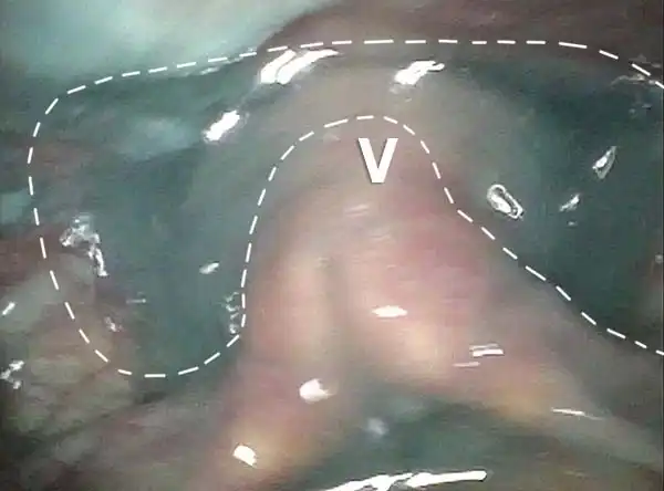
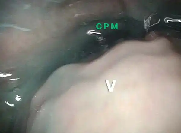
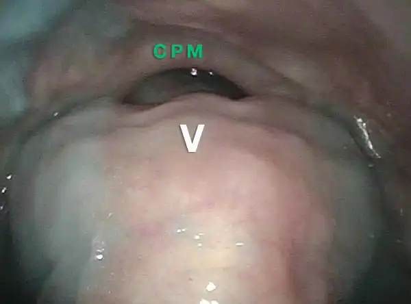
A-CPD, before, during, and after Myotomy.
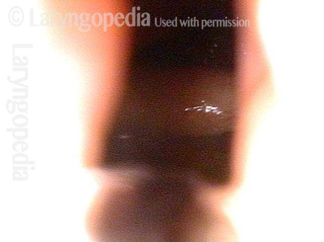
Non-relaxing cricopharyngeus muscle (1 of 4)
Non-relaxing cricopharyngeus muscle (1 of 4)
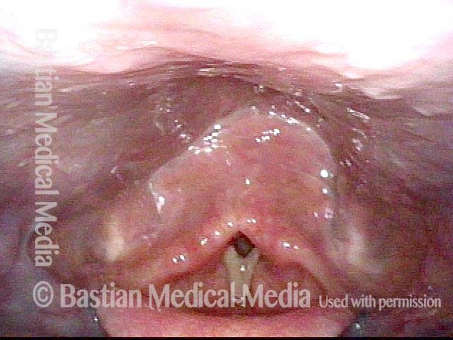
Opening the esophageal orifice (2 of 4)
Opening the esophageal orifice (2 of 4)
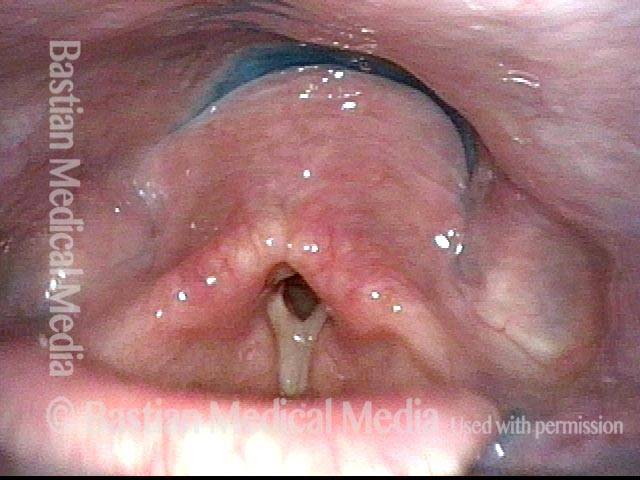
Laser cricopharyngeus myotomy (3 of 4)
Laser cricopharyngeus myotomy (3 of 4)
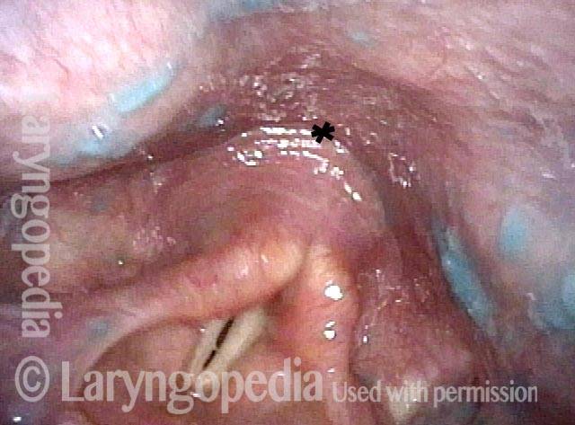
Cricopharyngeus myotomy nearly complete (4 of 4)
Cricopharyngeus myotomy nearly complete (4 of 4)
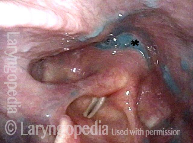
The Cricopharyngeus Muscle Seen During Swallowing
This person struggles to swallow due to a combination of prior tongue cancer surgery decades ago, and longterm radiation effects. Solid foods are the most problematic, and so this sequence shows an attempt to swallow water stained with blue food coloring.
Swallowing crescent (1 of 5)
Swallowing crescent (1 of 5)
Swallowing water (2 of 5)
Swallowing water (2 of 5)
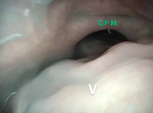
Cricopharyngeus muscle (3 of 5)
Cricopharyngeus muscle (3 of 5)
Relaxed CPM (4 of 5)
Relaxed CPM (4 of 5)
Partially open esophagus due to A-CPD (5 of 5)
Partially open esophagus due to A-CPD (5 of 5)
Antegrade Cricopharyngeal Dysfunction, Before and After Myotomy
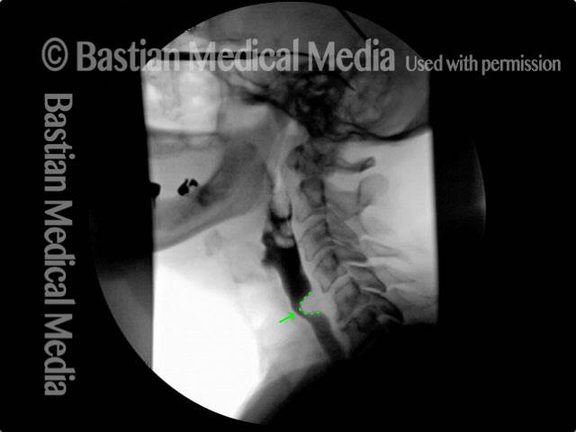
Cricopharyngeal dysfunction: before myotomy (1 of 2)
Cricopharyngeal dysfunction: before myotomy (1 of 2)
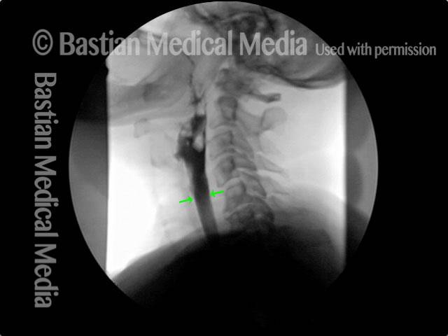
Cricopharyngeal dysfunction: after myotomy, resolved (2 of 2)
Cricopharyngeal dysfunction: after myotomy, resolved (2 of 2)
Example 2
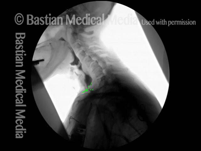
Cricopharyngeal dysfunction: before myotomy (1 of 2)
Cricopharyngeal dysfunction: before myotomy (1 of 2)
Cricopharyngeal dysfunction: after myotomy, resolved (1 of 2)
Cricopharyngeal dysfunction: after myotomy, resolved (1 of 2)
Very High-pitched Voice Elicits the Same Pharynx Contraction as Swallowing
Secretions (1 of 4)
Secretions (1 of 4)
Contracted pharynx (2 of 4)
Contracted pharynx (2 of 4)
Cracker residue (3 of 4)
Cracker residue (3 of 4)
Pharyngeal walls (4 of 4)
Pharyngeal walls (4 of 4)
Reflux Into Hypopharynx, Characteristic of Antegrade Cricopharyngeal Dysfunction
Reflux into hypopharynx (1 of 3)
Reflux into hypopharynx (1 of 3)
Water flows into the swallowing crescent (2 of 3)
Water flows into the swallowing crescent (2 of 3)
Larynx opens up (3 of 3)
Larynx opens up (3 of 3)
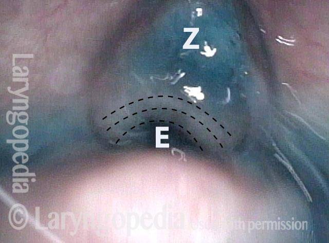
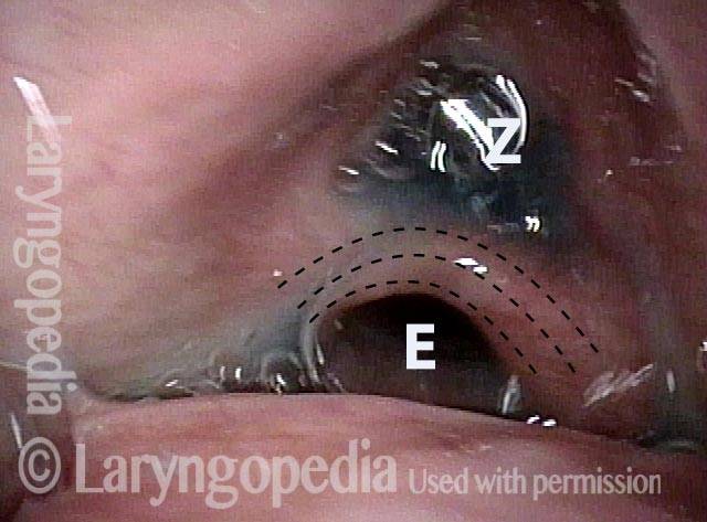
Cricopharyngeus Non-Relaxation and Zenker’s Sac Seen During VESS
Immediately after swallow (1 of 4)
Immediately after swallow (1 of 4)
One second later (2 of 4
One second later (2 of 4
Un-relaxed cricopharyngeus muscle (3 of 4)
Un-relaxed cricopharyngeus muscle (3 of 4)
More water (4 of 4)
More water (4 of 4)
Having Trouble Swallowing Foods and Pills? A-CPD Can Be Treated with Cricopharyngeal Myotomy
A small percentage of (mostly) older people develop A-CPD. They have difficulty initially with solid foods and pills. As the months and years pass, the tendency for food to lodge in the throat gradually increases.
Eventually, they must limit their diets to softer and “easier” things more and more like “baby food.” Special focus is placed on an effective endoscopic (through the mouth) laser procedure
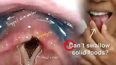
Cricopharyngeal Dysfunction: Difficulty Swallowing, Especially Solid Foods
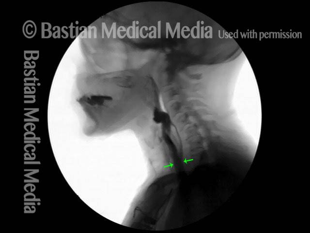
Dr. Bastian explains this progressive swallowing problem and presents options for treatment. Cricopharyngeal dysfunction is caused by failure of relaxation of the upper esophageal sphincter—cricopharyngeus muscle—during eating. Typically it is solid foods that tend to lodge in the mid-neck area where this muscle is located.

Cricopharyngeal Dysfunction: Before and After Cricopharyngeal Myotomy
This video shows x-rays of barium passing through the throat, first with a narrowed area caused by a non-relaxing upper esophageal sphincter (cricopharyngeus muscle), and then after laser division of this muscle. Preoperatively, food and pills were getting stuck at the level of the mid-neck, and the person was eating mostly soft foods. After the myotomy (division of the muscle), the patient could again swallow meat, pizza, pills, etc. without difficulty.
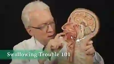
Swallowing Trouble 101
This video gives an overview of how swallowing works, how it can sometimes go wrong (presbyphagia or A-CPD), and possible ways to treat those problems (swallowing therapy or cricopharyngeal myotomy).
