A disorder in which the laryngeal saccule is inflated and becomes abnormally enlarged. A common symptom of a laryngocele is hoarseness.
How it develops
The laryngeal saccule, or laryngeal appendix, is a very small blind sac—a dead-end corridor, so to speak—which is located just above the vocal cords, one on each side, and is lined with glands that supply lubrication to the cords. When a person makes voice, it is possible for a little bit of the air being pushed up out of the trachea to slip into this saccule. If over time enough air enters the saccule with enough force, the saccule may begin to be inflated and stretched out, leading to a laryngocele.
In some cases, the air that slips into and inflates the laryngocele will slip back out again as soon as the person stops making voice, so that the laryngocele abruptly inflates and deflates with each start and stop of speech or voice-making. (The photos and video below are an example of this.) In other cases, the air cannot exit as easily, but it may be reabsorbed slowly during quiet times or during sleep—only to be inflated again at the next instance of more active speaking.
Laryngocele vs. saccular cyst
A much more common disorder of the laryngeal saccule (compared with a laryngocele) is a saccular cyst, which can occur if the entrance to the laryngeal saccule becomes blocked. In this scenario, air is absorbed, but secretions build up and gradually expand the saccule.
Symptoms and treatment for laryngocele
A common symptom is hoarseness, because while the saccule is inflated, it may press press down on the vocal cords, not allowing them to vibrate freely, or it may block the laryngeal vestibule just above the cords and partially muffle the sound produced by the cords. Standard treatment is surgical removal, through one of two approaches: a small incision on the neck that leads into the larynx from the outside, or a laryngoscope that is inserted through the mouth and down into the larynx so that the laryngocele can be removed using a laser.
Laryngocele (1 of 5)
Before phonation begins: the laryngocele is not visible.
The laryngocele is again fully deflated and hidden from view.
Bilateral Laryngocele, Before and After Removal
Bilateral laryngocele (1 of 8)
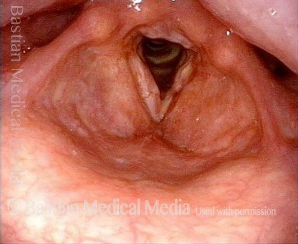
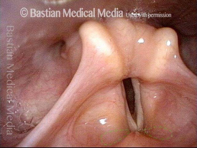
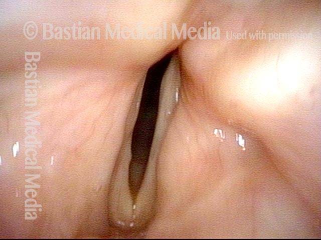
Vocal cords approaching point of best closure possible (due to left cord paresis). Faint dotted lines outline the approximate boundary of each laryngeal saccule, which not yet inflated.
Vocal cords approaching point of best closure possible (due to left cord paresis). Faint dotted lines outline the approximate boundary of each laryngeal saccule, which not yet inflated.
Bilateral laryngocele (2 of 8)
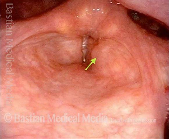
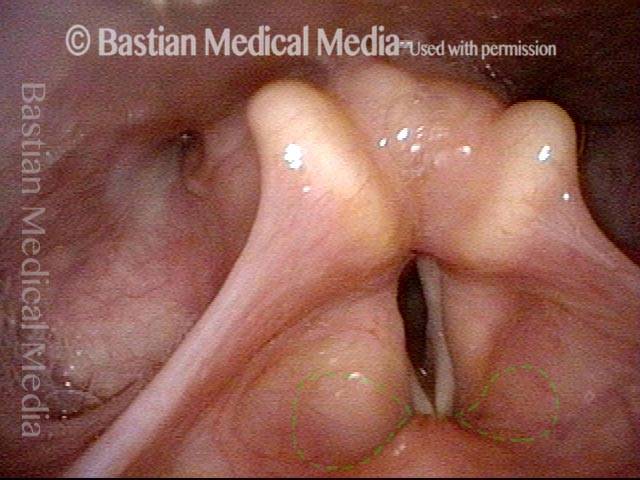
As air just begins coming upward between the cords, one can see subtle inflation (dotted lines), particularly of the right saccule (left of image).
As air just begins coming upward between the cords, one can see subtle inflation (dotted lines), particularly of the right saccule (left of image).
Bilateral laryngocele (3 of 8)
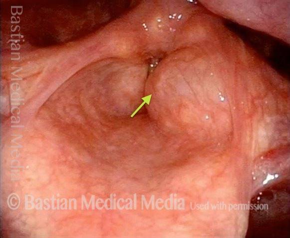
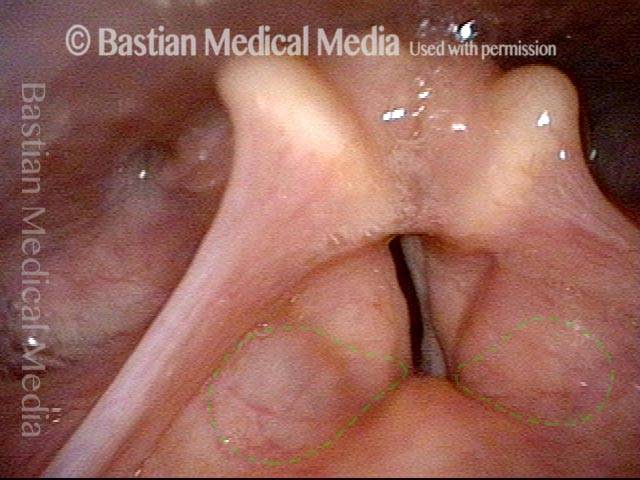
As phonation continues, inflation of the (now diagnosable) laryngocele becomes obvious, and the left laryngocele (right of image) is now more obviously inflated than before, again indicated by the dotted lines.
As phonation continues, inflation of the (now diagnosable) laryngocele becomes obvious, and the left laryngocele (right of image) is now more obviously inflated than before, again indicated by the dotted lines.
Bilateral laryngocele (4 of 8)
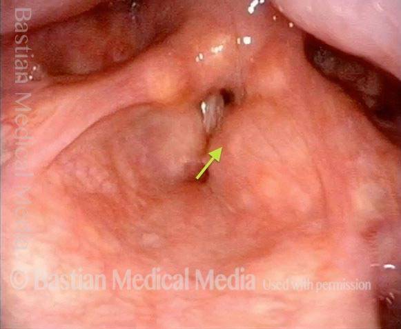
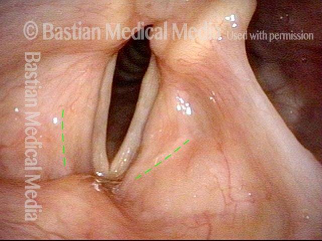
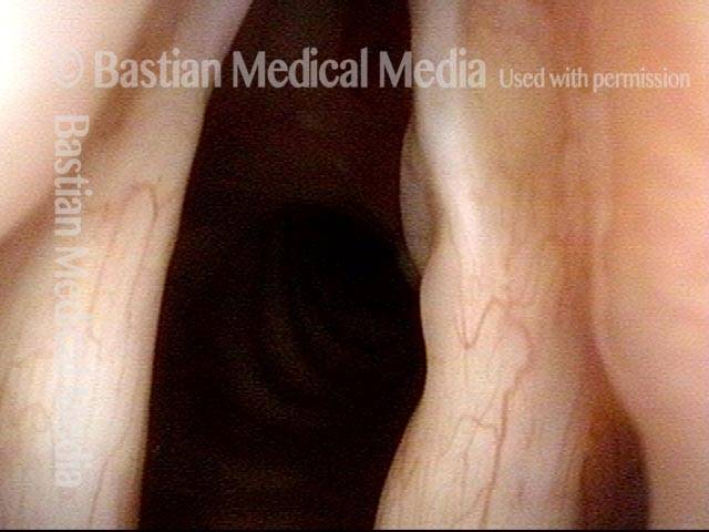
Near the end of a sustained period of voicing, maximum inflation of the laryngoceles is seen (dotted lines). On the right side (left of image), the stretching mucosa is so thinned as to appear translucent.
Near the end of a sustained period of voicing, maximum inflation of the laryngoceles is seen (dotted lines). On the right side (left of image), the stretching mucosa is so thinned as to appear translucent.
Bilateral laryngocele, after removal (5 of 8)
Same patient, breathing position, 12 weeks after complete removal of the bilateral laryngoceles via false cord incisions (lines of incision shown by dotted lines). This patient also has long-standing paralysis of the right vocal cord (left of image) and limited mobility of the left cord, so the cords don’t open fully for breathing.
Same patient, breathing position, 12 weeks after complete removal of the bilateral laryngoceles via false cord incisions (lines of incision shown by dotted lines). This patient also has long-standing paralysis of the right vocal cord (left of image) and limited mobility of the left cord, so the cords don’t open fully for breathing.
Bilateral laryngocele, after removal (6 of 8)
Phonatory position. Note the lack of inflation of the now-absent laryngoceles, and compare that with photos 3 and 4 of this series.
Phonatory position. Note the lack of inflation of the now-absent laryngoceles, and compare that with photos 3 and 4 of this series.
Bilateral laryngocele, after removal (7 of 8)
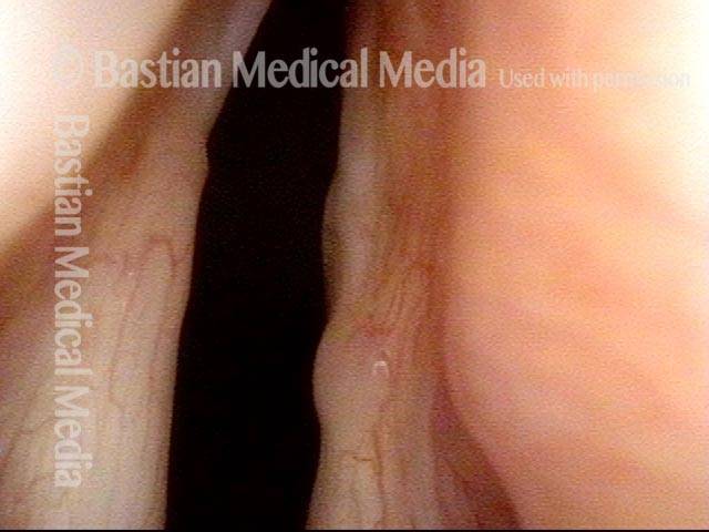
Closer view of the posterior ends of the true vocal cords during maximal abduction for breathing. Space between the vocal cords is an estimated 50% of normal, because of the paralyzed right cord and the limited mobility of the left cord.
Closer view of the posterior ends of the true vocal cords during maximal abduction for breathing. Space between the vocal cords is an estimated 50% of normal, because of the paralyzed right cord and the limited mobility of the left cord.
Bilateral laryngocele, after removal (8 of 8)
Same close-up view, but during phonation. The left vocal cord (right of image) has shifted slightly toward the midline, but the cords do not actually close and, thus, the patient cannot produce glottic (true vocal cord) voice. An implant could help to close this gap, but the patient will first try developing a “false cord voice.”
Same close-up view, but during phonation. The left vocal cord (right of image) has shifted slightly toward the midline, but the cords do not actually close and, thus, the patient cannot produce glottic (true vocal cord) voice. An implant could help to close this gap, but the patient will first try developing a “false cord voice.”
A laryngocele is a disorder of the saccule, or laryngeal appendix, in which air abnormally expands it. Watch this video to see how a laryngocele behaves in real-time, and why that can affect the voice.


