The use of only two narrow wave bands of light (blue and green) during an endoscopic examination, so as to make blood vessels and other tissues in the mucosa more visible. Blood vessels become more visible under narrow-band illumination because both of the wave bands are easily absorbed by hemoglobin in the blood. In addition, the blue and green wave bands in narrow-band illumination each penetrate the tissue to differing degrees, so that blood vessels near the mucosal surface appear as a different color from blood vessels deeper in the tissue.
In the field of laryngology, narrow-band illumination can help an examining clinician to identify the vascular changes that characterize a range of disorders, including recurrent respiratory papillomatosis, capillary ectasia, glottic sulcus, ulcerative laryngitis, and others. It can also help to identify subtle, hazy leukoplakia. The technology, developed by Olympus, is officially called Narrow Band Imaging™. KayPentax offers a different, software-based method for highlighting vascularity and other tissue characteristics, which is called iScan™.
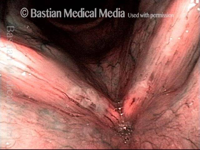
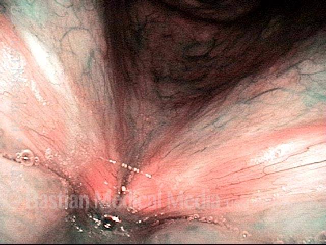
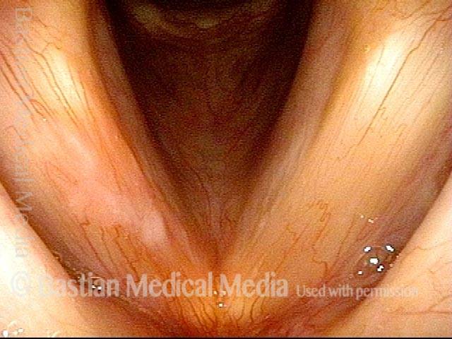
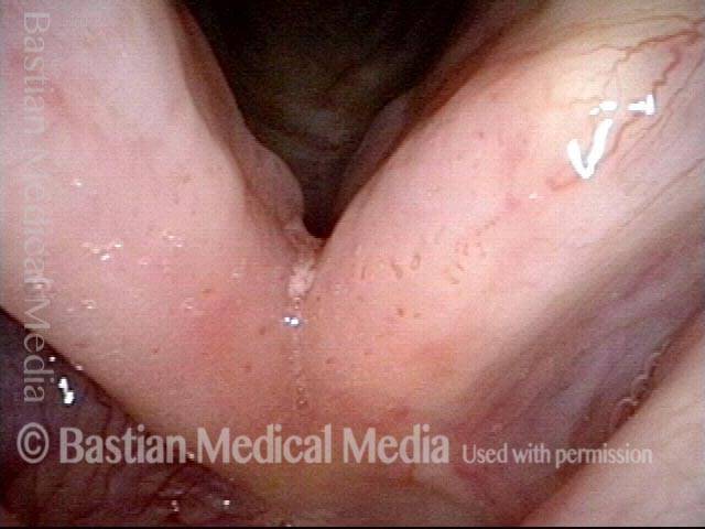
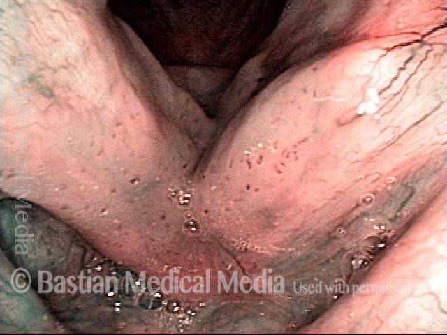
Narrow-band vs. Standard Light
Narrow-band vs. standard light (1 of 2)
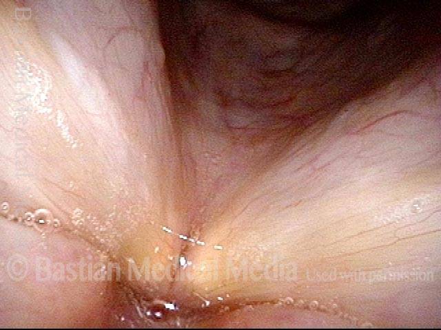
Narrow-band vs. standard light (2 of 2)
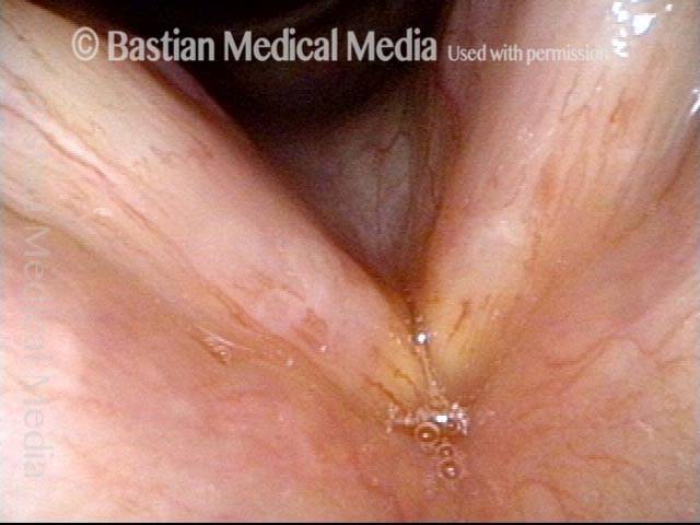
Narrow-band vs. Standard Light: Leukoplakia
Narrow-band vs. standard light: leukoplakia (1 of 2)
Narrow-band vs. standard light: leukoplakia (2 of 2)
Example 2
Narrow-band vs. standard light: leukoplakia (1 of 2)

Narrow-band vs. standard light: leukoplakia (2 of 2)
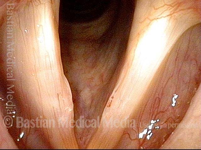
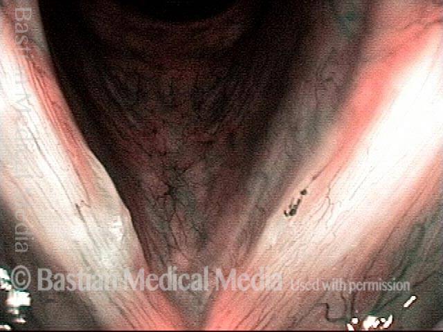
Narrow-band vs. Standard Light: Capillary Ectasia
Narrow-band vs. standard light: capillary ectasia (1 of 2)
Narrow-band vs. standard light: capillary ectasia (2 of 2)
Narrow-band vs. Standard Light: HPV-induced Lesions
Narrow-band vs. standard light: HPV-induced lesions (1 of 2)
Narrow-band vs. standard light: HPV-induced lesions (2 of 2)
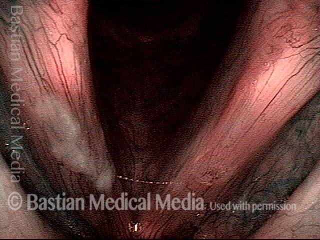
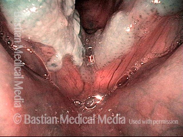
Narrow-band vs. Standard Light: Candida
Narrow-band vs. standard light: candida (1 of 2)
Narrow-band vs. standard light: candida (2 of 2)