SLAD-R (Selective laryngeal adductor denervation-reinnervation) is a surgical option for adductory spasmodic dysphonia. This procedure was introduced by Dr. Gerald Berke of UCLA in the late 1990’s. The concept is to sever the anterior branch of the recurrent laryngeal nerve. This denervates the spasming laryngeal adductors (particularly thyroarytenoid and lateral cricoarytenoid muscles).
The squeezed, strained quality and/ or “catching, cutting out, stopping” of the voice are replaced initially with an extremely breathy and weak voice. This initially weak voice is analogous to what one might sound like after a Botox injection that is far too high a dose.
To return strength to the voice, a branch of the ansa cervicalis nerve that normally supplies some relatively “unimportant” neck muscles is anastomosed (connected) to the severed nerve. It takes 3 months to a year for tone to begin to return to the adductory muscles. Since the “unimportant” neck muscles were not affected by the dystonia, the hope is that the new nerve supply to the laryngeal muscles may not be affected by dystonia.
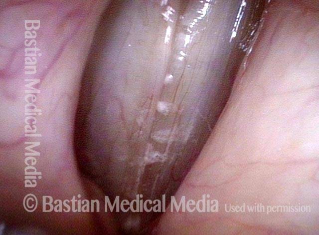
Six years post SLAD-R (1 of 4)
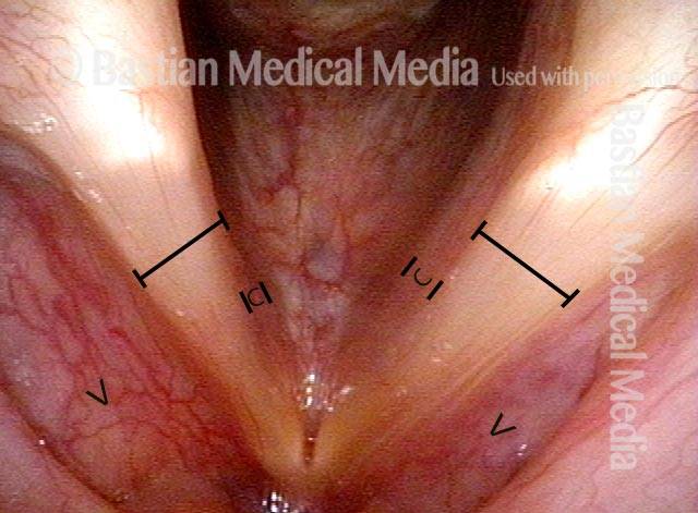
TA + LCA muscles (2 of 4)
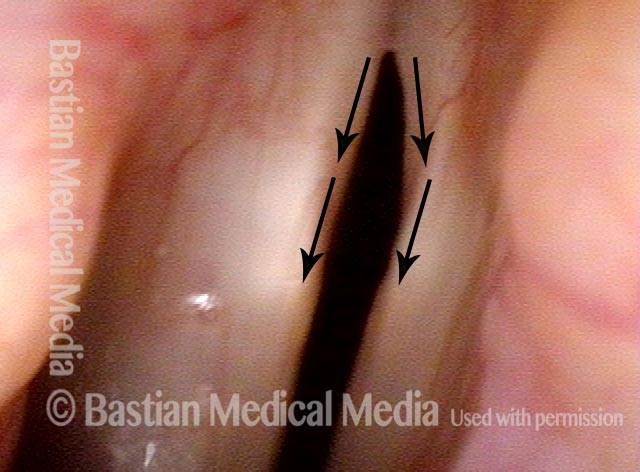
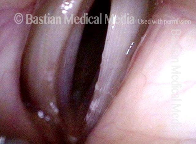
Greater amplitude of right cord (3 of 4)
Patient has returned to Botox (4 of 4)