A rare inflammatory disease of small arteries and veins (vasculitis) that classically involves vessels supplying the tissues of the lungs, nasal passages (sinuses), and kidneys.
At our practice, we see a large population of individuals with what we believe to be a forme fruste (incomplete expression) of Wegener’s granulomatosis* causing subglottic and tracheal stenosis.
*Newer terminology is granulomatosis with polyangiitis (GPA)
Subglottic Stenosis, Due to Wegener’s Granulomatosis
Subglottic stenosis, due to Wegener’s (1 of 2)
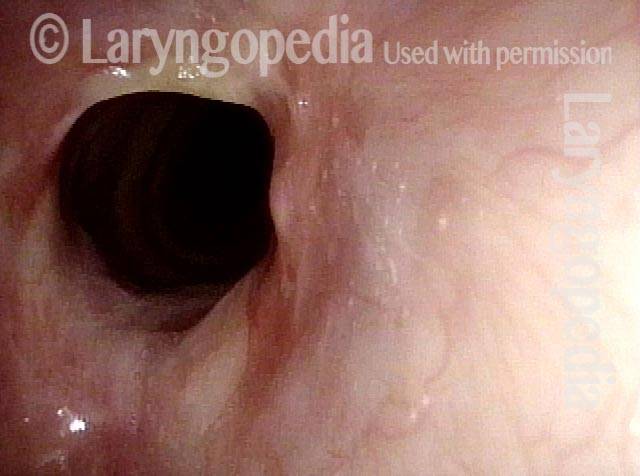
This person has Wegener’s granulomatosis, confirmed by anti-neutrophil cytoplasmic antibodies (ANCA) testing. Here, looking from above the vocal cords, one can see an estimated 50% narrowing of the subglottic and high tracheal passageway.
Subglottic stenosis, due to Wegener’s (2 of 2)
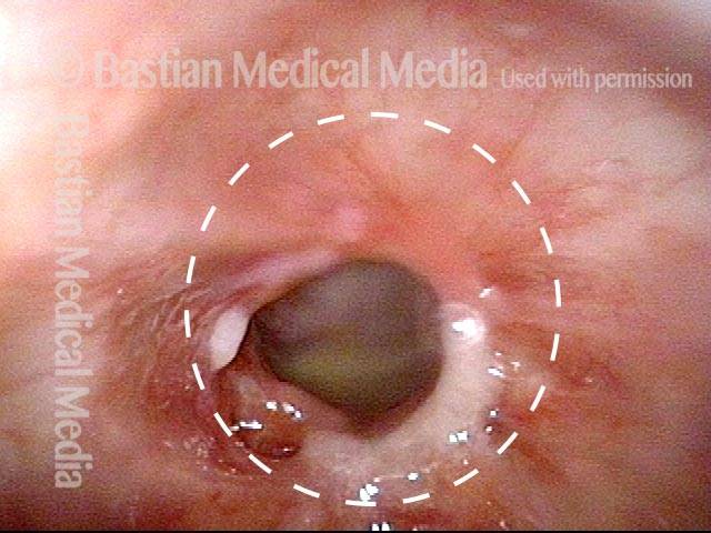
Viewed from within the subglottis, one can see more clearly the inflammatory nature of this stenosis. A dotted oval estimates what the normal caliber or width of this airway would be. This patient has been managed with systemic medication, but also occasional dilation, steroid injection, and Mitomycin C application.
Airway Stenosis Caused By Wegener’s Granulomatosis, Before and After Dilations
Airway stenosis (1 of 5)
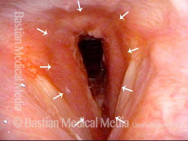
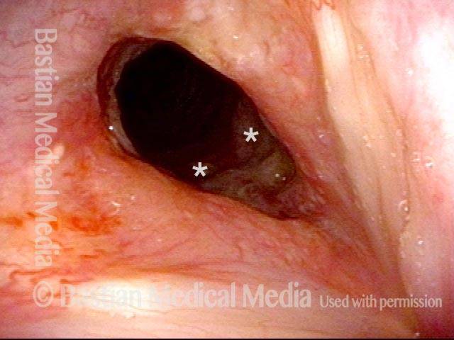
Marked inflammatory narrowing in the immediate subglottis. Within the ring of arrows is the inflamed, reddened tissue, which is narrowing the airway into the shape of a slit. This man needs to be active for his work, but notices shortness of breath and noisy breathing with exertion.
Airway stenosis, after dilation (2 of 5)
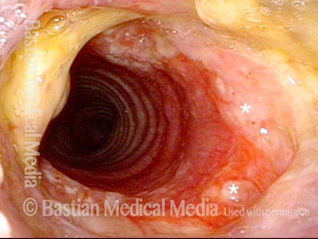
Nine days after a dilation procedure, with local steroid injection and painting with Mitomycin C. The airway has widened, so that it is more oval-shaped and less slit-like. Compare with photo 1. Although a degree of stenosis remains, symptoms have subsided dramatically. For reference, asterisks mark the same points in the subglottis in this photo and the next photo.
Airway stenosis, after dilation (3 of 5)
Same exam, looking beyond the immediate subglottis. There is an inflammatory response that involves several centimeters of the upper trachea. Inflammatory areas often “trap” mucus, as seen here.
Airway stenosis, before another dilation (4 of 5)
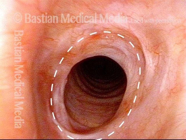
Now five months after the dilation procedure mentioned in photos 2 and 3. The patient has been receiving systemic treatment with methotrexate and prednisone. General appearance of the inflammation has decreased. In spite of this, as expected, the stenosis has persisted (dotted oval shows the estimated caliber or width of a normal airway) and symptoms have gradually increased. Thus, another dilation was scheduled for the next day.
Airway stenosis, after another dilation (5 of 5)
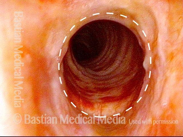
A week after photo 4, following the most recent dilation. There is expected immediate postoperative inflammation and an increase in the airway’s caliber or width by an estimated 30% (dotted oval again shows the estimated caliber or width of a normal airway; compare with photo 4). Symptoms are again abolished.
Vascular Manifestations of Wegener’s-related Septum Changes, and Subglottic Stenosis Indistinguishable from Forme Fruste Wegener’s
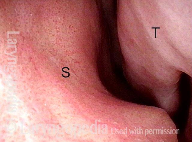
View inside left nostril (1 of 4)
This man has Wegener’s Granulomatosis, with sino-nasal, subglottic, and pulmonary effects, and is on immunosuppressive therapy with very good clinical results. This view is just inside his left nostril and our focus – seen better in the next photo – is the stippled vascular pattern sometimes seen in auto-immune disorders. (S = septum, and T = inferior turnbinate.)
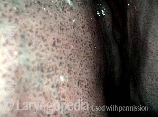
Narrow band light (2 of 4)
Under narrow band light, the unusual vascular pattern of both septum and turbinate becomes much more obvious.
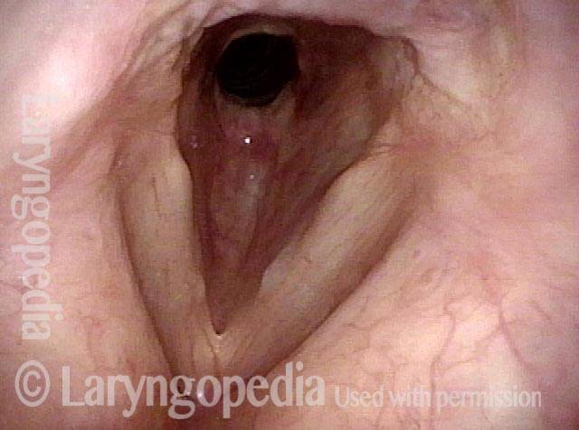
Distant view (3 of 4)
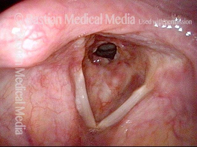
In this distant view, his subglottic stenosis looks just like the many other examples in Laryngopedia of forme fruste Wegnener’s. The stenosis seen with both entities are visually indistinguishable.
Closer view (4 of 4)
A closer view of the stenosis reveals more clearly the adherent mucus that is so difficult for such patients to cough out due to the “speed bump” interruption of the mucociliary blanket at the stenosis.