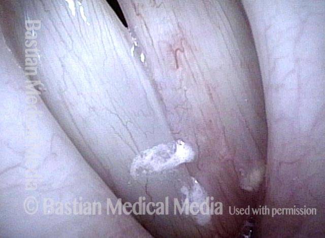
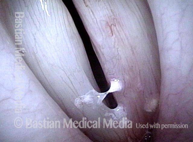
Pedunculated, meaning attached by a stalk; the opposite of sessile.
Pedunculation Defined in Pictures
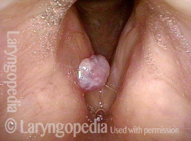
Polyp (1 of 7)
This large polyp resulted from an episode of extremely aggressive voice use six months earlier. In this photo, one cannot tell if the point of attachment covers the same area of the circumference of the lesion, or if it is smaller.
Polyp (1 of 7)
This large polyp resulted from an episode of extremely aggressive voice use six months earlier. In this photo, one cannot tell if the point of attachment covers the same area of the circumference of the lesion, or if it is smaller.
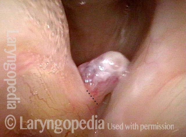
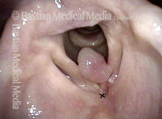
Inspiration (2 of 7)
Here, the examiner has elicited rapid inspiration. The rush of air inward pulls the polyp inward and downward, revealing its stalk or peduncle. The attachment is indicated by the dotted line.
Inspiration (2 of 7)
Here, the examiner has elicited rapid inspiration. The rush of air inward pulls the polyp inward and downward, revealing its stalk or peduncle. The attachment is indicated by the dotted line.
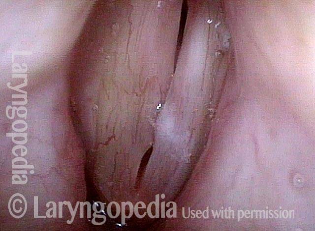
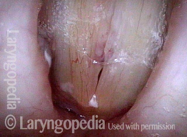
Closed position (3 of 7)
The vocal cords are closed while continuing to draw air in. The polyp is now hidden below the point of closure of the cords.
Closed position (3 of 7)
The vocal cords are closed while continuing to draw air in. The polyp is now hidden below the point of closure of the cords.
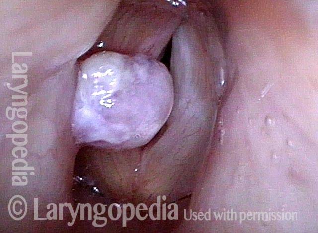
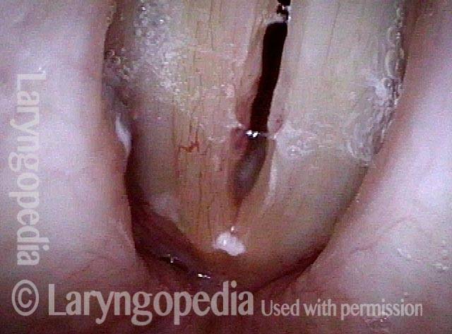
Phonatory view (4 of 7)
When voice is produced, the polyp flips upwards between the colds and now lies on the upper surface of the vocal colds. None of this movement could happen if the polyp were not pedunculated.
Phonatory view (4 of 7)
When voice is produced, the polyp flips upwards between the colds and now lies on the upper surface of the vocal colds. None of this movement could happen if the polyp were not pedunculated.
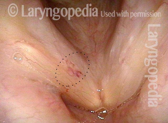
After removal (5 of 7)
A week after removal of the polyp. Compare with photo 1 to see that the “wound” (area of vascularity) is far smaller than the diameter of the original polyp (dotted line), showing in a second way the idea that the attachment had “pinched in” to a stalk.
After removal (5 of 7)
A week after removal of the polyp. Compare with photo 1 to see that the “wound” (area of vascularity) is far smaller than the diameter of the original polyp (dotted line), showing in a second way the idea that the attachment had “pinched in” to a stalk.
Vocal Cord “Tear” and Granuloma
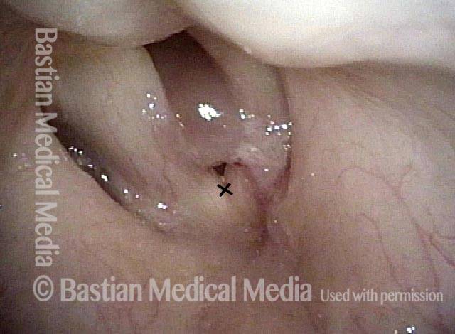
Intubation injury (1 of 8)
Gross hoarseness was immediately evident after a surgical procedure involving endotracheal intubation. After a few months, this granuloma is evident. It appears to be pedunculated and attached only where indicated by the dotted line. The small "X" is for reference with photos 2 and 4.
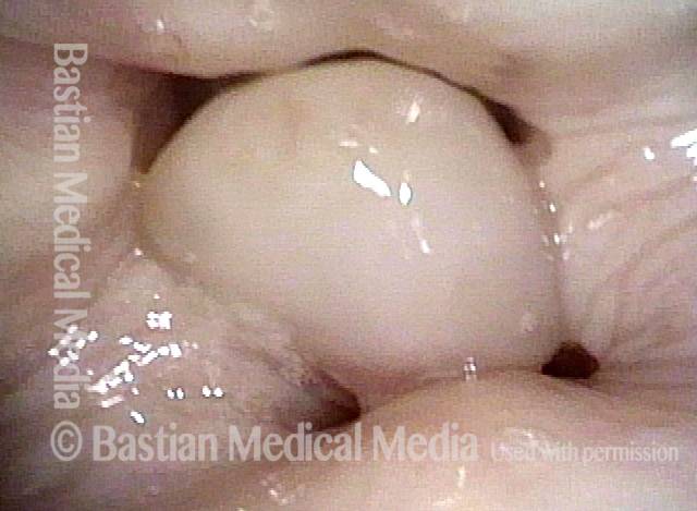
Granuloma drawn into glottis (2 of 8)
Here, the granuloma is drawn downward into the glottis by the inspiratory airstream. The "X" is for reference with photos 1 and 4.
Granuloma drawn into glottis (2 of 8)
Here, the granuloma is drawn downward into the glottis by the inspiratory airstream. The "X" is for reference with photos 1 and 4.
Phonation (3 of 8)
During phonation, seen at closer range, the granuloma rides upward and nearly fills the laryngeal vestibule.
Phonation (3 of 8)
During phonation, seen at closer range, the granuloma rides upward and nearly fills the laryngeal vestibule.
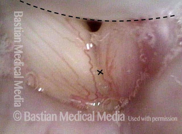
Anterior commissure (4 of 8)
Closeup at the anterior commissure. Dotted line indicates anterior edge of the granuloma. The "X" is for reference with photos 1 and 2.
Anterior commissure (4 of 8)
Closeup at the anterior commissure. Dotted line indicates anterior edge of the granuloma. The "X" is for reference with photos 1 and 2.
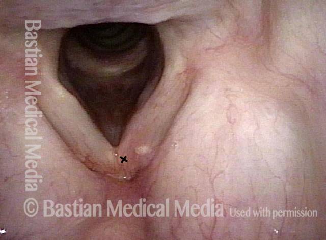
Granuloma detached (5 of 8)
A few months later, voice has improved. The granuloma has spontaneously detached. Pinkness remains.
Granuloma detached (5 of 8)
A few months later, voice has improved. The granuloma has spontaneously detached. Pinkness remains.
Vocal cord blurring (6 of 8)
During voicing under standard light, note that there is vocal cord blurring on the right cord (left of photo) far more than on the left (blurring is indicated by thin, black lines). This suggests that the left side (right of photo) is not vibrating well.
Vocal cord blurring (6 of 8)
During voicing under standard light, note that there is vocal cord blurring on the right cord (left of photo) far more than on the left (blurring is indicated by thin, black lines). This suggests that the left side (right of photo) is not vibrating well.
Closed phase (7 of 8)
Low in the female range, at A3 (220 Hz), closed phase of vibration.
Closed phase (7 of 8)
Low in the female range, at A3 (220 Hz), closed phase of vibration.
Open phase (8 of 8)
Open phase of vibration at the same pitch, showing that only the right cord (left of photo) vibrates, because the tear of the left cord has scarred and stiffened it.
Open phase (8 of 8)
Open phase of vibration at the same pitch, showing that only the right cord (left of photo) vibrates, because the tear of the left cord has scarred and stiffened it.
Tagged Diagnosis, Other useful terms
