An encyclopedia about voice, swallowing, airway, coughing, & other head + neck disorders.

Bronchoscopy

Bronchoscopy is a procedure that directly visualizes the trachea (windpipe) and bronchial tree using a flexible or rigid bronchoscope. The larynx is also visualized in passing.

2 Types of Bronchoscopy

Flexible Bronchoscopy

  • Performed most often by pulmonologists in a procedure suite or operating room, typically with topical anesthesia and intravenous sedation.
  • In ENT/laryngology practice, for screening and functional evaluation, flexible distal chip scopes allow awake, office-based examination of the larynx, subglottis, trachea, and mainstem bronchi, using only topical anesthesia.
  • Screening bronchoscopy by laryngologists is particularly useful for dynamic functional assessment (e.g., observing airway collapse, secretion clearance, or cough response), and also to assess subglottic and tracheal stenosis.

Rigid Bronchoscopy (typically by thoracic surgeons and laryngologists when needed)

  • Performed less frequently in the modern era since flexible bronchoscopes now provide excellent visualization and working channels.
  • Indicated when a larger working channel is required, such as for foreign body removal or certain therapeutic interventions.
  • Requires general anesthesia in the operating room.

Indications

Bronchoscopy may be performed to:

Technique

Flexible Bronchoscopy (Office-based, dynamic or screening assessment by a laryngologist):

  1. Topical anesthesia is applied to nasal passages, larynx, subglottis, and trachea.
  2. With the patient seated, the scope is passed transnasally, through the vocal cords, into the trachea and mainstem bronchi.
  3. The examiner can prompt maneuvers (rapid inspiration, coughing) to evaluate airway motion and secretions.

Flexible Bronchoscopy (Procedure room, formal diagnostic/therapeutic use by a pulmonologist):

  1. Topical anesthesia applied to mouth, throat, larynx, trachea, and bronchi.
  2. The patient lies on a stretcher under IV sedation.
  3. Secretions may be suctioned for culture, biopsies obtained, and foreign bodies or lesions removed.

Risks

  • Common: mild discomfort, coughing, gagging.
  • Rare: bleeding, transient laryngospasm, or airway irritation.

Summary

Bronchoscopy is an essential tool in both laryngology and pulmonology, allowing direct evaluation of airway structure and function, with or without therapeutic intervention (suction, biopsy, dilation, etc.). Office-based flexible bronchoscopy offers quick, safe, dynamic assessment of stenosis, functional collapse, and nonorganic airway disorders, while carrying minimal risk to patients.

Photo Essay of Bronchoscopy

Wheezing, Induced by Bronchial Abnormality

Visual Portfolio, Posts & Image Gallery for WordPress

Wheezing, induced by bronchial abnormality (1 of 5)

This patient has noticed faint wheezing for most of her life. Here is a panorama of the entrance to her larynx, which is normal—wide open and unobstructed—and does not yet explain her wheezing. For reference, an asterisk marks the upper surface of the cricoid ring.

Wheezing, induced by bronchial abnormality (2 of 5)

Further down, now just below the vocal cords; an asterisk again indicates the anterior cricoid ring. The upper tracheal rings here are normal. Still no explanation for her wheezing.

Wheezing, induced by bronchial abnormality (3 of 5)

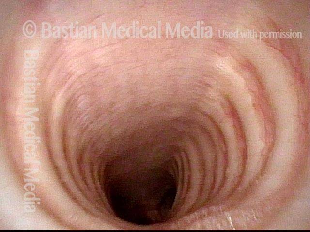
Further down yet, now into the mid-trachea. Still no sign of any anatomical cause for the wheezing.

Wheezing, induced by bronchial abnormality (4 of 5)

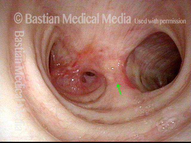
Further down still, viewing the distal trachea; an arrow marks the carina. The source of the wheezing now becomes evident: the take-off of the right main stem bronchus (left of image) is extremely abnormal and narrowed. The left main stem bronchus appears to have a subtle stenosis, which is a red herring.

Wheezing, induced by bronchial abnormality (5 of 5)

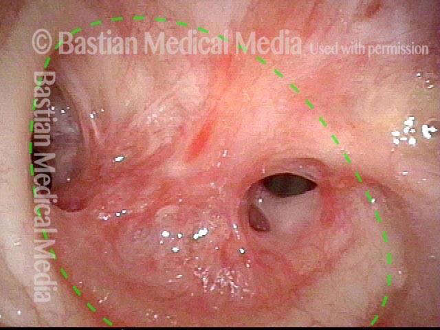
Close-up on the abnormal take-off for the right main stem bronchus, which should be wide open. Instead, there are accessory bronchi exiting what is probably a congenital malformation. A dotted line indicates the expected size of this passageway, were it not malformed.

Tracheoesophageal Party Wall with Wheezing

Visual Portfolio, Posts & Image Gallery for WordPress

Abducted breathing position (1 of 5)

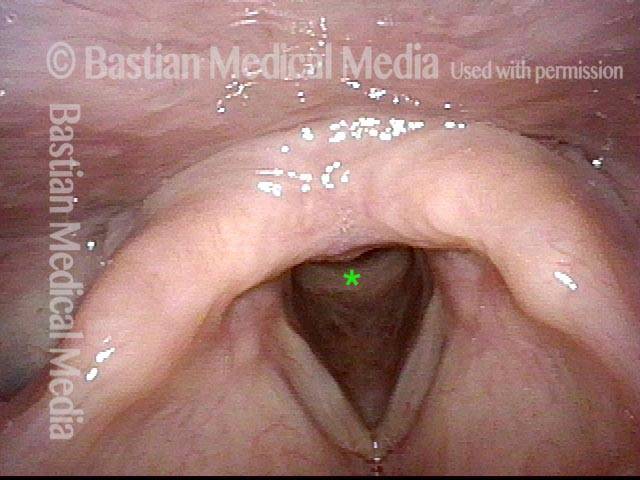
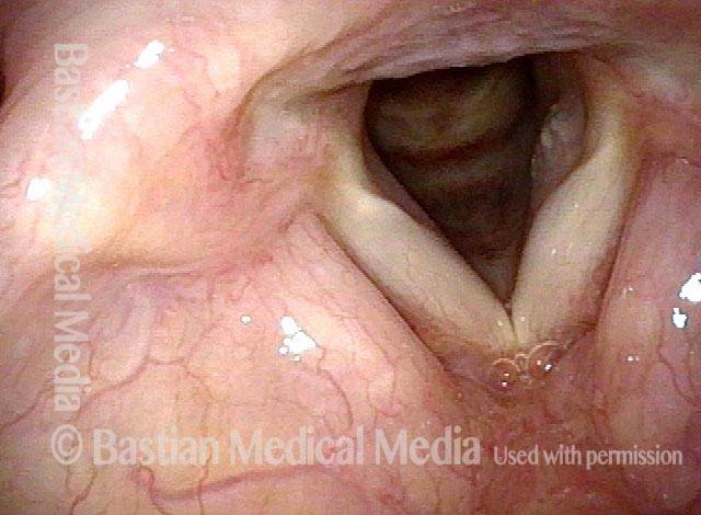
Normal laryngeal entrance, with vocal cords in abducted (breathing) position.

View of mid-trachea (2 of 5)

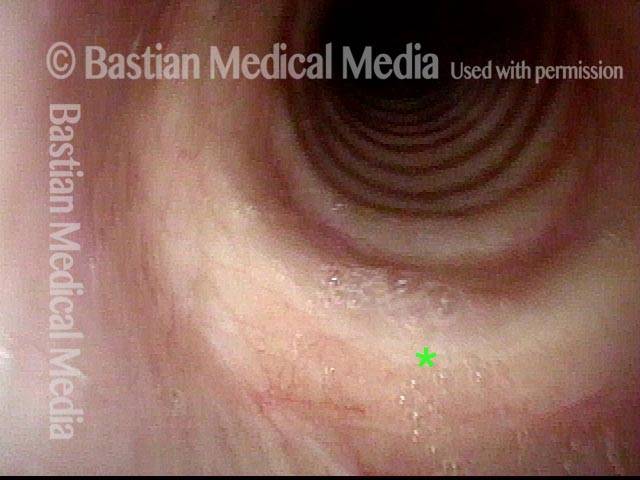
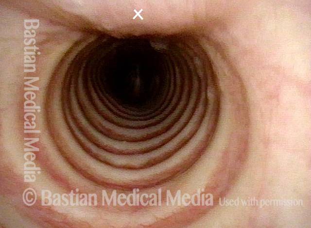
View in normal mid-trachea. Note that the cartilaginous rings make up approximately 2/3 of the circumference and that the membranous trachea (upper photo at ‘X’) is more flat.

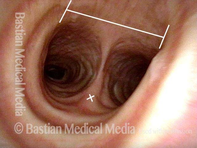
View just above the carina (3 of 5)

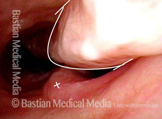
View just above the carina, where the distal trachea splits into left and right mainstem bronchi. Anterior take-off of carina at the ‘X’. The straight line delimits the membranous (flexible) tracheal wall.

Wheezing begins (4 of 5)

With Valsalva maneuver to accentuate patient’s functional expiratory wheezing. Note that the membranous tracheal and bronchial walls bulge inward on a functional basis to narrow the airway. Wheezing begins to be heard. The ‘X’ again marks the anterior take-off of the carina. Compare with Photo 3.

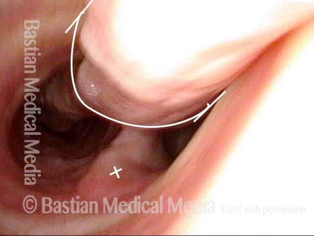
Left bronchus blocked (5 of 5)

As bulging inward continues, the left mainstem bronchus is particularly blocked. This explains why, on auscultation of the chest, wheezing sounds louder on the left than the right. Compare with photos 3 and 4.

Upper Airway Wheezing, as a Kind of “Skill”

Visual Portfolio, Posts & Image Gallery for WordPress

At the level of the vocal cords (1 of 8)

Vocal cords, in breathing position, in a person with wheezing who has been diagnosed elsewhere with asthma. Nothing seen here to explain the wheezing.

In the upper trachea: Valsalva maneuver (2 of 8)

Now descended into the upper trachea. As the patient performs (upon request) a semi-Valsalva manuever, one can see mild inward bulging of the membranous trachea (top- left of photo). This is not sufficient to explain the patient’s wheezing.

In the mid-trachea: quiet breathing (3 of 8)

Descended further, into the mid-trachea, with the carina (dotted line) seen in the distance, as the patient breathes quietly.

In the mid-trachea: Valsalva maneuver (3 of 8)

Same view as photo 3, but during another semi-Valsalva maneuver. The membranous tracheal wall bulges inward again, but much more noticeably here than in the upper trachea (photo 2), especially down by the carina (which is now hidden from view).

In the lower trachea: quiet breathing (5 of 8)

Down further yet, almost to the level of the carina (dotted line). On each side is the entrance to each of the mainstream bronchi.

In the lower trachea: Valsalva maneuver (6 of 8)

Same view as photo 5, but during another semi-Valsalva maneuver. The membranous tracheal wall bulges inward again to partly block the airway, especially the right mainstem bronchus. This patient’s wheezing sounds loudest over the sternal notch and manubrium; softer wheezing is also heard in the distal lung fields, and as expected from this photo, is louder on the right than the left.

In the right mainstem bronchus: quiet breathing (7 of 8)

Further down yet, now looking directly into the right mainstem bronchus.

In the right mainstem bronchus: Valsalva maneuver (8 of 8)

Same view as photo 7, but during another semi-Valsalva maneuver. Note compression as well of lobar bronchi, also partly responsible for the patient’s wheezing.

Share this article

Voice, Swallowing, Airway Disorder?

Still have questions? Schedule a call
with Dr. Bastian via Zoom.

Table of Contents