The esophagus is the passageway that connects the throat or pharynx to the stomach. Technically, it begins at the upper esophageal sphincter and ends at the lower esophageal sphincter.
Three Views of the Entrance to the Esophagus From Far Away to Close-up
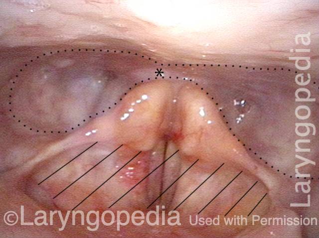
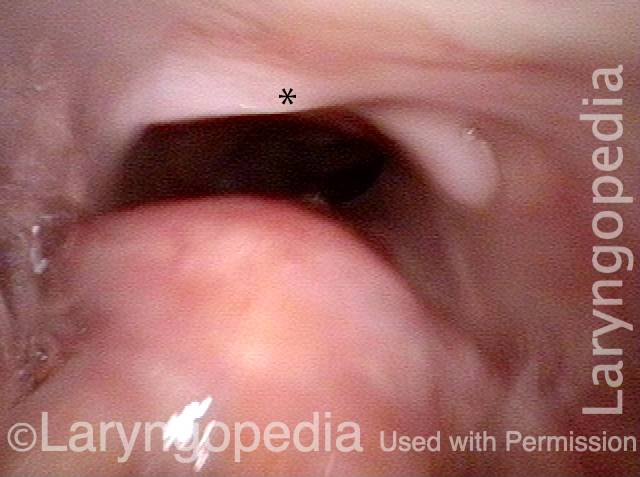
Swallowing Crescent (1 of 3)
During swallowing, the “the swallowing crescent”—outlined by the dotted line—receives swallowed food or liquid in order to funnel it into the esophagus (not open in this view). The asterisks are reference points to compare all three photos. One does not want any material to enter the laryngeal vestibule (hashed lines).
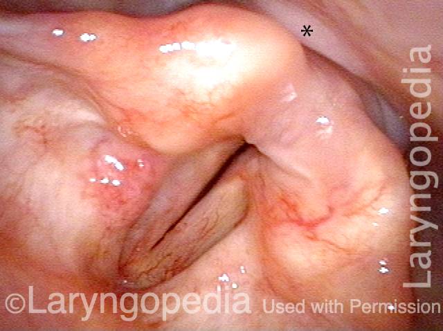
Closed esophagus (2 of 3)
A closer view. The esophagus is still not open in this view. Compare asterisk with prior and following photo.
Open Esophagus (3 of 3)
At the moment of a dry swallow, the esophagus opens as shown here. Again, the asterisks allow comparison with photos 1 and 2.
Esophagus, After Total Laryngectomy
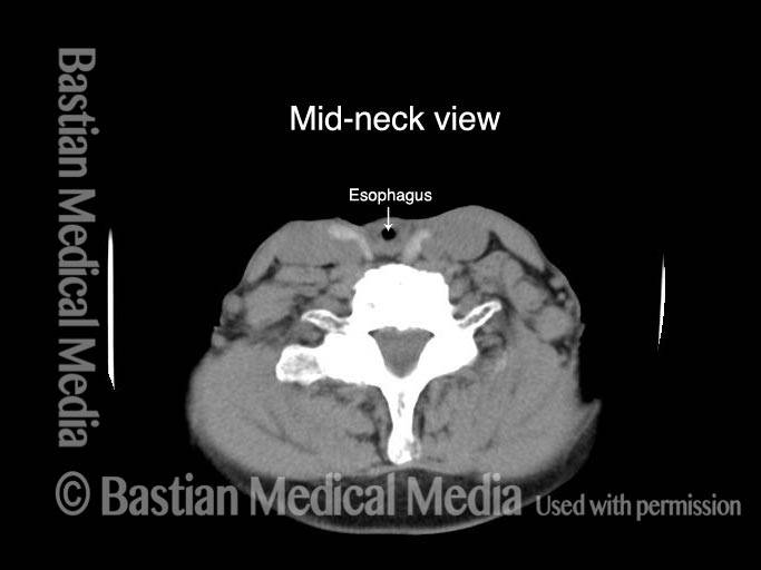
Esophagus, after total laryngectomy (1 of 4)
After total laryngectomy. At the level where larynx was, there is no airway seen. The small air collection (at arrow) is within the reconstructed esophagus, at the level of the neck.
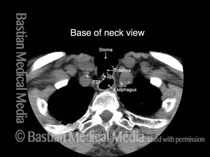
Esophagus, after total laryngectomy (2 of 4)
At the base of neck, the trachea has been sutured forward to neck skin. In this view, the tracheoesophageal prosthesis (TEP) can be seen traversing the shared tracheoesophageal party wall. In this view, the esophagus is very dilated with air.
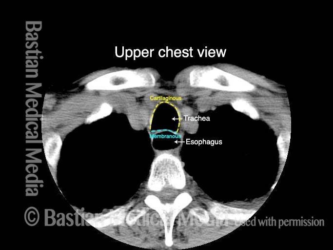
Esophagus, after total laryngectomy (3 of 4)
Below the tracheostome, the entire trachea is again seen. The horseshoe-shaped anterior segment of the trachea’s wall, two-thirds of the total circumference, is the trachea’ s cartilaginous component. The posterior one-third is the membranous trachea, which also constitutes the anterior one-third of the esophagus, and is also called the tracheoesophageal party wall. The esophagus is again dilated with air here.
Esophagus, after total laryngectomy (4 of 4)
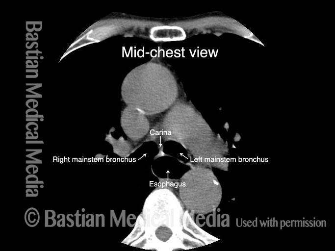
At the mid-thoracic level, the trachea is splitting into the two mainstem bronchi, and the esophagus is still seen due to dilation with swallowed air.
Endoscopic View of Esophageal (Acid) Reflux
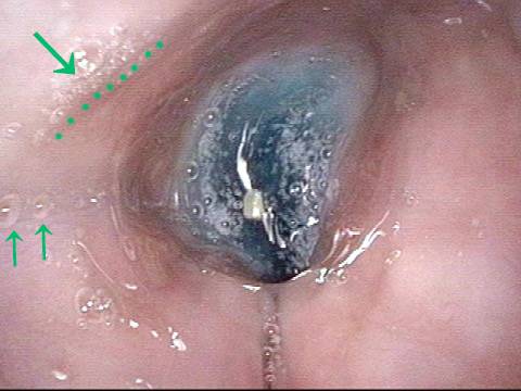
Liquid in the lower esophagus (1 of 2)
After swallowing blue food-colored water, it sits momentarily in the lower esophagus waiting to enter the stomach. The saliva bubbles indicated by arrow and dotted lines are for reference with the next photo.
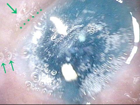
Acid reflux in the lower esophagus (2 of 2)
A moment later, without effort or gag, the blue water refluxes (zooms upwards from the lower esophagus and stomach) towards the stationary camera chip. If this occurred with acidic stomach contents, the esophagus would suffer chemical irritation and the patient might experience “heartburn.”
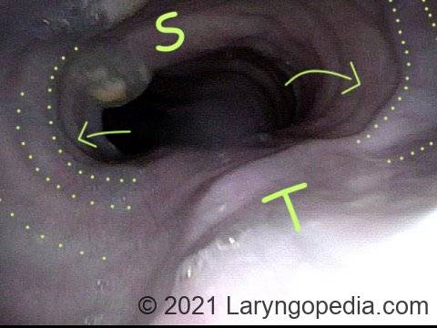
Dramatic Dilation of the Esophagus in A Person with R-CPD Due to Buildup of Swallowed Air that He Cannot Belch to Get Rid Of
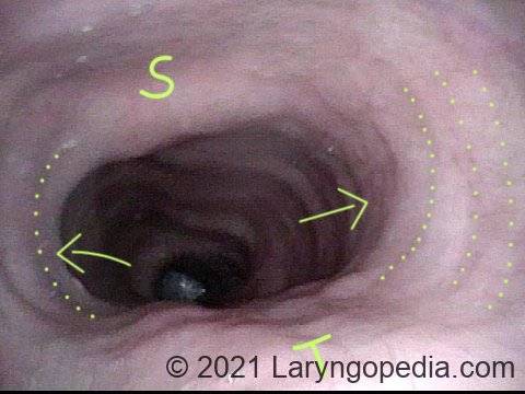
View of the mid-esophagus (1 of 2)
This view in the mid-esophagus was obtained with a 3.6mm scope without an air channel. The dilation is from the patient’s own unbelchable air. Note quite major lateral dilation of the esophagus, indicated by concentric dotted lines and arrows. Dilation is not possible in the direction of unyielding spine (S) and trachea (T).
View of the mid-esophagus (2 of 2)
A view that shows more clearly the indentation of trachea (T). Persons with this much dilation of esophagus often complain as much of chest pressure as they do abdominal bloating. This man has experienced “large” reduction of R-CPD symptoms after botulinum toxin injection into his upper esophageal sphincter (cricopharyngeus muscle).