Idiopathic Subglottic Stenosis (Inflammatory Stenosis)

Idiopathic (unknown cause) subglottic stenosis is narrowing in a lumen or passageway caused by an inflammatory process. It is a frustrating breathing problem seen mostly in women. It appears to be an autoimmune disorder that we have thought for many years to be an incomplete expression (forme fruste) of Wegener’s Granulomatosis, aka Granulomatosis with Polyangiitis (GPA).

Treatment

In a caseload of perhaps 125 patients, it appears that standard of care remains occasional outpatient dilation, steroid injection, and mitomycin C application. Most patients go a year or more between dilations.

The rub: occasionally, dilations need to be done more frequently in an “active” case, or there may be reasons to avoid the brief general anesthesia required for dilation such as: morbid obesity, difficult jaw/tooth/neck anatomy, or even a phobia of general anesthesia.

Visual Portfolio, Posts & Image Gallery for WordPress

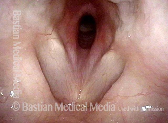
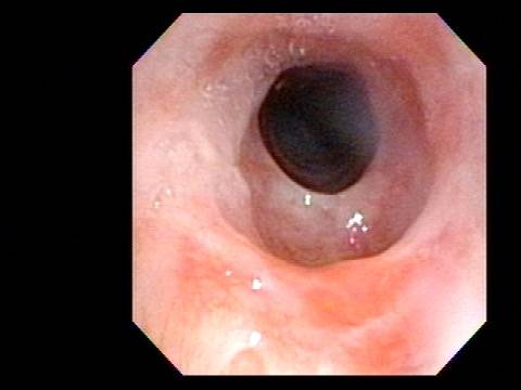
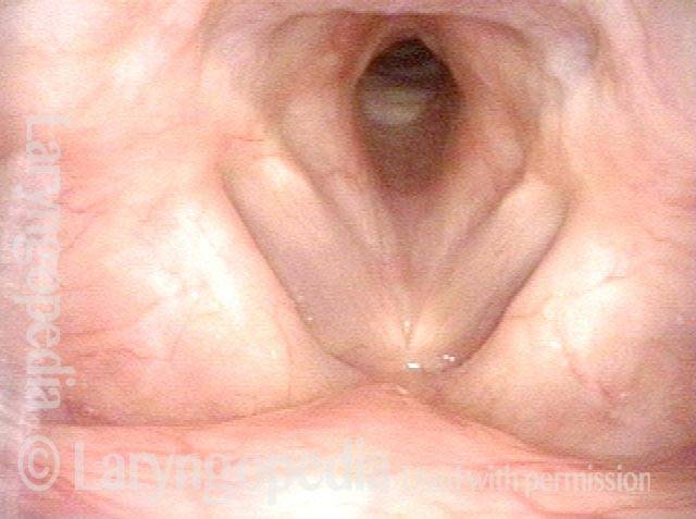
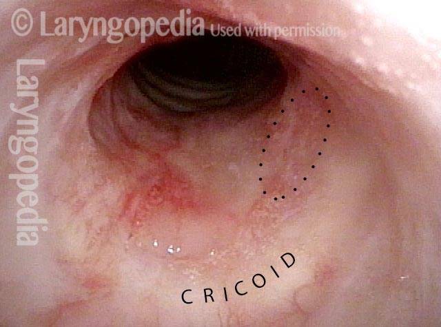
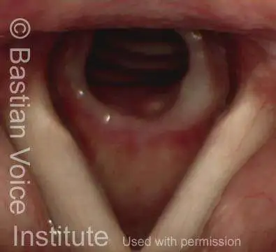
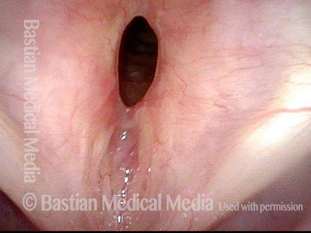
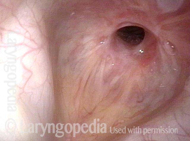
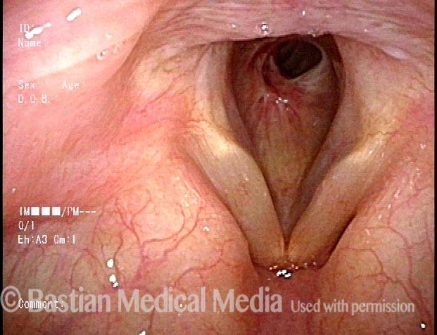
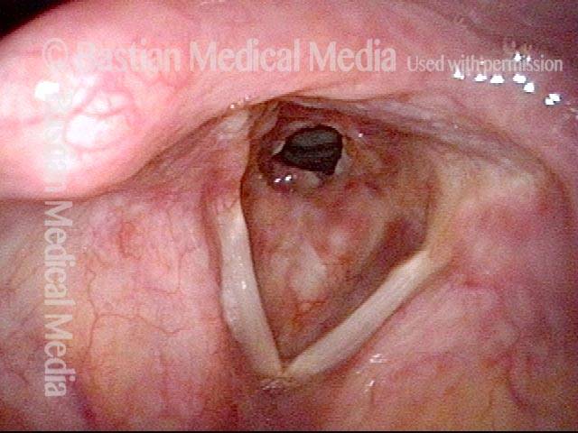
Rheumatoid arthritis (1 of 4)

Young woman diagnosed several years earlier with rheumatoid arthritis, but 8 months ago noted noisy breathing and exercise intolerance. This medium-range view shows inflammatory subglottic narrowing.

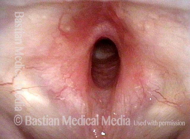
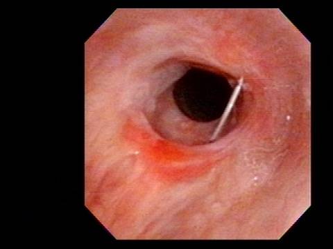
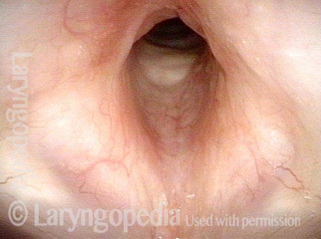
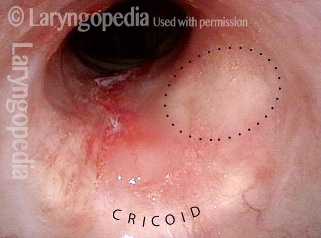
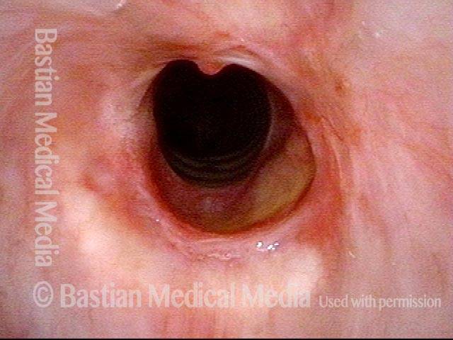
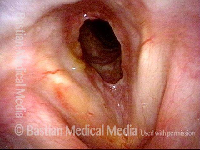
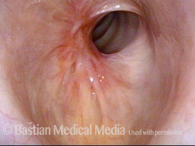
Inflammatory subglottic stenosis (2 of 4)

At closer range, this inflammatory subglottic stenosis is seen better.

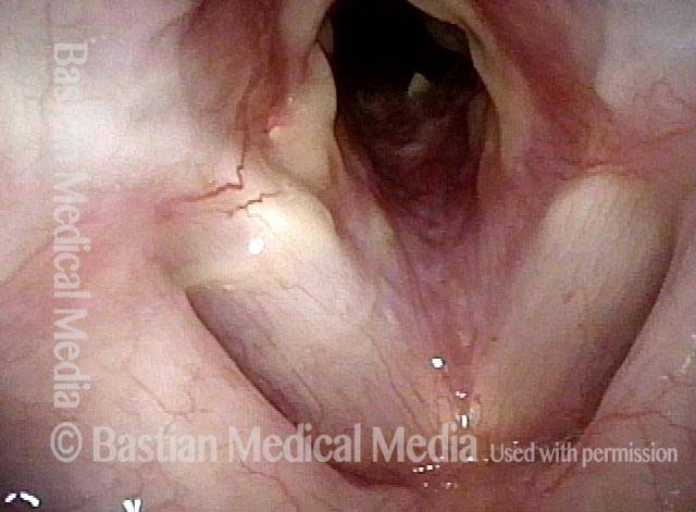
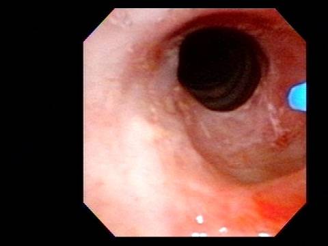
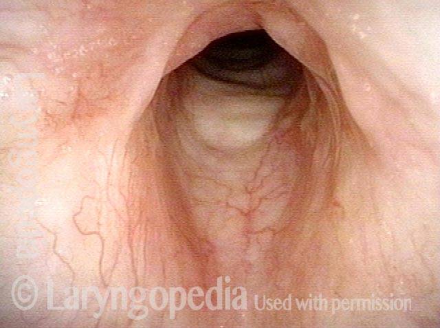
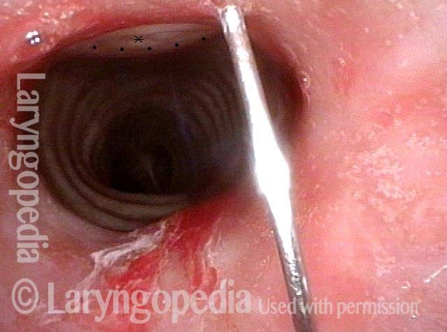
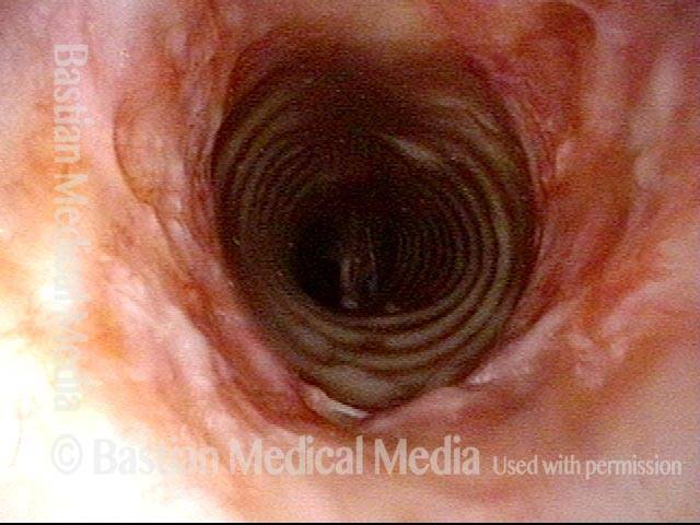
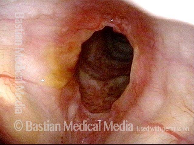
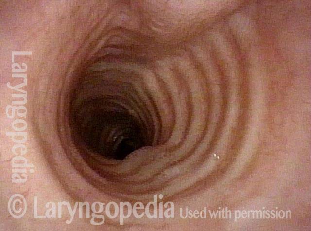
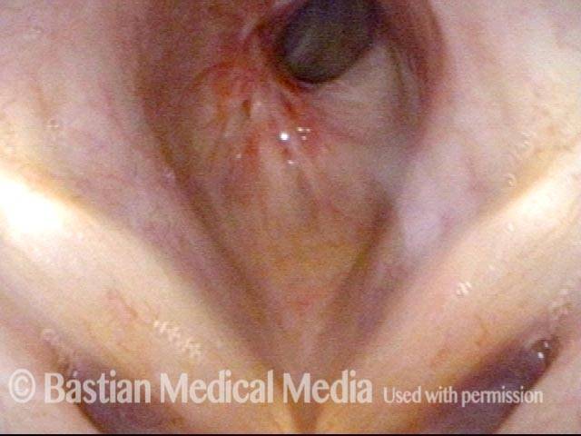
One week after dilation (3 of 4)
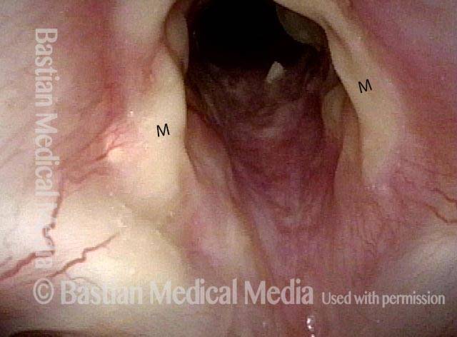
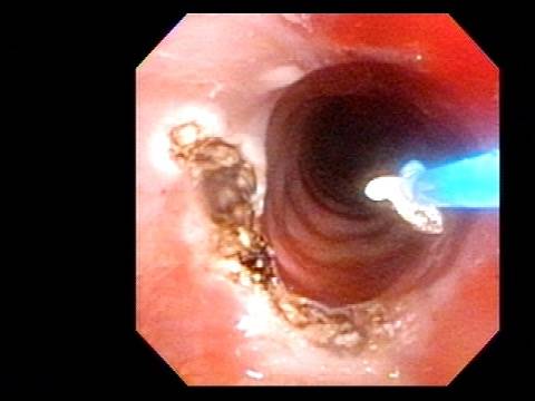
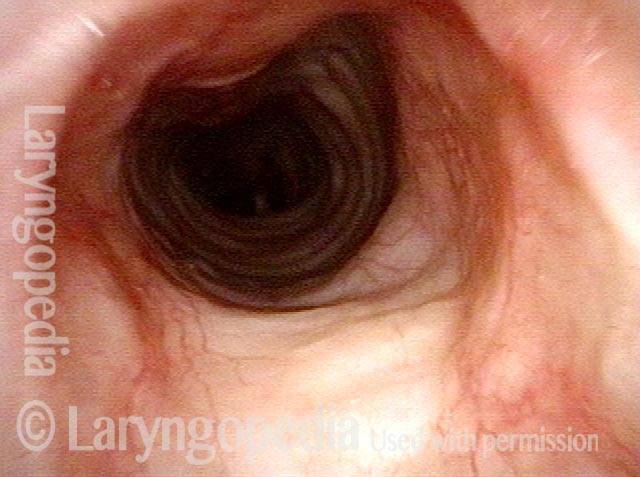
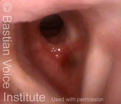
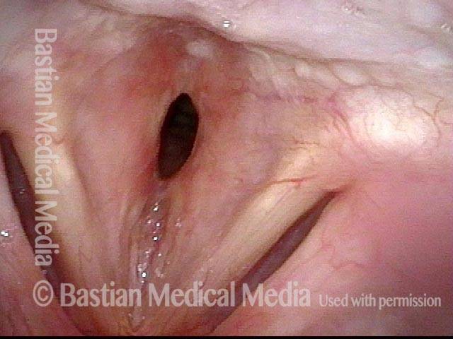
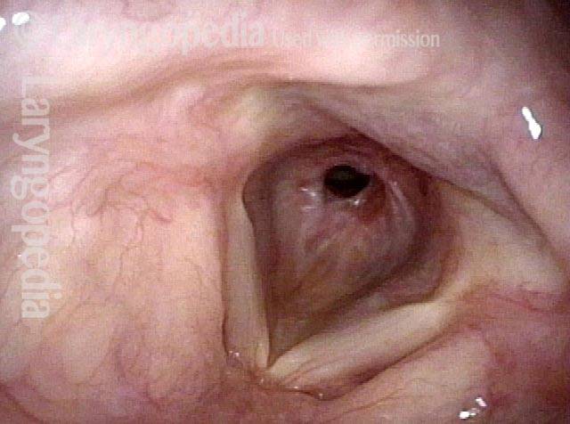
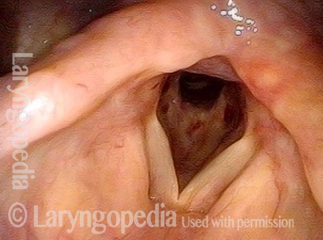
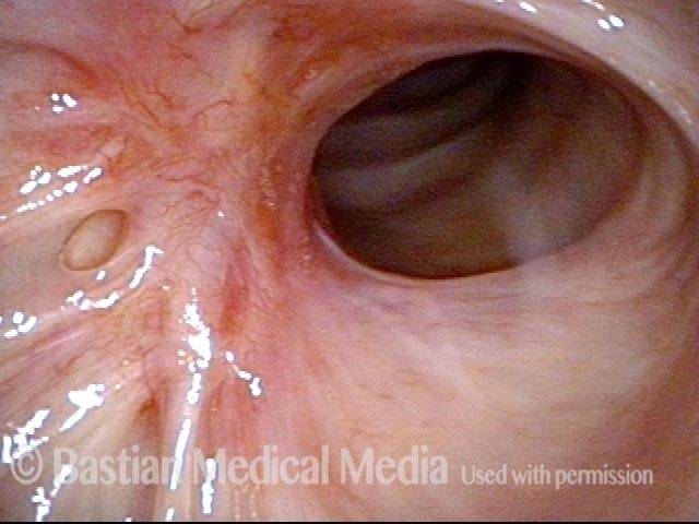
A week after outpatient dilation, steroid injection, the patient’s symptoms are dramatically reduced. Adherent mucus makes the caliber look smaller than it is. Compare with photo 1.

Mucus (4 of 4)

At closer range, the lumen is much enlarged, especially if adherent mucus were not present (‘M’ stands for mucus). Compare with photo 2.

“Office” Treatment Option for Inflammatory Subglottic Stenosis

In such situations, one naturally casts about for alternatives. Low dose methotrexate has not been very effective in our experience. And we work with a motivated rheumatologist open to considering rituximab, even with a negative ANCA study. Or the laryngologist might consider an office-based airway procedure as illustrated in the photos below. This particular person only requires dilation every 18 months or so, but it represents a major stressor for her given her fears, her size, and somewhat challenging anatomy. She is one of several offered the treatment shown below: steroid injection in the voice laboratory, followed by laser treatment.

Our thought is that in some, it may be the ridges and air turbulence that creates the feeling of airway restriction, or that these rings of stenosis “catch” mucus. In the case below, the clinician was almost dubious that what was accomplished would make that much difference to the person’s breathing. But upon calling her a week after the procedure below, and asking if the improvement was nil, subtle, small, medium, or large, she replied “large…a huge improvement.”

Visual Portfolio, Posts & Image Gallery for WordPress

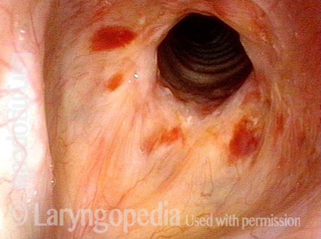
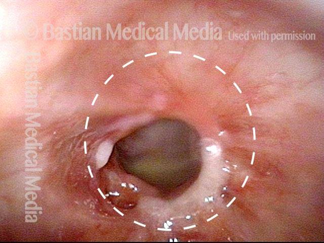
60% Stenosis (1 of 5)

Estimated at a 60% stenosis, using the vocal cords as a reference.

Narrowing & erythema (2 of 5)

At closer range, the examiner sees concentric ridges of narrowing and erythema characteristic of this disorder.

Triamcinolone is injected (3 of 5)

With patient sitting in the chair with topical anesthesia, a needle is passed from anteriorly just below the cricoid. Here, triamcinolone is being injected into the posterior component of the stenosis.

Laser ablation (4 of 5)

The blue laser fiber (thulium laser) is seen just prior to beginning laser ablation. Note in particular the circular shape of the area of greatest stenosis, to compare with the next photo.

Stenosis is coagulated (5 of 5)

The anterior half of the ring of greater stenosis has been coagulated. Note that the shape of the opening is changed to more of an anterior-posteriorly oriented oval. Distal trachea is also more easily seen. Again, the patient reported that this improved her breathing to a large degree.

Idiopathic Subglottic Stenosis Has Different Levels

Visual Portfolio, Posts & Image Gallery for WordPress

Post dilation (1 of 4)

Six months after dilation of idiopathic (inflammatory) subglottic stenosis, the patient has noted only slight deterioration, and breathing ability remains acceptable to her.

Closer view (2 of 4)

At closer range, the inflammatory component appears more evident.

Rich vascular pattern (3 of 4)

The rich vascular pattern accompanying the lesion is seen better and is a visual finding of inflammation.

“Sharing” the airway (4 of 4)

Here, the scope has been passed through the area of maximal narrowing and the patient becomes acutely aware of greater difficulty breathing. “Sharing the airway” is a way of ‘measuring’ it functionally. Note again the congested capillaries.

Another way to Inject Idiopathic Subglottic Stenosis

Visual Portfolio, Posts & Image Gallery for WordPress

Idiopathic inflammatory subglottic stenosis (1 of 3)

This patient is about to receive a triamcinolone (steroid) injection into her idiopathic inflammatory subglottic stenosis, while sitting in a chair under topical anesthesia. Dotted circle is for reference with Photo 2.

Priot to injection (2 of 3)

A needle has been passed through anterior neck skin and its tip rests out of sight, submucosally just inferior to the anterior cricoid ring. Note that the milky white medication has been infused submucosally within the dotted ring.

Injection (3 of 3)

Here, the 27-gauge needle traverses the trachea in order to inject the posterior tracheal wall. The submucosal white medication appears at the *.

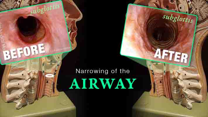
Subglottic Stenosis, after Treatment

Visual Portfolio, Posts & Image Gallery for WordPress

Subglottic stenosis, before treatment (1 of 2)

Subglottic and high tracheal stenosis, inflammatory, idiopathic (Lab).

Subglottic stenosis, after treatment (2 of 2)

Same patient, a few days after dilation and steroid injection (Lab).

Subglottic Stenosis, before and after Dilation

Visual Portfolio, Posts & Image Gallery for WordPress

Subglottic stenosis, before dilation (1 of 2)

This individual has undergone at least a dozen prior dilations, each of which provides dramatic relief from noisy breathing and exercise intolerance. Here the patient is halfway to needing re-dilation, due to the typical inflammatory stenosis that is seen. Compare with photo 2.

Subglottic stenosis, after dilation (2 of 2)

One week after one of this patient’s dilations (with Kenalog injection and topical Mitomycin C), showing a dramatic widening of her airway; compare with photo 1. After a number of years, inflammatory lesions such as this sometimes “burn out,” and the interval between dilations increases.

Example 2

Visual Portfolio, Posts & Image Gallery for WordPress

Subglottic stenosis (1 of 5)

Middle-aged woman with unexplained shortness of breath and noisy breathing, due to this idiopathic inflammatory and very high subglottic stenosis. The patient initially declined dilation due to her anxiety. She also had granularity of the nasal septum and a positive ANCA profile for Wegener’s granulomatosis.

Subglottic stenosis, worsened (2 of 5)

Five months later, the symptoms became intolerable, and the stenosis was noted to be slightly narrower and with a greater posterior component. The patient agreed to dilation.

Subglottic stenosis, worsened (3 of 5)

Same exam as photo 2; this close-up view shows more clearly the inflammatory nature of this stenosis.

Subglottic stenosis, after dilation (4 of 5)

Five days after outpatient dilation, triamcinolone injection, and topical mitomycin C application. The patient’s symptoms have vanished, the harsh inspiratory noise is no longer heard, and the size of the airway, though still not normal, is more than doubled. Compare with photo 2 of this series.

Subglottic stenosis, after dilation (5 of 5)

Same exam as photo 4, close-up view. Intensification of the inflammatory changes of this stenosis are expected so early after dilation. Compare size of the stenosis with photo 3 of this series.

Stenosis Before and After Dilation for Forme Fruste Wegener’s

Visual Portfolio, Posts & Image Gallery for WordPress

Subglottic stenosis (1 of 5)

High-grade subglottic stenosis in a woman diagnosed syndromically with forme fruste Wegener’s granulomatosis-related subglottic stenosis. With a narrowing less than 50%, she is very symptomatic. Her breathing is noisy, and her forced inspiration time is about 3 seconds. Compare with photo 4.

Inflammation (2 of 5)

Closer visualization reveals the inflammatory nature of the stenosis.

Flexible scope (3 of 5)

Here the distal chip flexible scope has passed through the narrowing not only to see into distal trachea, but also as a breathing test. The patient becomes very aware of reduced space and this indicates a marginal airway.

Post-dilation (4 of 5)

A week after dilation, triamcinolone injection and mitomycin C application, the patient says breathing is now normal, and forced inspiration time is only a second in duration–normal. Compare with photo 1.

Post-operative bruising (5 of 5)

Closer visualization with early postoperative bruising. The trachea is now easily visible through the larger opening. Compare with photo 2.

Subglottic / Tracheal Stenosis

Visual Portfolio, Posts & Image Gallery for WordPress

Wegener’s granulomatosis (1 of 4)

Inflammatory subglottic/tracheal stenosis, thought to be the result of an incomplete expression of Wegener’s granulomatosis (no history of trauma).

Subglottic / Tracheal stenosis (2 of 4)

Close view, from vocal cord level.

Subglottic / Tracheal stenosis (3 of 4)

Close view, from just above vocal cords.

Inflammed Stenosis (4 of 4)

Close view, showing the inflammatory nature of the stenosis.

Subglottic Stenosis, Due to Wegener’s Granulomatosis

Visual Portfolio, Posts & Image Gallery for WordPress

Subglottic stenosis, due to Wegener’s (1 of 2)

This person has Wegener’s granulomatosis, confirmed by anti-neutrophil cytoplasmic antibodies (ANCA) testing. Here, looking from above the vocal cords, one can see an estimated 50% narrowing of the subglottic and high tracheal passageway.

Subglottic stenosis, due to Wegener’s (2 of 2)

Viewed from within the subglottis, one can see more clearly the inflammatory nature of this stenosis. A dotted oval estimates what the normal caliber or width of this airway would be. This patient has been managed with systemic medication, but also occasional dilation, steroid injection, and Mitomycin C application.

Share this article

Stenosis Summary YouTube Thumbnail
Play Video about Stenosis Summary YouTube Thumbnail

Inflammation Subglottic Stenosis

Are you experiencing gradual onset of noisy, even effortful breathing? One uncommon but important explanation is idiopathic, inflammatory subglottic stenosis. That just means “unknown cause, likely autoimmune narrowing just below the vocal cords and at the highest part of the windpipe.”

This video explains the disorder and its management, shows “before” and “after” photos, and reassures viewers that if yours is the usual case, there doesn’t seem to be any effect on general health or lifespan.

Table of Contents

Subscribe
Notify of

0 Comments
Newest
Oldest Most Voted
Inline Feedbacks
View all comments
0
Click to see all comments.x