The term “long-haulers” has been used to refer to persons with lingering systemic symptoms after successful initial recovery from Covid-19. Symptoms such as coughing, fatigue, loss of taste and smell, brain fog, etc. occur weeks or months after first falling ill. Even some whose bout with Covid-19 seemed mild can experience this “long-haul” phenomenon. For more, here’s a link to a CDC publication describing this scenario before the words “long-haul” were attached to this syndrome.

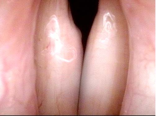
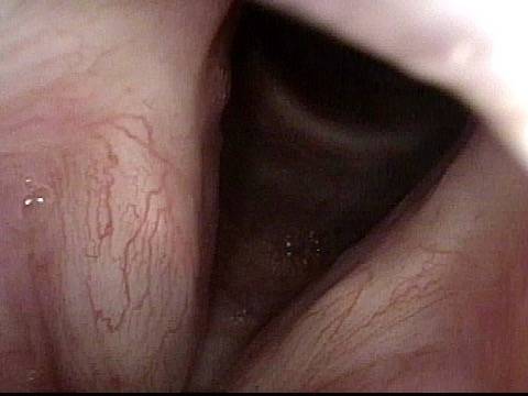
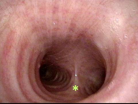
Laryngologists are now seeing patients weeks or months after their recovery from severe Covid-19 infection that required hospitalization and intubation/ventilation. These patients seem to be presenting primarily for chronic breathing and voice complaints. We are finding what could be called “long-haul” injuries from the breathing tube used while they were on ventilators. These injuries can be unavoidable when it is necessary to leave these breathing tubes in place for days to weeks due to grave illness. Such injuries in non-Covid patients are documented on Laryngopedia (see, for example: Intubation injury – Laryngopedia). Below are photos showing tracheal stenosis (narrowing), and post-intubation phonatory (voice) insufficiency, in “long-haul” breathing tube injury Covid-19 patients.

Wheezing after Covid-19 can also be Large Airway Wheezing

Visual Portfolio, Posts & Image Gallery for WordPress

Vocal Cords (1 of 4)

This person was experiencing some shortness of breath on exertion. She was not wheezing at rest and had no personal or family history of asthma. Still, auscultation of the lungs by a prior physician while requesting that she exhale forcefully revealed wheezing sounds. She was sent for evaluation of vocal cord dysfunction. At the beginning of airway examination, during forced exhalation and audible wheezing these widely separated vocal cords tell us the source of the sound is not the vocal cords.

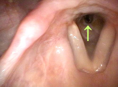
Trachea & Anterior Carina (2 of 4)

Suspecting large airway wheezing, topical anesthesia was used to obtain this view of the trachea in an office setting. The anterior carina is designated in this and following photos with a *.

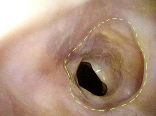
Trachea Bulges Inward (3 of 4)

Here, the patient has just begun exhaling forcefully. The membranous tracheal wall begins to bulge inward, not as an anatomical abnormality, but as a functional phenomenon that might occur in virtually any person.

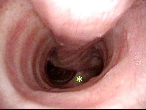
Trachea Bulges Inward (4 of 4)

At forceful end-expiration, however, the patient demonstrates unusually good ability to bulge her membranous trachea inward and nearly obstruct both mainstem bronchi. The result? Very audible wheezing. To distinguish this from actual asthma, the examiner need only listen over the manubrium and then peripheral lung fields. If this comparison reveals that wheezing is much louder centrally than peripherally, the explanation in my experience has always been large airway wheezing and not asthma. Of course, asthma AND large airway wheezing can occur together.

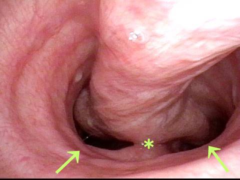
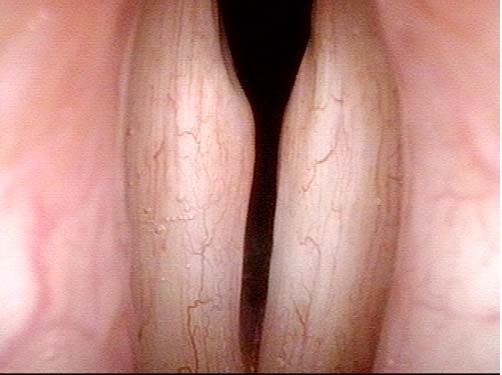
Tracheal stenosis as a complication of Covid-19 treatment

Visual Portfolio, Posts & Image Gallery for WordPress

Narrowing in the Trachea > 50% (1 of 2)

This patient was in hospital for Covid-19 infection and was intubated for approximately 7 days. Now, 4 months later, she is well in general, and lung damage is mostly repaired. Yet, she is still short of breath. A key clue to the explanation is that she has “noisy breathing.” In this photo, seen in the distance below her open vocal cords, is a > 50% narrowing in her trachea (arrow). Air “squeezing” through this narrowing makes her harsh breathing noise.

Expected Size of Tracheal Opening (2 of 2)

At much closer range, the dots outline the expected size of tracheal opening. This tracheal stenosis, has been caused by the combination of inflammation and infection coupled with the pressure of the sealing balloon of her breathing tube. It is possible that balloon had to be inflated more than preferred to handle high pressure ventilation (though we do not have this information).

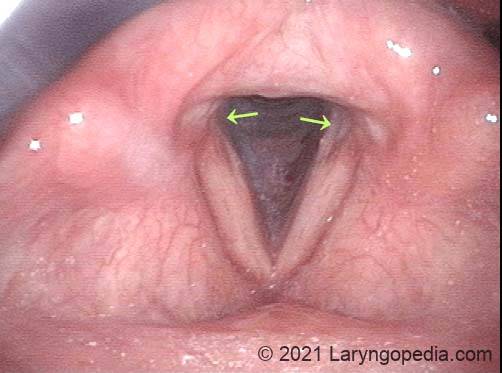
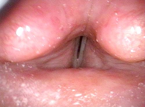
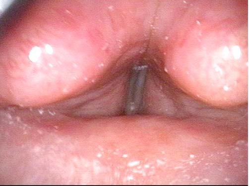
Injury to the vocal cords causing voice change, as a complication of Covid-19 treatment

Visual Portfolio, Posts & Image Gallery for WordPress

Erosions in the Posterior Vocal Cords (1 of 5)

This second patient was also intubated for Covid-19 and now, months later, continues to experience a drastic voice change. Initial examiners were unable to explain this voice change. Here, the vocal cords are fully apart, and the experienced eye can see erosions in the posterior vocal cords (arrows). Erosions like these do not cause the magnitude of hoarseness this patient is experiencing. A more likely is tissue loss + cricoarytenoid joint injury (as we shall see is the case…)

Pre-phonatory Instant (2 of 5)

This is the “pre-phonatory instant.” The vocal cords have been put into position for making voice, but vibration has not yet commenced. The cords seem to come together fairly well, and so again, the reason for her very severe hoarseness is not yet evident.

Phonatory Instant (3 of 5)

Vibration has now commenced, producing an extremely hoarse voice, the cause for which is not yet seen in these “distant” views.

Close Up of Vocal Cords (4 of 5)

Now at much closer range, we again view the pre-phonatory instant, exactly as seen more distantly in photo 4. We see here that the gap between the cords is greater than appreciated from “afar.” And the erosions seen in photo 1 are actually significant divots, caused by pressure necrosis of the endotracheal tube.

Insufficient compression of the cords (5 of 5)

Vibration commences, exactly as in photo 3. But there is tremendous air-wasting through the keyhole created by the divots, and there is insufficient compression between the vibrating parts of the cords. Hence, the patient’s inability to say a normal number of words on one breath, and her severely degraded “breathy-pressed” voice quality.