

Biopsy
Biopsy refers to both the process of removing a bit of tissue from a person so that it can be studied and “diagnosed” by the pathologist, and also to the piece of tissue itself. “The surgeon biopsied (verb) the vocal cord and then sent the biopsy (noun) to the pathologist for examination under the microscope.”
Photo Example
HIV Testing (1 of 2)
2-millimeter forceps being used to scrape the area of leukoplakia to retrieve cells for HPV testing. In this case, it was negative.
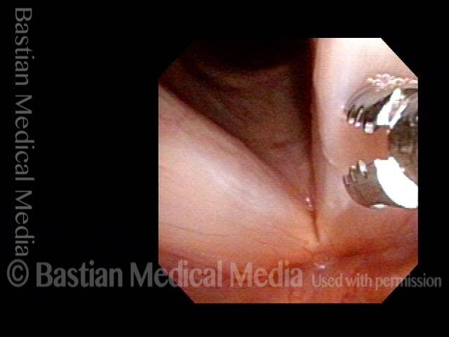
HIV Testing (1 of 2)
2-millimeter forceps being used to scrape the area of leukoplakia to retrieve cells for HPV testing. In this case, it was negative.
Biopsy (2 of 2)
Cup forceps closed. Note that there is no biopsy, per se, but just scraping of the surface of the cords.
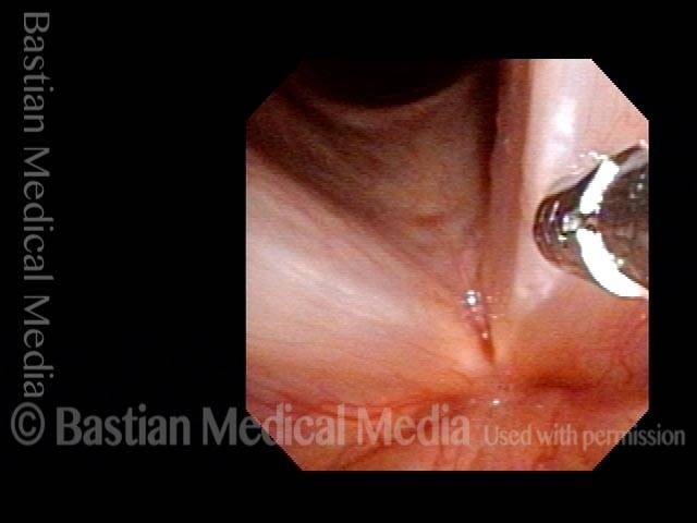
Biopsy (2 of 2)
Cup forceps closed. Note that there is no biopsy, per se, but just scraping of the surface of the cords.
Example 2
Biopsy, epiglottis (1 of 1)
Biopsy of lesion involving the petiole (low laryngeal surface of epiglottis). The pathology report revealed squamous cell carcinoma, usually caused by smoking.
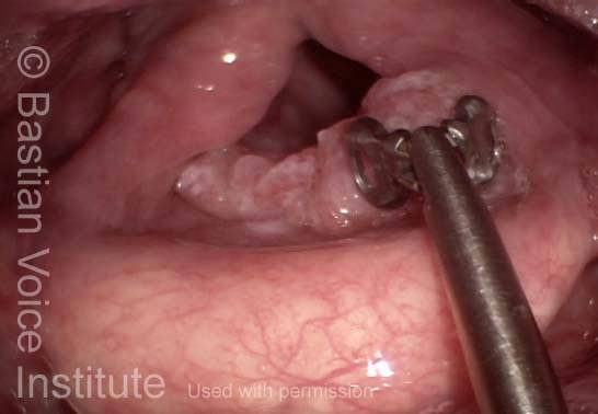
Biopsy, epiglottis (1 of 1)
Biopsy of lesion involving the petiole (low laryngeal surface of epiglottis). The pathology report revealed squamous cell carcinoma, usually caused by smoking.
Biopsy of Early Vocal Cord Cancer
Chronic hoarseness (1 of 3)
Examination of this man for chronic hoarseness reveals diffusely abnormal-looking tissue on his vocal cords. The area on the upper surface of the right vocal cord (enclosed in dotted circle) appears to be most likely to be diagnostic. Note the bulk, and aberrant blood vessels.
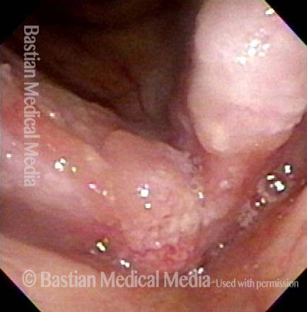
Chronic hoarseness (1 of 3)
Examination of this man for chronic hoarseness reveals diffusely abnormal-looking tissue on his vocal cords. The area on the upper surface of the right vocal cord (enclosed in dotted circle) appears to be most likely to be diagnostic. Note the bulk, and aberrant blood vessels.
Just before biopsy (2 of 3)
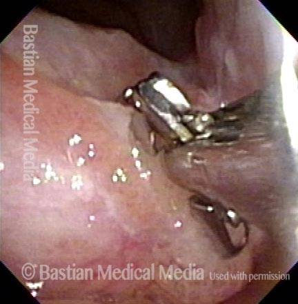
A 2 millimeter cup forceps has been passed through a channel scope and is planted on this area in open position, just before the biopsy.
Just before biopsy (2 of 3)
A 2 millimeter cup forceps has been passed through a channel scope and is planted on this area in open position, just before the biopsy.
Cancer finding (3 of 3)
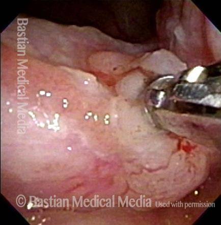
Cup forceps have now been closed to encompass a small fragment of tissue. The pathologist’s answer after looking at this sample under the microscope: cancer
Cancer finding (3 of 3)
Cup forceps have now been closed to encompass a small fragment of tissue. The pathologist’s answer after looking at this sample under the microscope: cancer
Tumor in Trachea
Biopsy (1 of 4)
Tumor growing through wall of trachea, from a paratracheal lymph node. Biopsy forceps are about to close to take a tissue fragment for study.
Biopsy (1 of 4)
Tumor growing through wall of trachea, from a paratracheal lymph node. Biopsy forceps are about to close to take a tissue fragment for study.
After biopsy (2 of 4)
After biopsy was taken at arrow. The result: squamous cell carcinoma thought to be an unusual metastasis from unusually aggressive larynx cancer.
After biopsy (2 of 4)
After biopsy was taken at arrow. The result: squamous cell carcinoma thought to be an unusual metastasis from unusually aggressive larynx cancer.
Tumor gone (3 of 4)
Soon after radiation therapy, the tumor has melted away, leaving a depression in the tracheal wall.
Tumor gone (3 of 4)
Soon after radiation therapy, the tumor has melted away, leaving a depression in the tracheal wall.
Slow return (4 of 4)
Eighteen months later, the patient has experienced a fairly durable response, with very slow return of tumor.
Slow return (4 of 4)
Eighteen months later, the patient has experienced a fairly durable response, with very slow return of tumor.
Share this article
Support Laryngopedia
Are you a patient or doctor who loves Laryngopedia? If we've helped you, help us help others too!
Tagged Diagnosis, Photos, Procedures & tools