 Cancer
Cancer is a malignant growth or tumor caused by abnormal and uncontrolled cell division. The hallmark of cancer is its potential ability to invade neighboring tissue or to spread (metastasize) to other parts of the body through the lymphatic system or the bloodstream.
Early cancers may have done neither, remaining localized to the tissue of origin. The majority of cancers in the head and neck are classified as carcinomas.
Laser Removal of Vocal Cord Cancer with Bilateral Disease
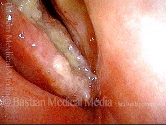
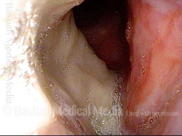
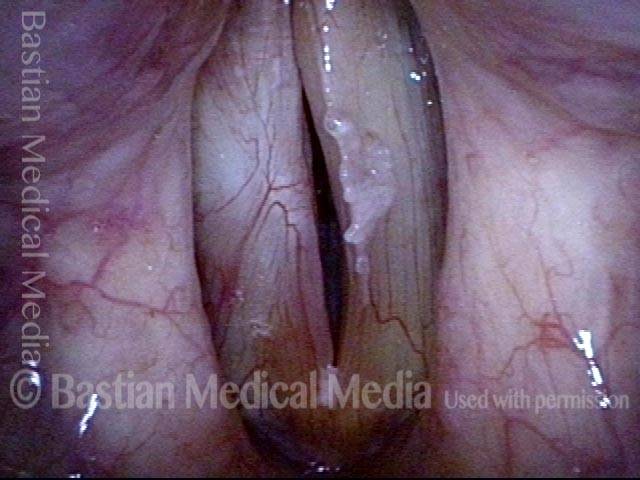
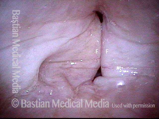
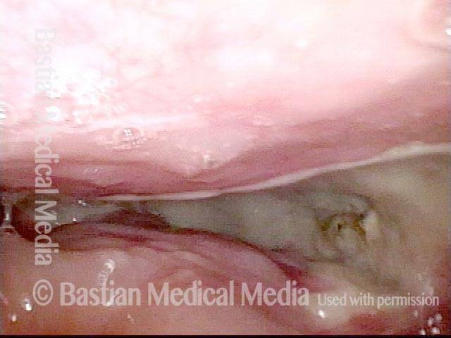
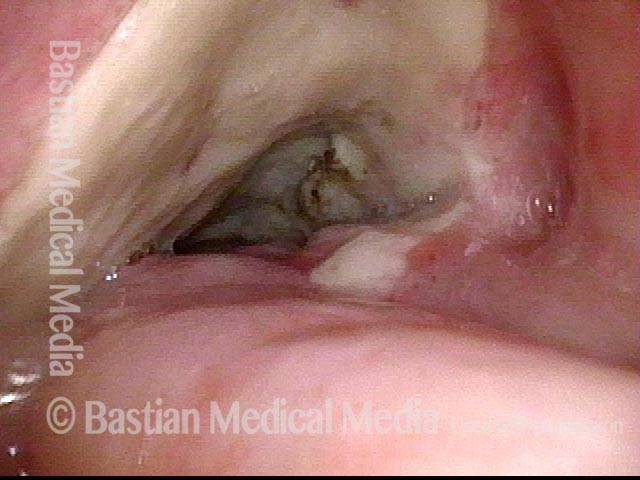
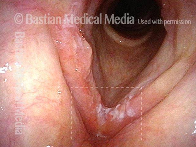
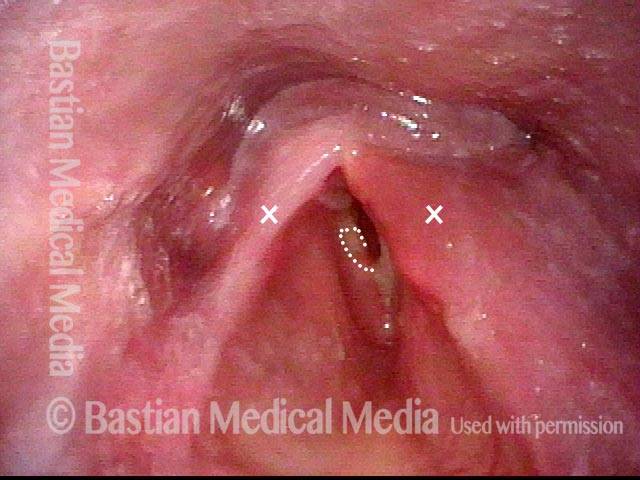
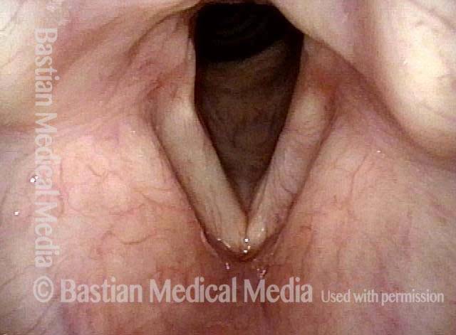
For treatment of early vocal cord cancer, both laser excision and radiotherapy are in competition as good treatment modalities. See also Early Vocal Cord Cancer: Remove with a Laser, or Radiate? Often, radiation is used when disease is bilateral, in the interest of preserving voice.
This is an example of the ability to do fairly extensive laser surgery bilaterally, yet preserving good voice. This man had a friend who had severe difficulty with radiation, and he was therefore opposed to that option.
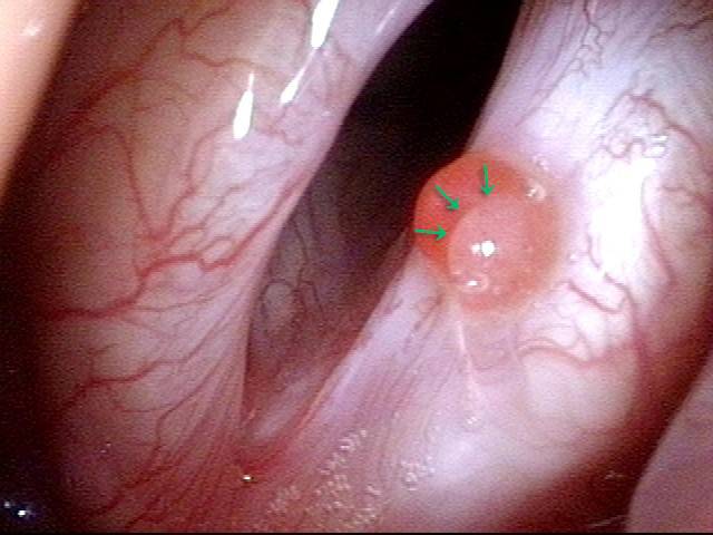
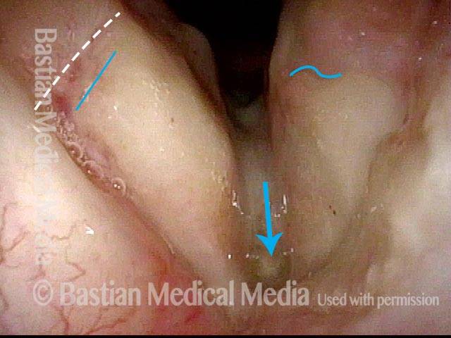
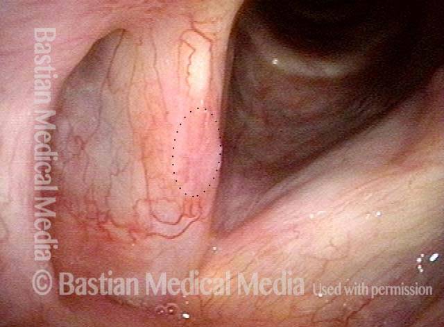
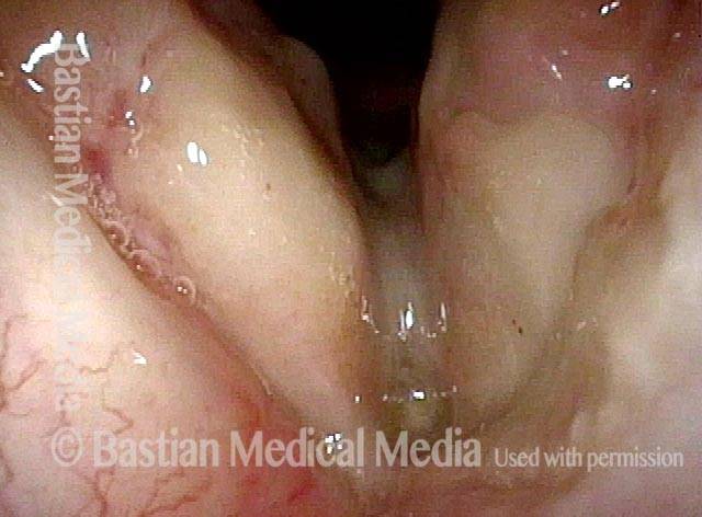
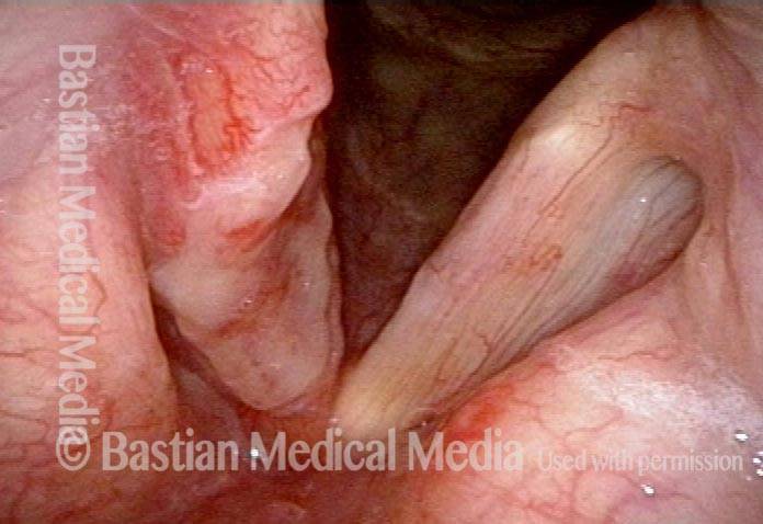
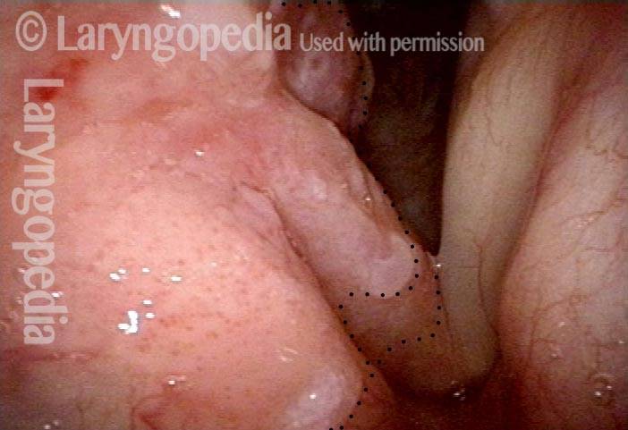
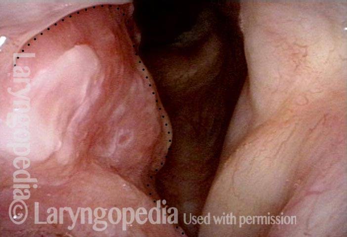
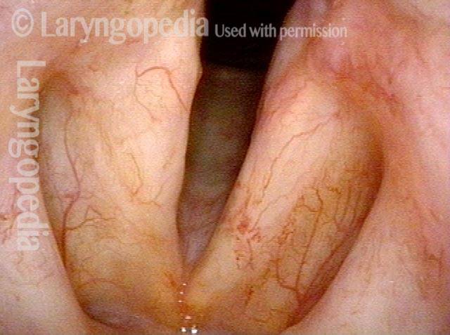
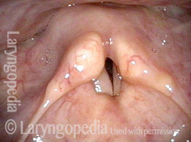
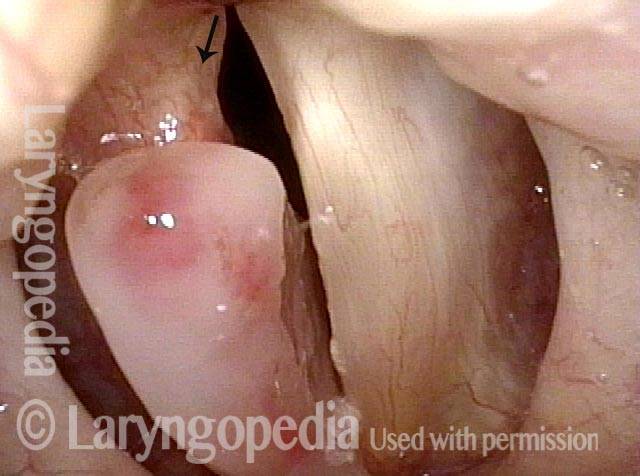
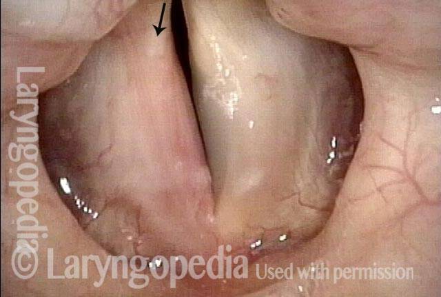
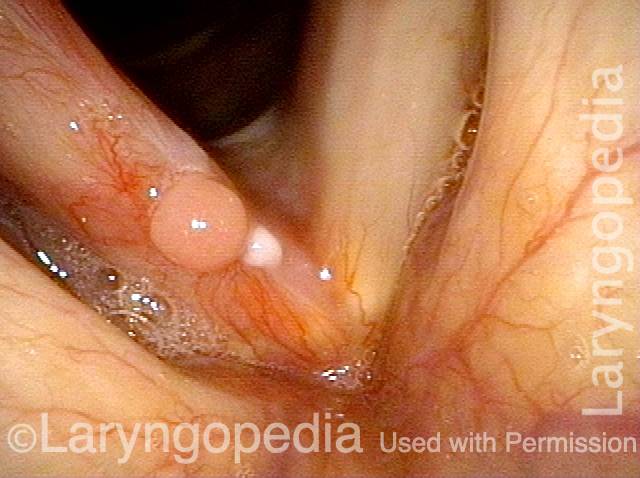
Vocal cord cancer (1 of 10)
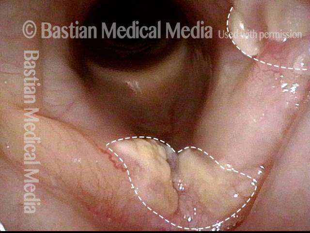
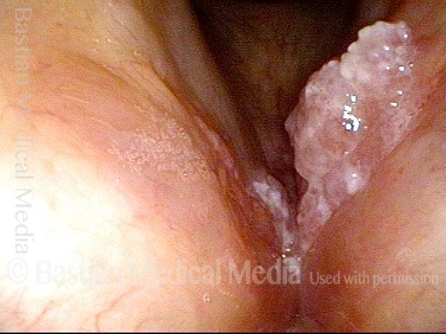
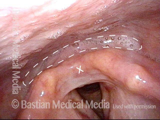
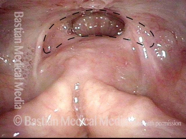
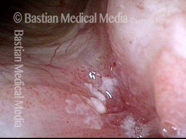
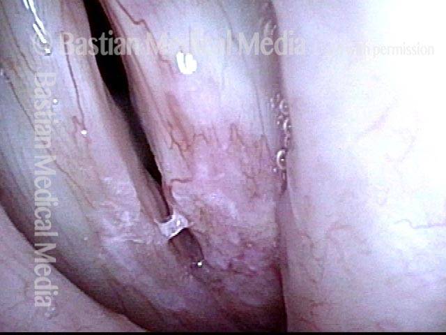
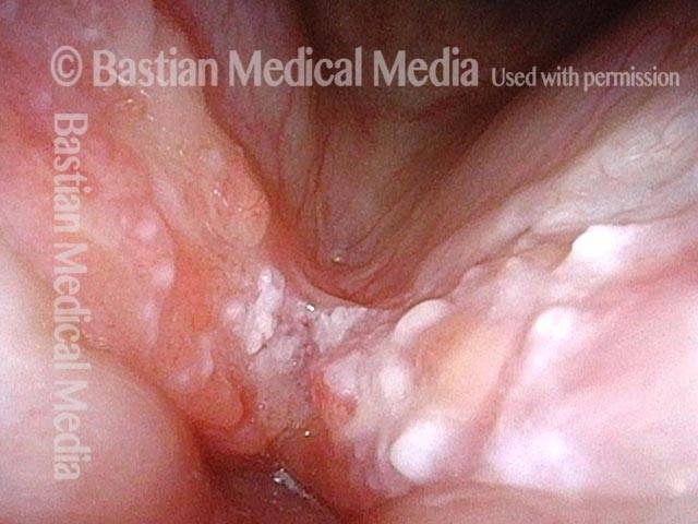
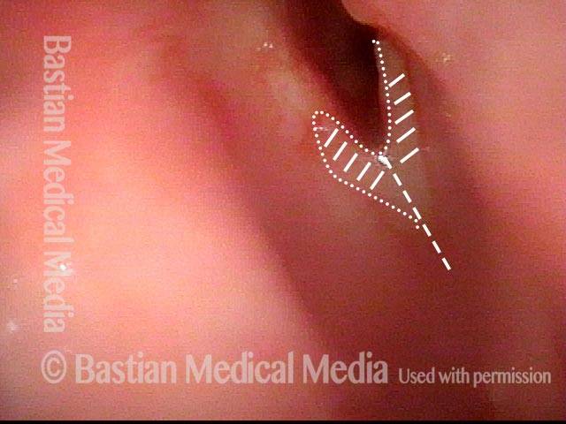
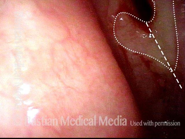
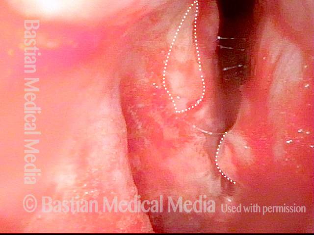
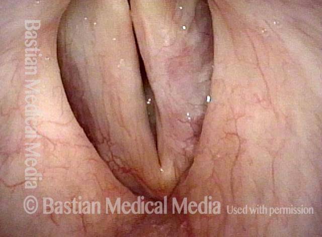
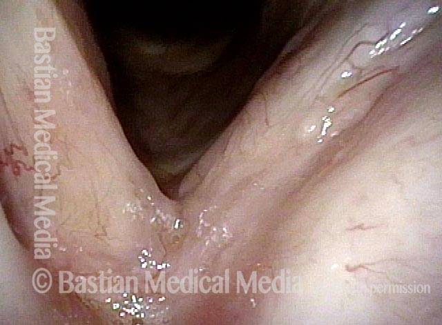
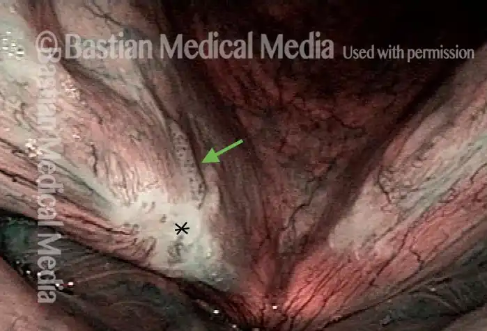
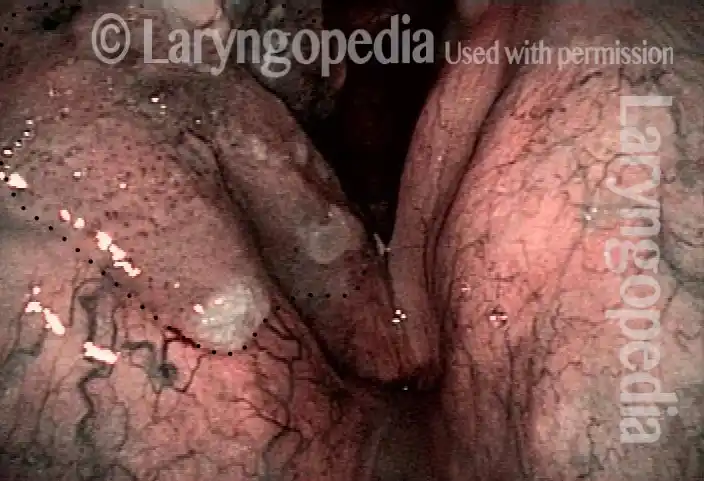
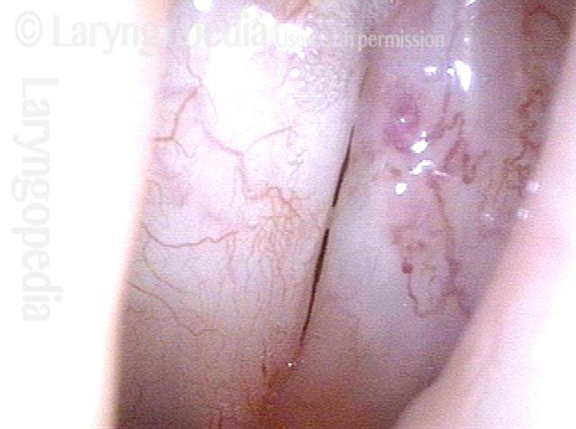
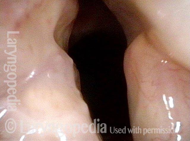
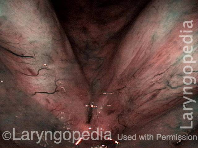
Stippling (2 of 10)
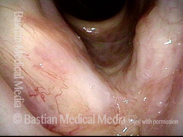
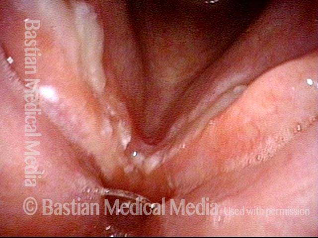
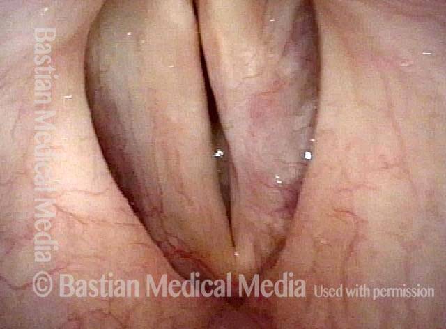
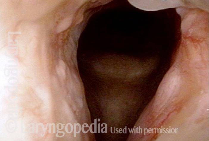
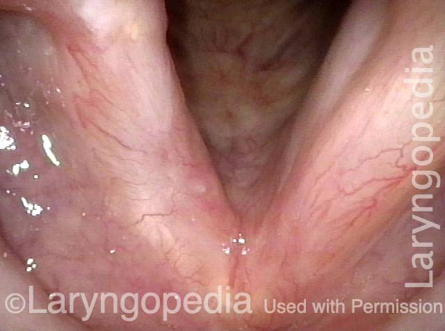
1 week after excision (3 of 10)
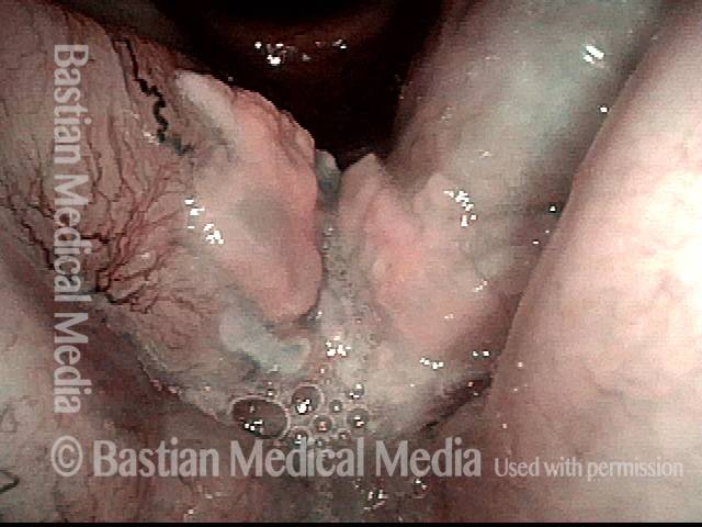
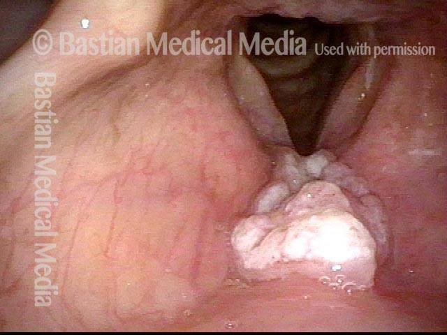
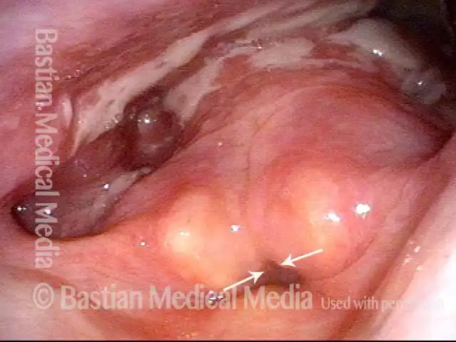
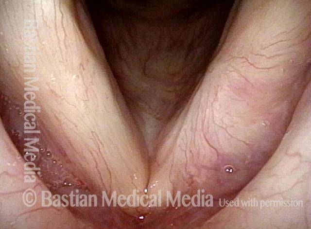
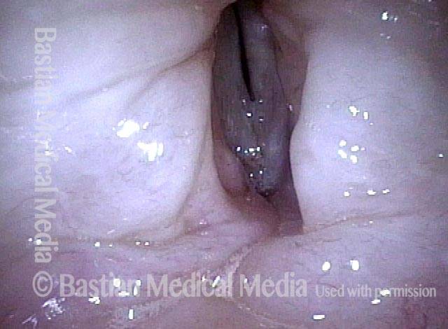
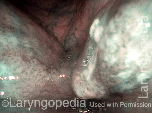
Reparative Granuloma emerges (4 of 10)

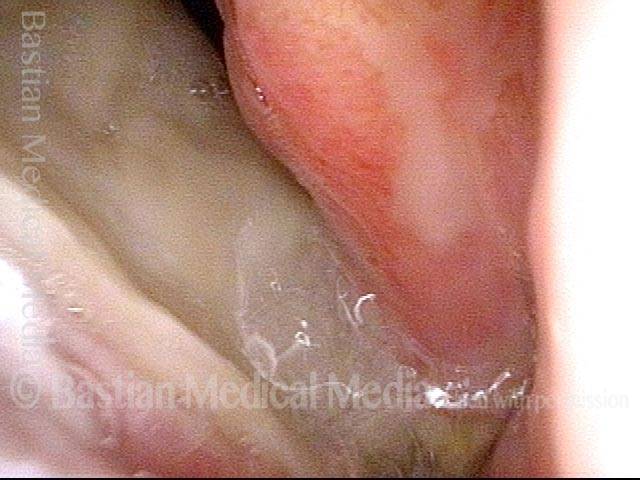
Granuloma interferes with voicing (5 of 10)
Granuloma fades away (6 of 10)
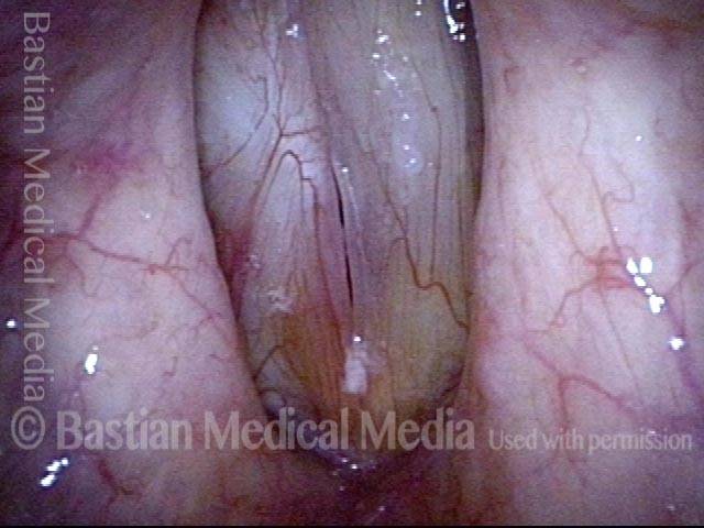
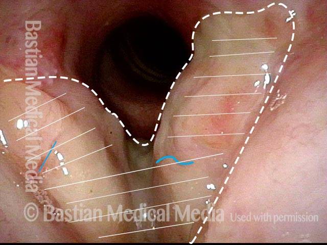
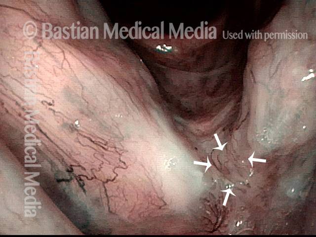
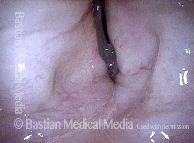
Closer view (7 of 10)
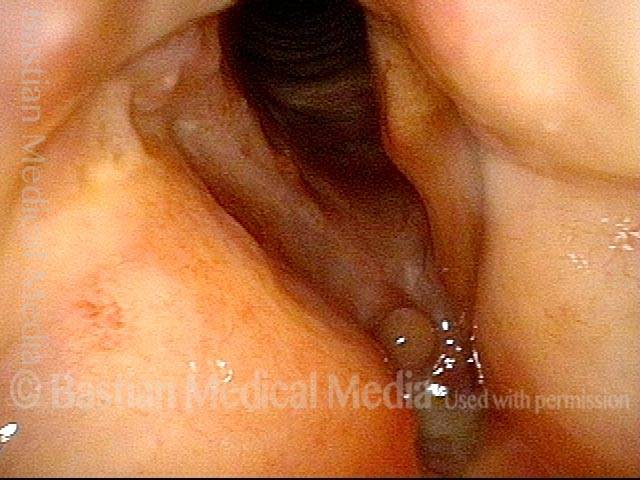
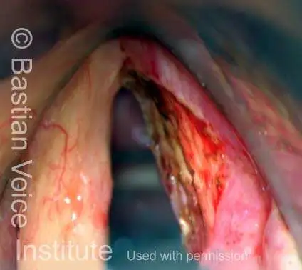
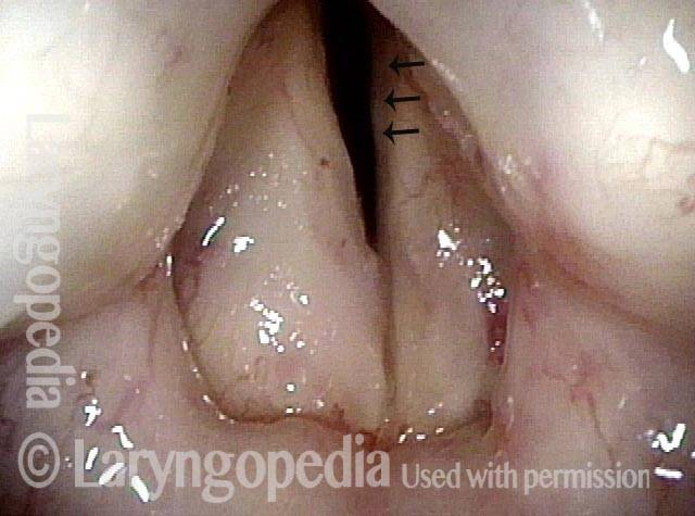
Granuloma cleft (8 of 10)
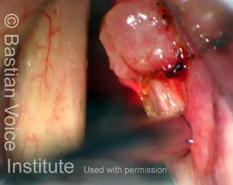
Blood tattoo (9 of 10)

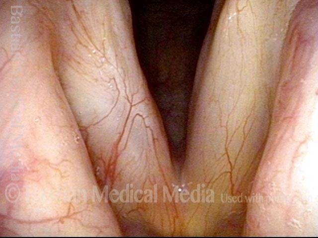
Voice is improved (10 of 10)
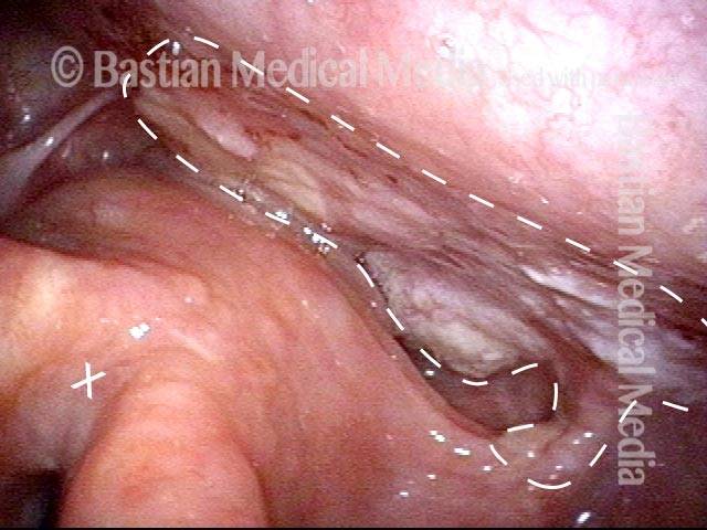
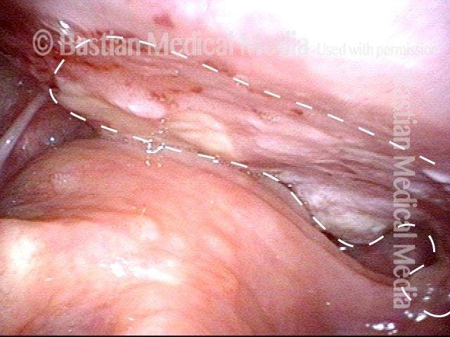
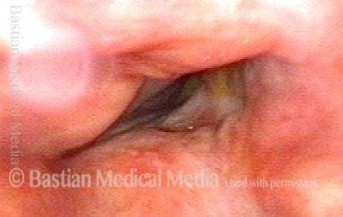
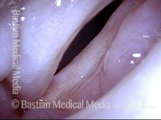
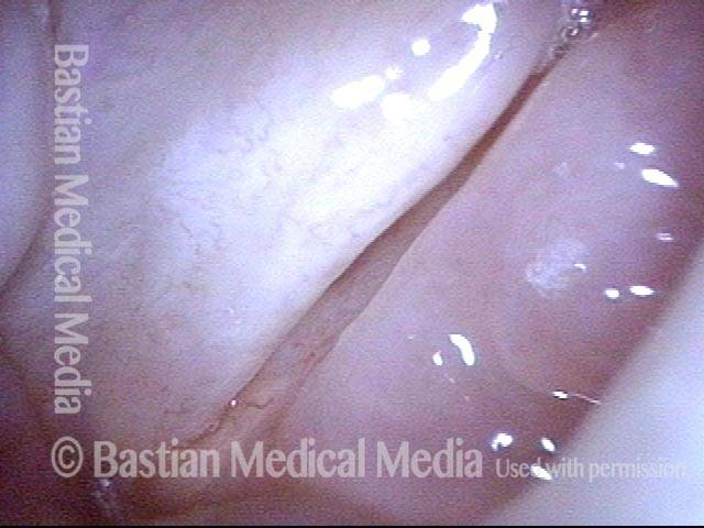
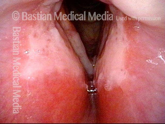
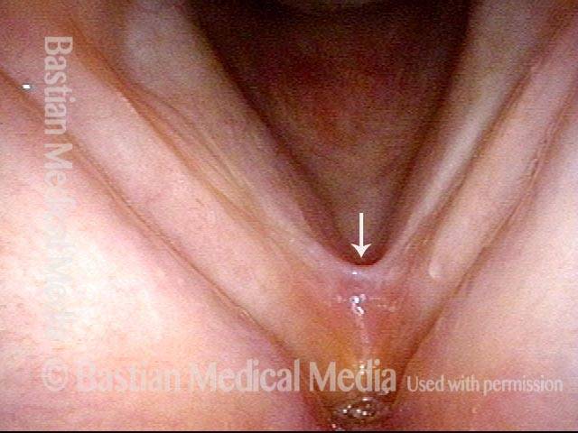
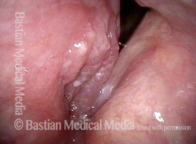
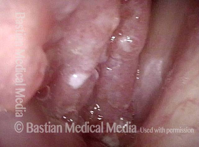
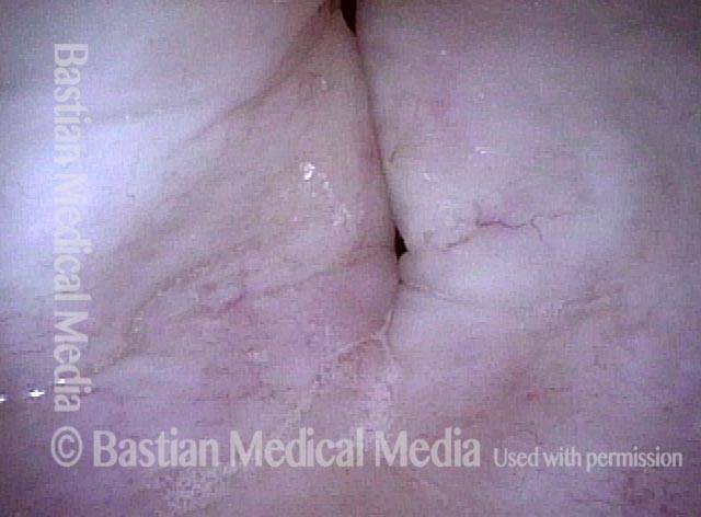
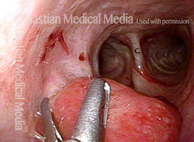
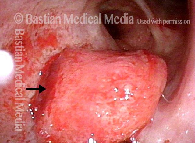
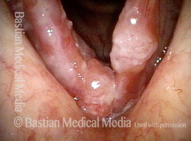
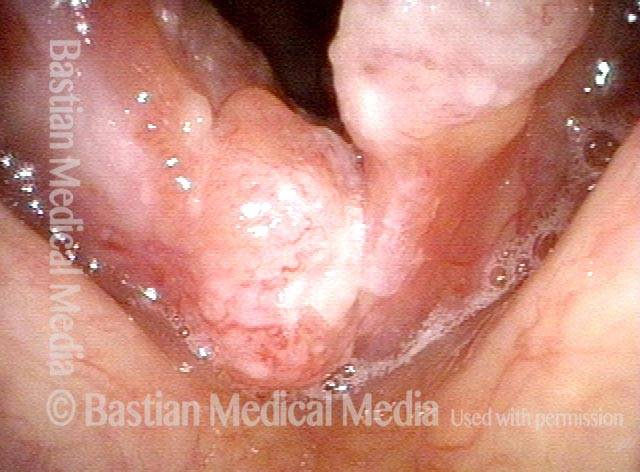
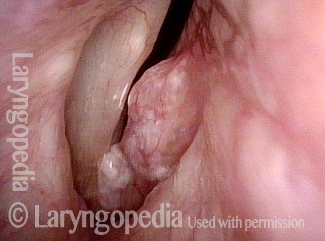
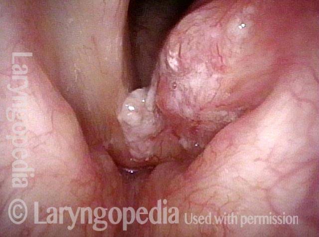
Laser Surgery for Bilateral Vocal Cord Cancer
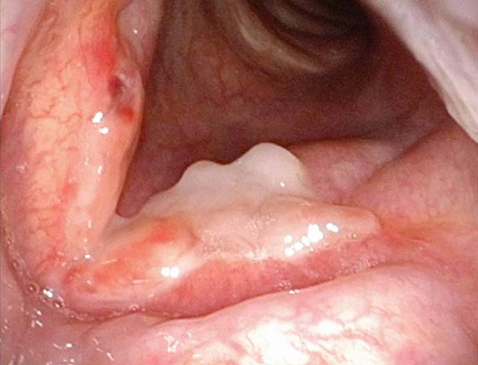
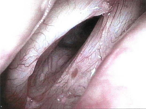
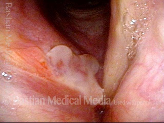
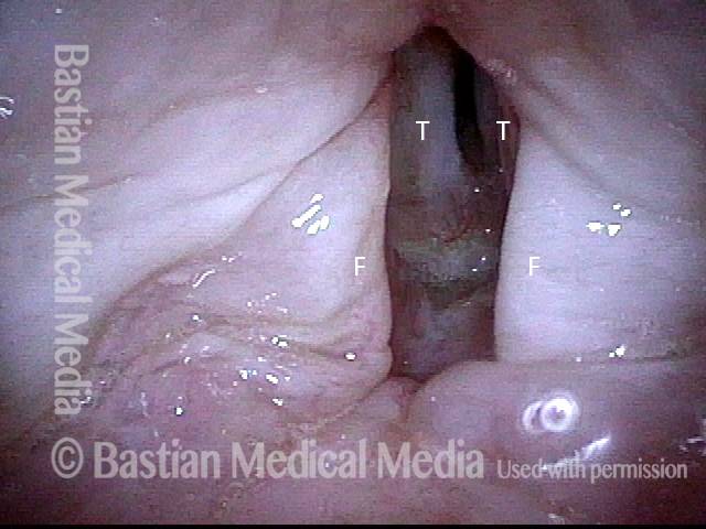
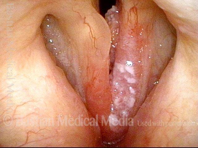
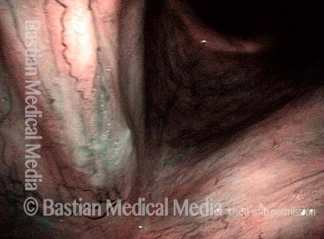
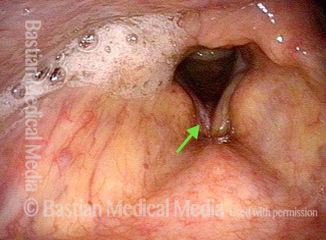
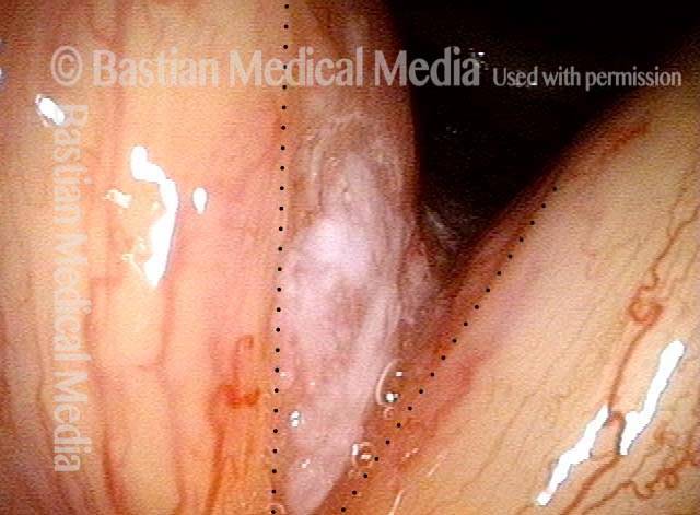
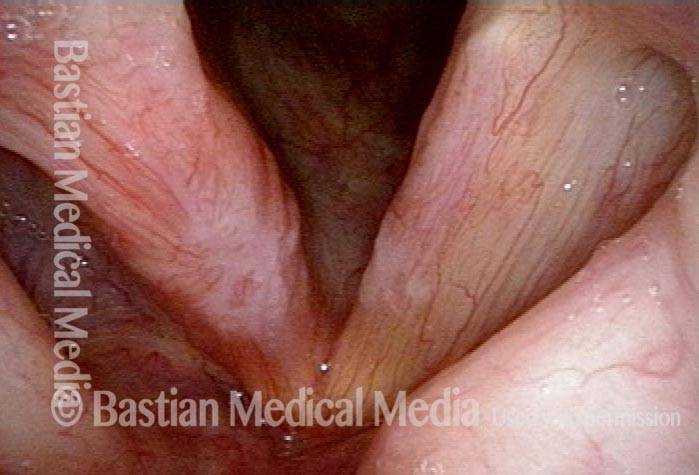
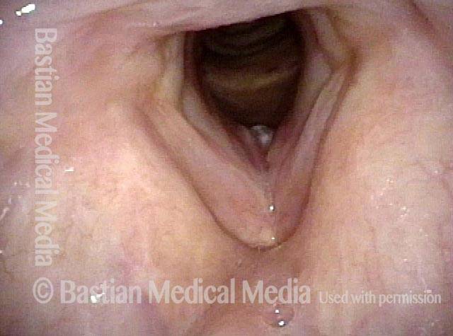
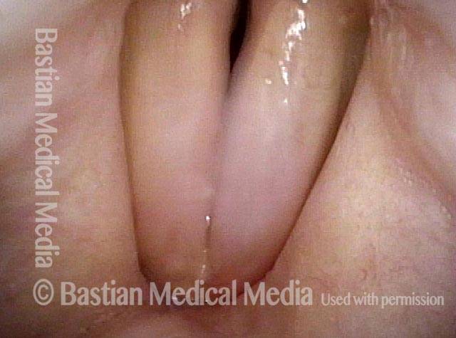
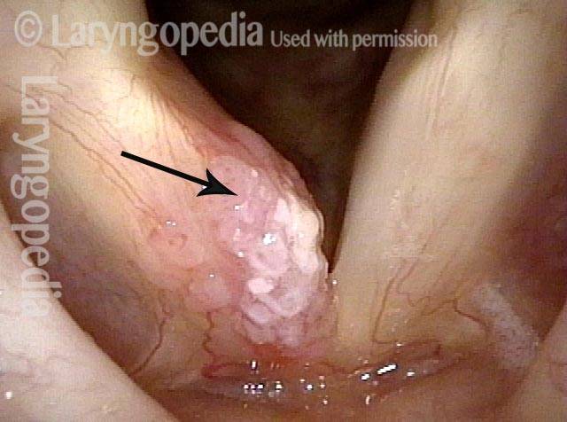
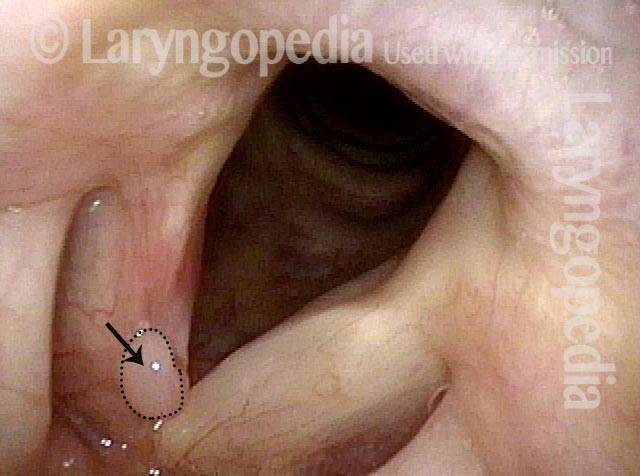
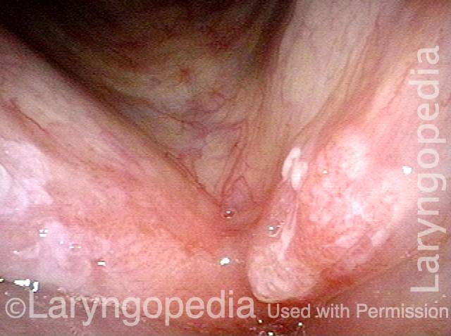
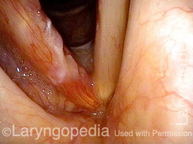
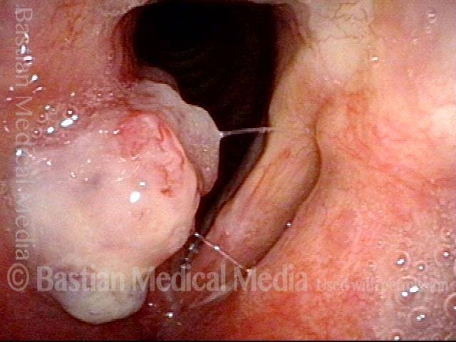
Squamous cell carcinoma (1 of 6)
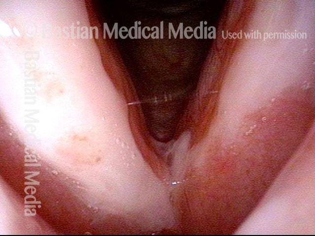
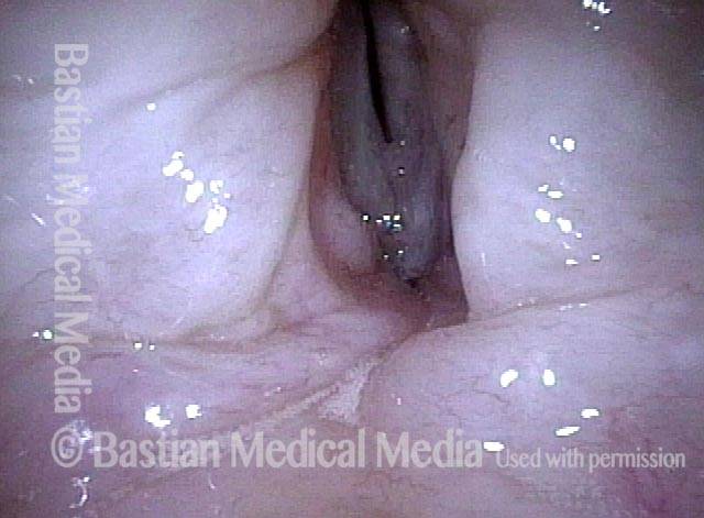
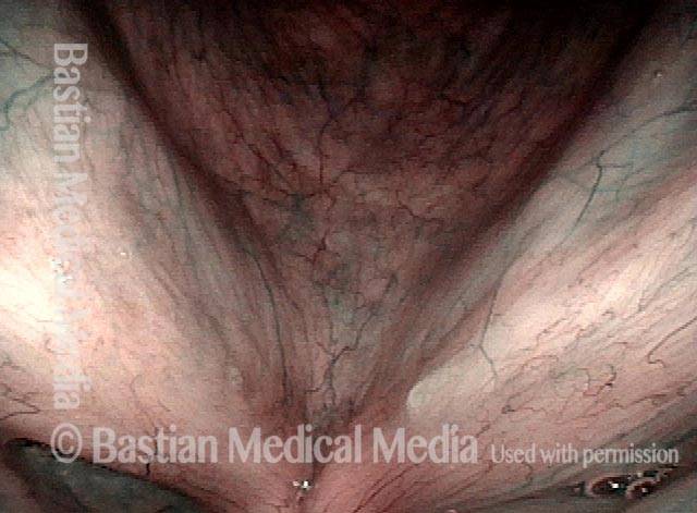
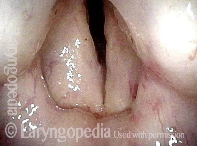
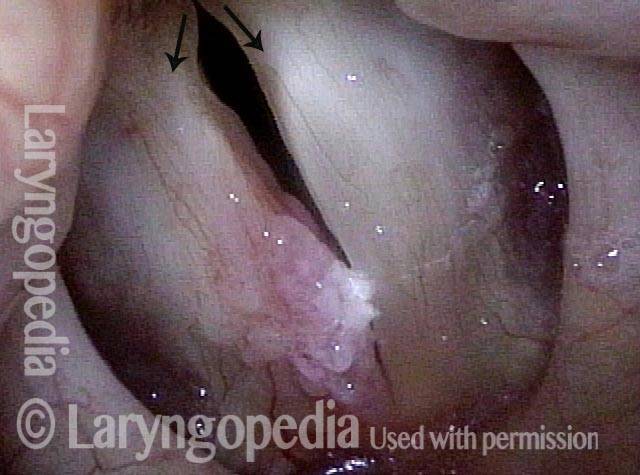
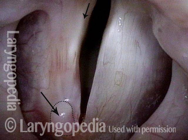
Tumor on the vocal cords (2 of 6)
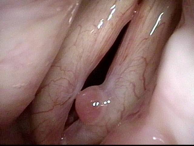
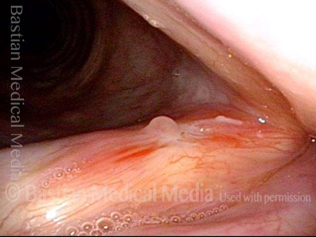
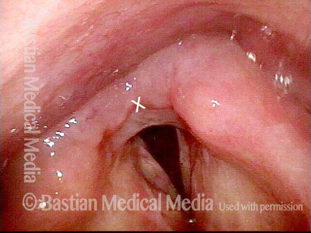
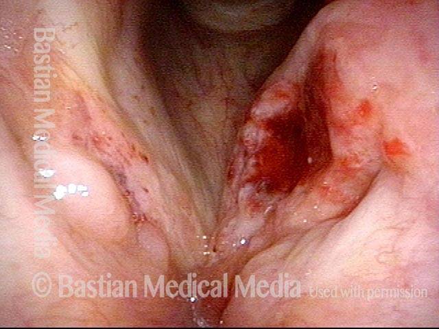
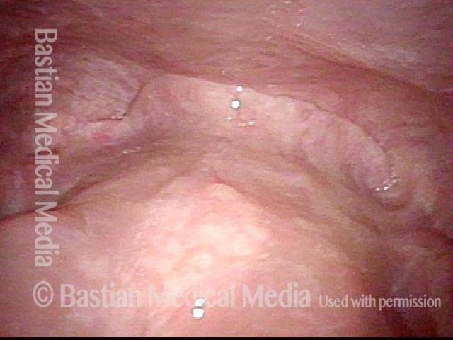
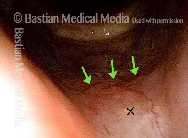
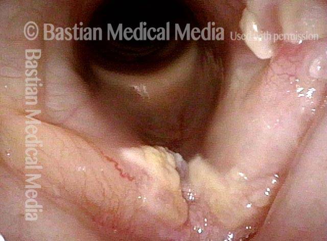
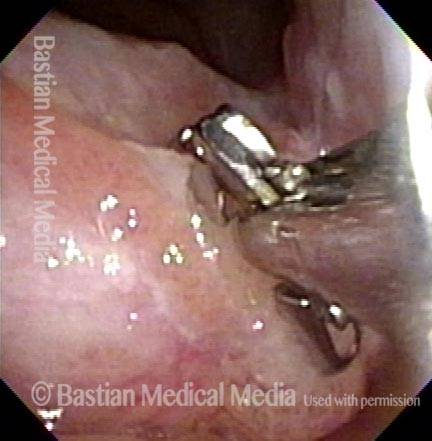
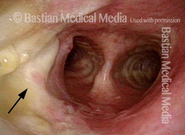
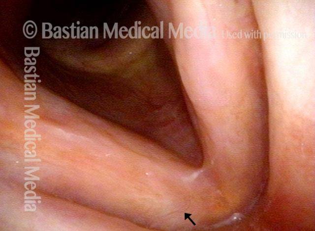
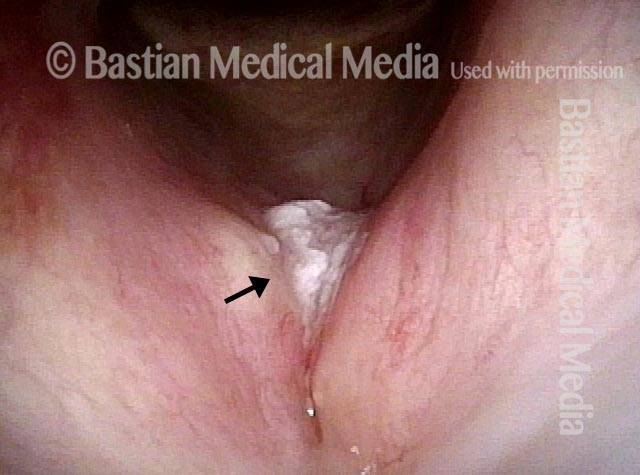
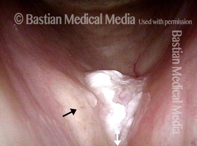
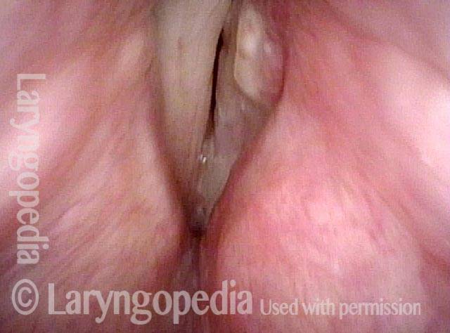
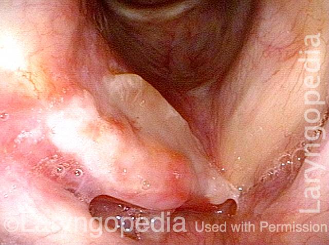
Granuloma delays voice recovery (3 of 6)
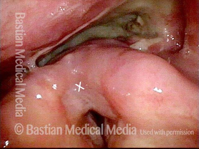
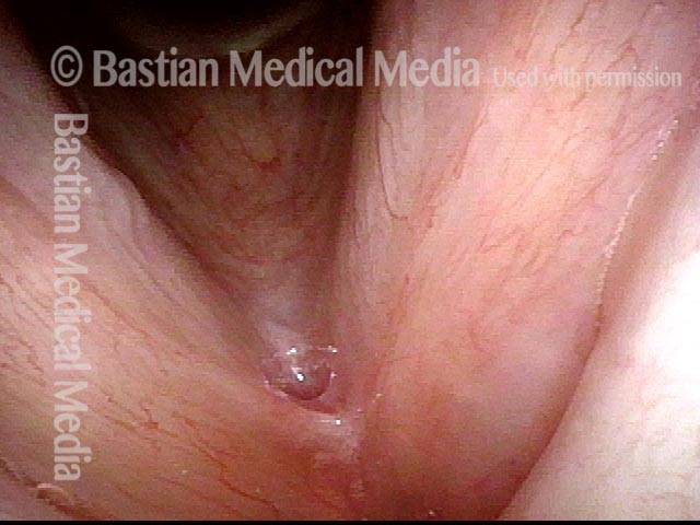
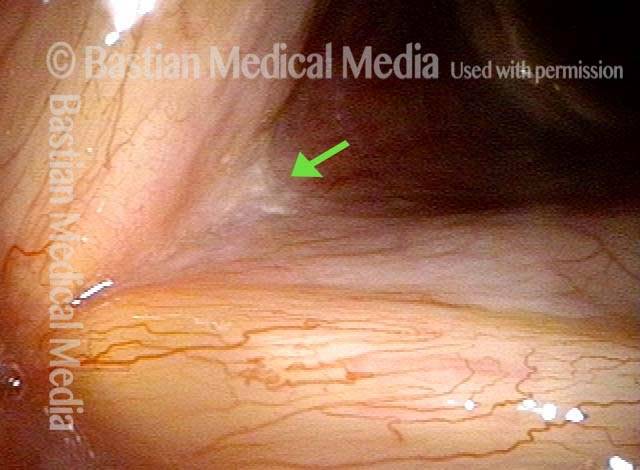
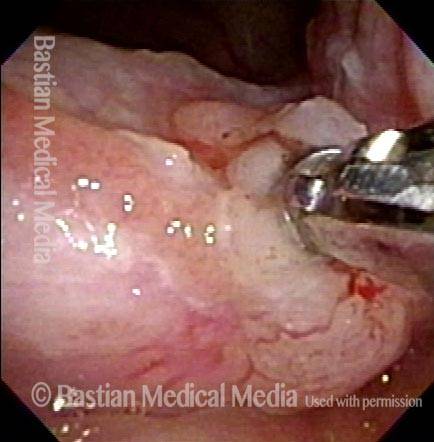
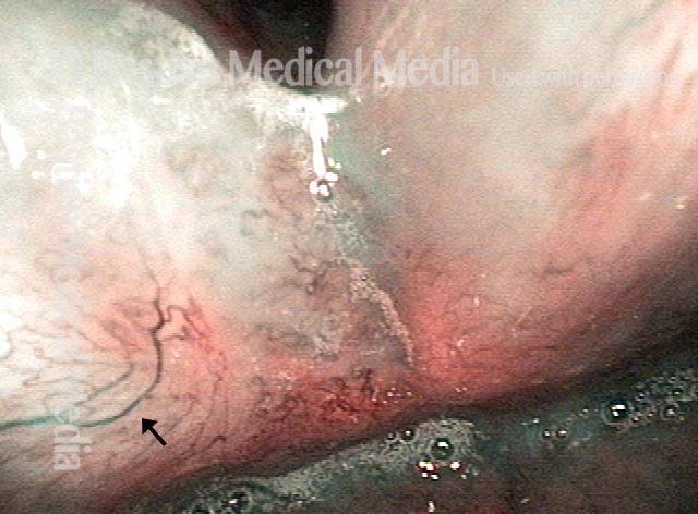
Closer view of granuloma (4 of 6)
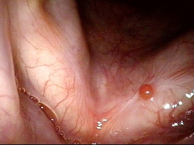
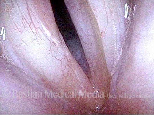
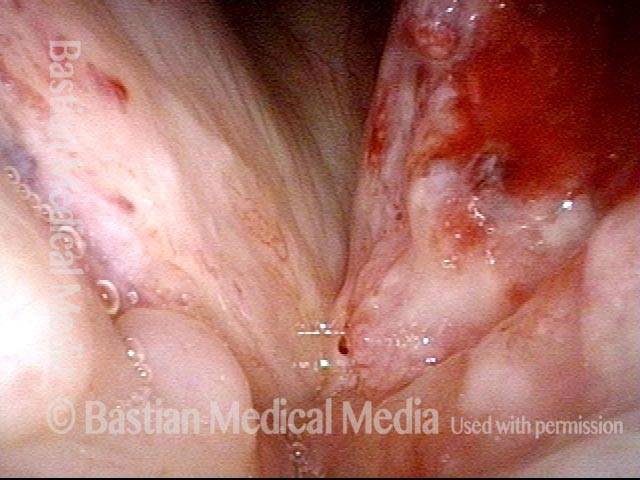
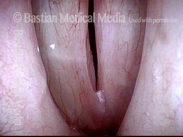
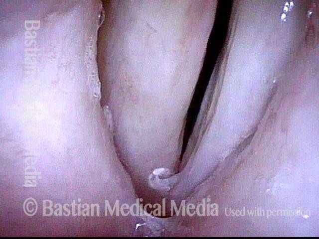
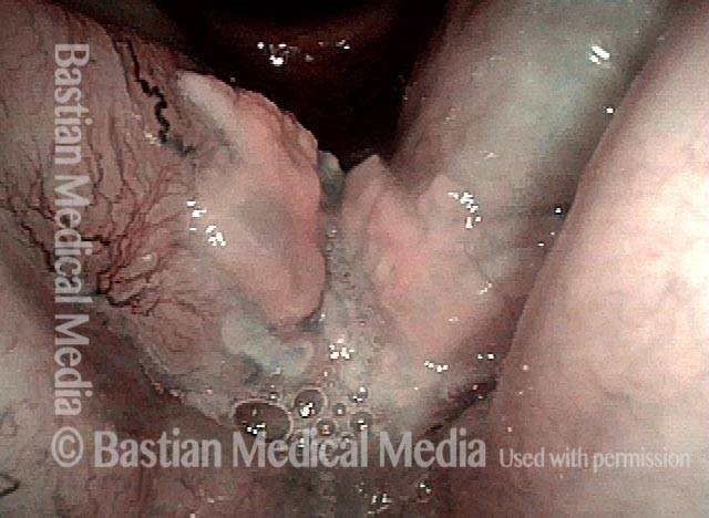
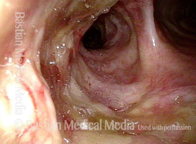
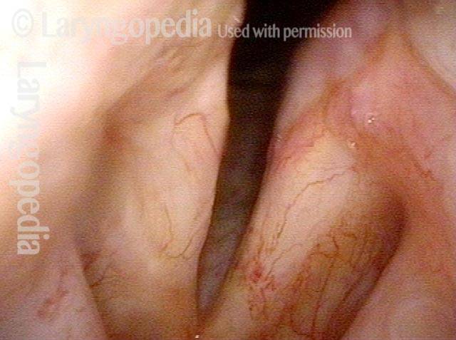
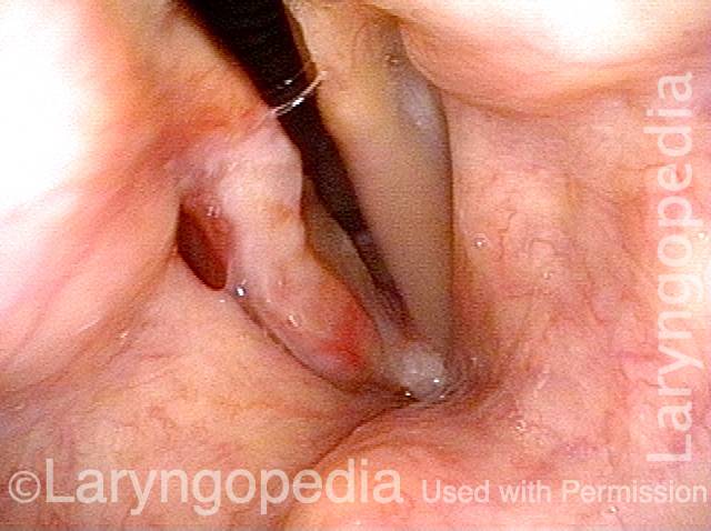
Granuloma is smaller (5 of 6)
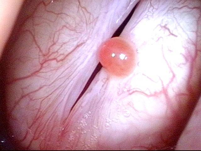
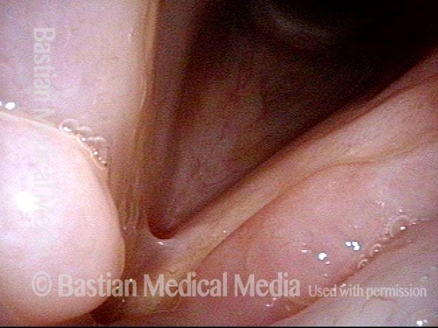
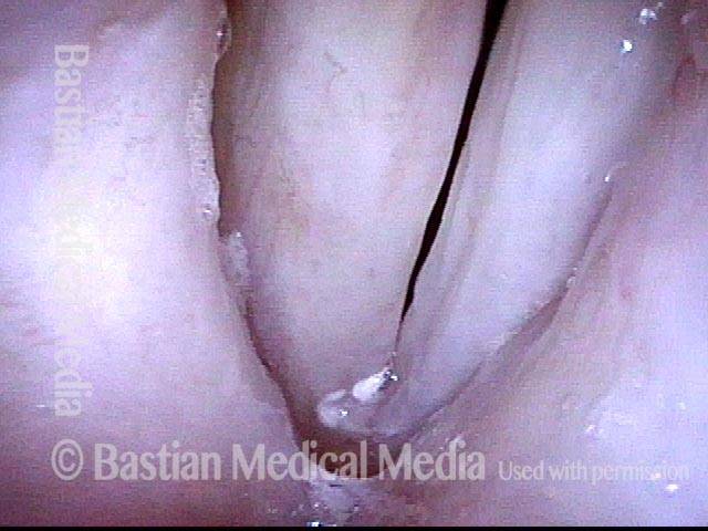
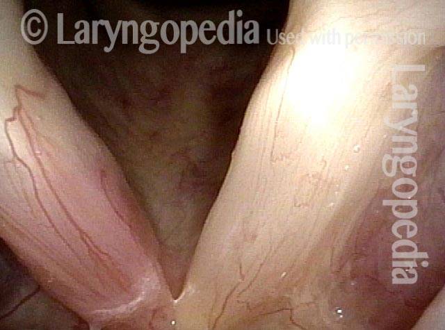
Granuloma doesn’t impede voice (6 of 6)

Vocal Cord Cancer, before and after Surgery
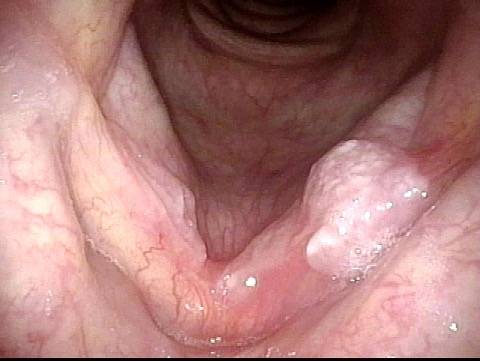
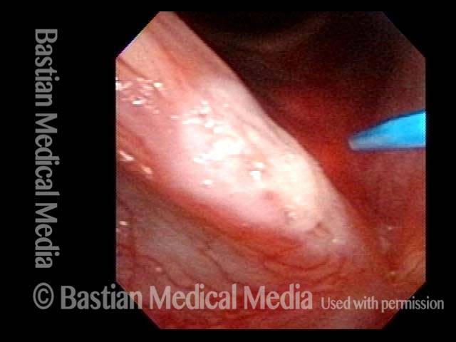
Vocal cord cancer (1 of 8)
Vocal cord cancer (2 of 8)
Vocal cord cancer, 1 week after surgery (3 of 8)
Vocal cord cancer, 1 week after surgery (4 of 8)
Vocal cord cancer, 7 weeks after surgery (5 of 8)
Vocal cord cancer, 7 weeks after surgery (6 of 8)
Vocal cord cancer, 7 weeks after surgery (7 of 8)
Vocal cord cancer, 7 weeks after surgery (8 of 8)
Example 2
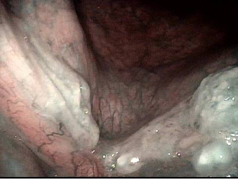
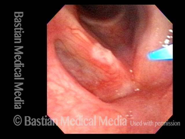
Vocal cord cancer (1 of 4)
Vocal cord cancer, 1 week after surgery (2 of 4)
Vocal cord cancer, 1 month after surgery (3 of 4)
Vocal cord cancer, after complete healing (4 of 4)
Glottic Cancer, Laser Removal
Glottic cancer, laser removal (1 of 3)
Glottic cancer, laser removal (2 of 3)
Glottic cancer, laser removal (3 of 3)
Glottic Cancer, After Surgery
Glottic cancer, after surgery (1 of 3)
Glottic cancer, after surgery (2 of 3)
Glottic cancer, after surgery (3 of 3)
Breaking cancer “rules” intelligently with use of laser
Post radiotherapy stage (1 of 8)
Laser surgery typically not acceptable (2 of 8)
Laser removal of tumor with careful followup (3 of 8)
Second view post laser surgery (4 of 8)
Six months post laser surgery (5 of 8)
Blood vessels stable two months post surgery (6 of 8)
Open phase of false vocal cord phonation (7 of 8)
Closed phase of false vocal cord phonation (8 of 8)
Glottic/vocal cord cancer
Glottic/vocal cord cancer (1 of 2)
Glottic/vocal cord cancer (2 of 2)
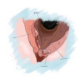
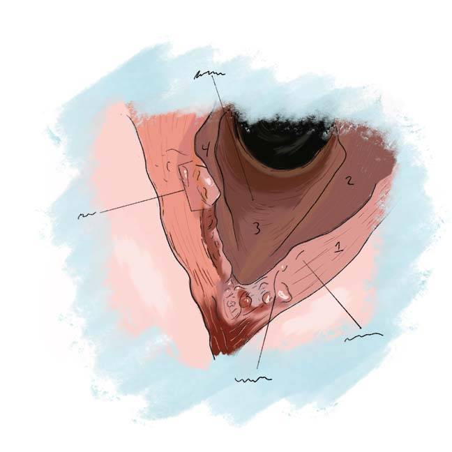
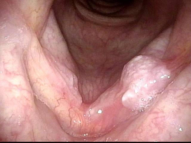
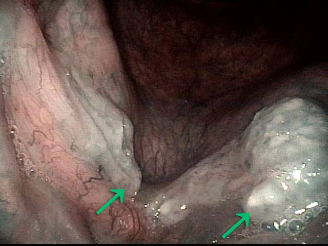
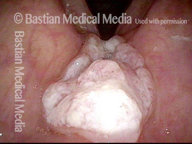
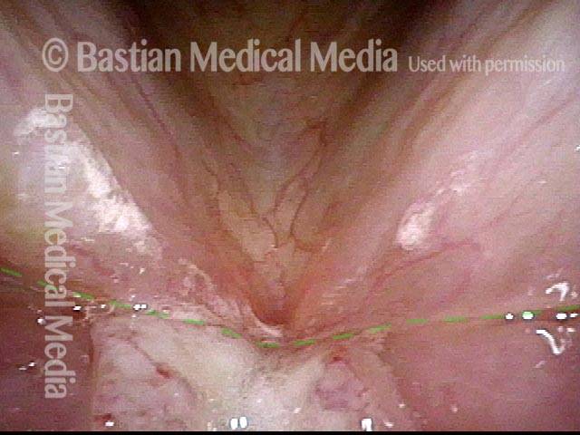
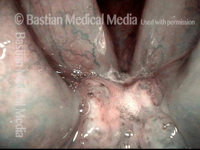
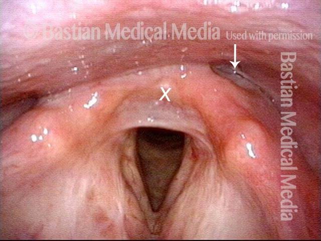
Supraglottic cancer
Supraglottic cancer (1 of 4)
Supraglottic cancer (2 of 4)
Supraglottic cancer (3 of 4)
Supraglottic cancer (4 of 4)
Hypopharyngeal Cancer, before and after Surgery
Hypopharyngeal cancer (1 of 10)
Hypopharyngeal cancer (2 of 10)

Hypopharyngeal cancer (3 of 10)
Hypopharyngeal cancer (4 of 10)
Hypopharyngeal cancer: 1 week after surgery (5 of 10)
Hypopharyngeal cancer: 1 week after surgery (6 of 10)
Hypopharyngeal cancer: 1 week after surgery (7 of 10)
Hypopharyngeal cancer: 1 week after surgery (8 of 10)
Hypopharyngeal cancer: several months after surgery (9 of 10)
Hypopharyngeal cancer: several months after surgery (10 of 10)
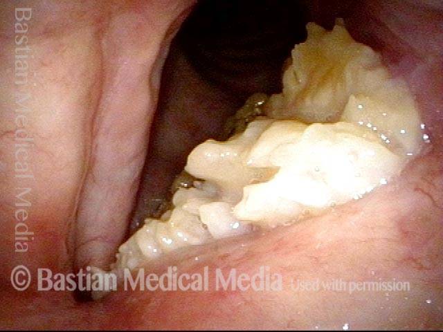
Verrucous Carcinoma, before and after Laser Treatment
Verrucous carcinoma (1 of 5)
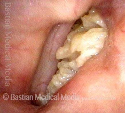
During voicing (2 of 5)
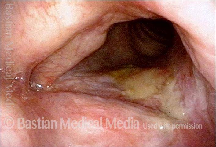
After laser treatment (3 of 5)
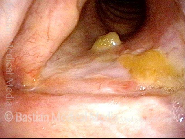
Mucus remains several weeks after laser treatment (4 of 5)
Much improvement several weeks after laser treatment (5 of 5)
Cancer, HPV Subtype 16, before and after Radiation
Cancer: HPV Subtype 16 (1 of 5)
Cancer: HPV Subtype 16 (2 of 5)
Cancer: HPV Subtype 16, after radiation therapy (3 of 5)
Cancer: HPV Subtype 16, after radiation therapy (4 of 5)
Cancer: HPV Subtype 16, after radiation therapy (5 of 5)
Vocal Cord Cancer, before, during, and after Radiation
Vocal cord cancer (1 of 7)

Vocal cord cancer (2 of 7)
Vocal cord cancer (3 of 7)
Vocal cord cancer, 3 weeks after radiotherapy (4 of 7)
Vocal cord cancer, 3 weeks after radiotherapy (5 of 7)
Vocal cord cancer, 2 months after radiotherapy (6 of 7)
Vocal cord cancer, 2 months after radiotherapy (7 of 7)
Example 2
Vocal cord cancer (1 of 8)
Vocal cord cancer (2 of 8)
Vocal cord cancer, during radiation (3 of 8)
Vocal cord cancer, during radiation (4 of 8)
Vocal cord cancer, 2 months after radiation (5 of 8)
Vocal cord cancer, 4 months after radiation (6 of 8)
Vocal cord cancer, 6 months after radiation (7 of 8)
Vocal cord cancer, 6 months after radiation (8 of 8)
Radiation Induced Web
Post radiation therapy (1 of 4)
Web formation (2 of 4)
Closer view of ulceration and web (3 of 4)
Flexible scope used to separate vocal cords (4 of 4)
Larynx Cancer Managed like “skin cancer”
Two years after excision (1 of 4)
Narrow-band lighting (2 of 4)
During thulium laser coagulation (3 of 4)
After thulium laser coagulation (4 of 4)
Vocal cord CA—a Case for Radiation Instead of Laser Resection
Long term smoker (1 of 4)
Tumor (2 of 4)
Radiation therapy suggested (3 of 4)
Two months later (4 of 4)
Scarring after Cancer Treatment but with Very Good Voice
Post laser excision (1 of 4)
Prephonatory instant (2 of 4)
Phonation (3 of 4)
Close-up view (4 of 4)
Cancer Beginning to Block Airway
Formerly heavy smoker (1 of 2)
Closer view (2 of 2)
Breaking the Rules in Larynx Cancer
Recurrent cancer (1 of 8)
Closer view (2 of 8)
One week post laser resection (3 of 8)
One year postop (4 of 8)
True cord phonation (5 of 8)
False vocal cords (6 of 8)
False vocal cord closure (7 of 8)
True cords (8 of 8)
Biopsy of Early Vocal Cord Cancer
Chronic hoarseness (1 of 3)

Just before biopsy (2 of 3)
Cancer finding (3 of 3)
Tumor in Trachea
Biopsy (1 of 4)
After biopsy (2 of 4)
Tumor gone (3 of 4)
Slow return (4 of 4)
HPV 31 Cancer Cure
Carcinoma in situ (1 of 4)
HPV subtype 31 (2 of 4)
Excisions (3 of 4)
Seven years later (4 of 4)
Before and after Radiation for Vocal Cord Cancer
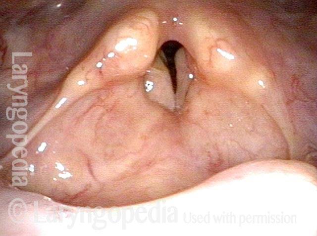
Bilateral vocal cord cancer (1 of 4)
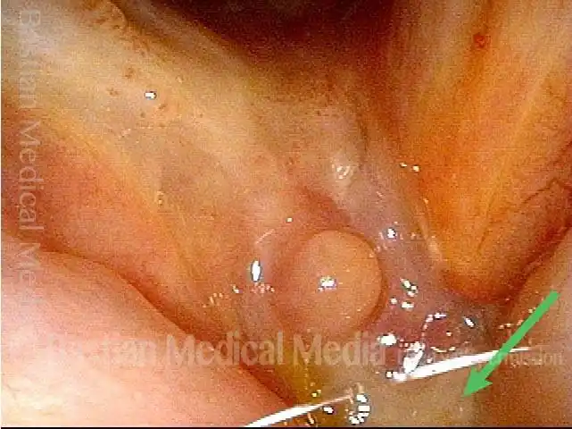
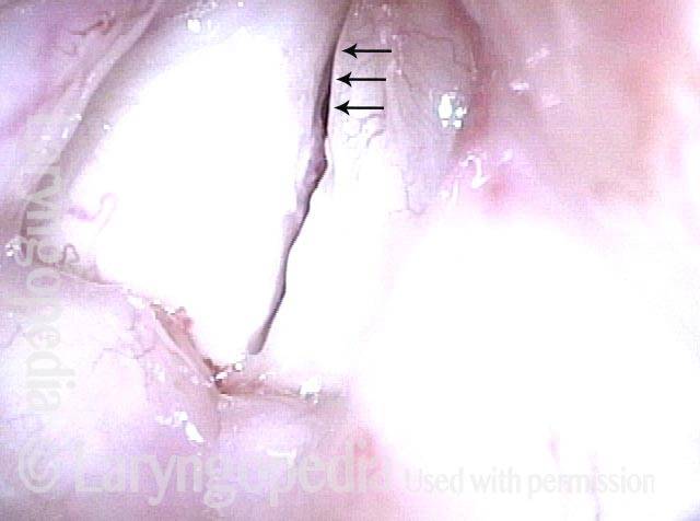
“Tumor vessels” (2 of 4)
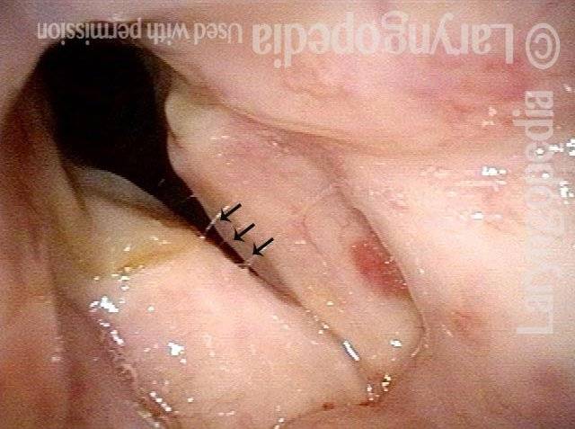
After radiotherapy (3 of 4)
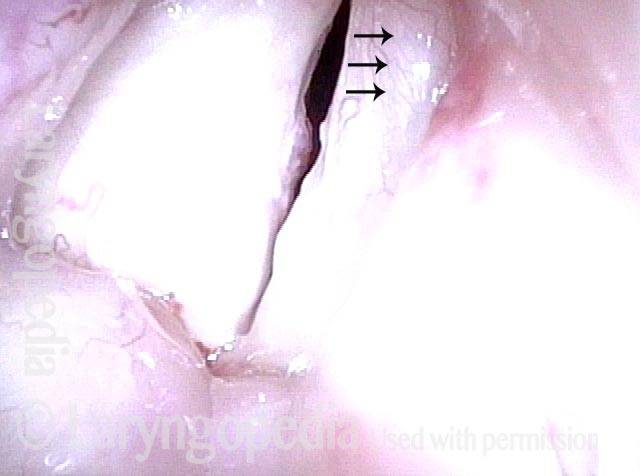
Vascular pattern (4 of 4)
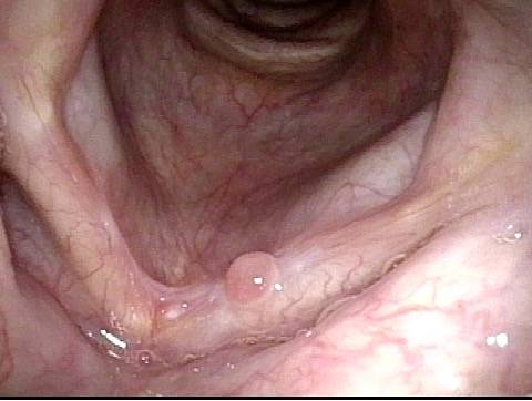
Small, but Dangerous!
Post radiotherapy (1 of 4)
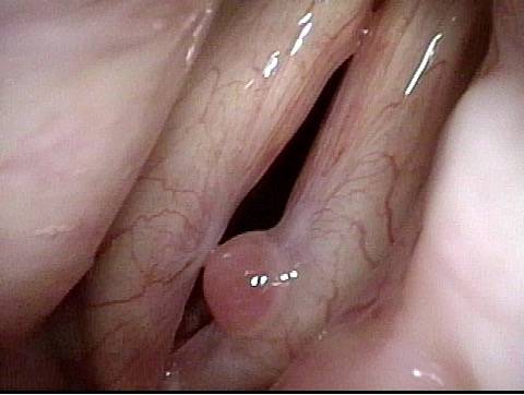
Normal voice (2 of 4)
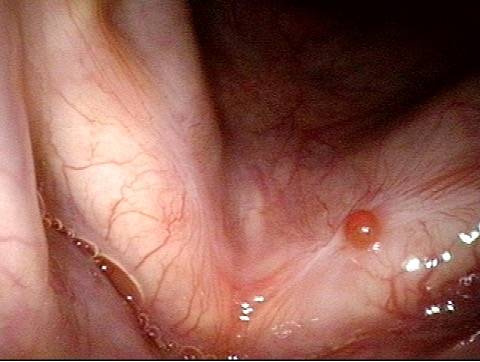
Persistent cancer (3 of 4)
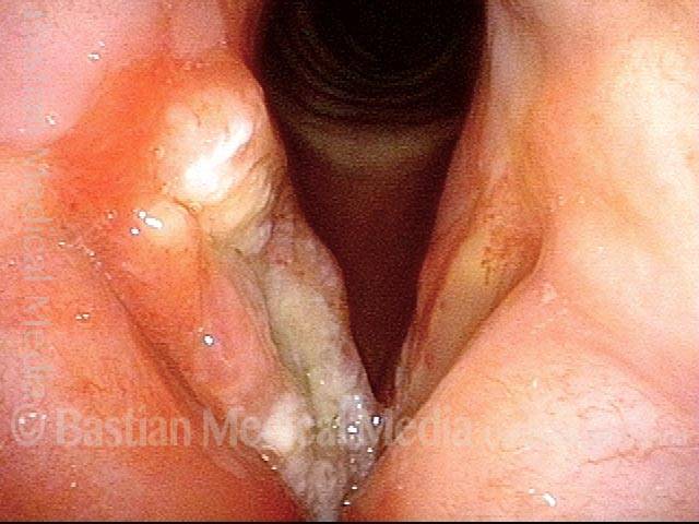
T4 tumor (4 of 4)
Unusual Posterior and Transglottic Epicenter for Larynx Cancer
Tumor (1 of 4)
Accentuation of the vascularity (2 of 4)
Posterior commissure (3 of 4)
A year later (4 of 4)
Nice try, but on to Radiation
Vocal cord lesion (1 of 4)
Closer view (2 of 4)
Removal of tumor (3 of 4)

Nice try, but on to radiation (4 of 4)
Progressive Cricoarytenoid Joint Fibrosis / Fixation as a Late Complication of Radiation
25 years post radiotherapy (1 of 4)
Fibrosis (2 of 4)
Closed phase (3 of 4)
Open phase (4 of 4)

Progressive Radiation Fibrosis Effects on the Larynx and a Solution to some of it
Forty years post-radiation (1 of 8)
Involuntary inspiratory voice (2 of 8)
Only capable of high pitch (3 of 8)
Open phase vibration (4 of 8)
One week post-commissuroplasty (5 of 8)
Rapid inhalation, closer view (6 of 8)
Three months post-surgery (7 of 8)
Closer view, post-surgery (8 of 8)
Evolution of the Wound after Laser Removal of a vocal Cord Cancer: Not Pretty at First, but Voice Result can be very Good
Vocal cord cancer (1 of 8)
Voice-making with tumor (2 of 8)
One week post-removal (3 of 8)

Voice-making, post-removal (4 of 8)
Six weeks post-op (5 of 8)
Voice-making, post-op (6 of 8)
Four months post-op (7 of 8)
Voice-making, four months post-op (8 of 8)
Laser can Beat Cancer and Spare Voice even after Radiotherapy Failure
Healing post-laser excision(4 of 4)
Post-laser excision (3 of 4)
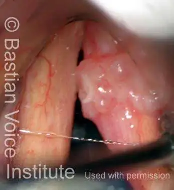
Leukoplakia and Stippled Vascularity(2 of 4)
Return of cancer (1 of 4)
How a Vocal Cord Heals after Laser Removal of a Cancer
squamous cell carcinoma (1 of 6)
1 week after laser excision (2 of 6)
Difficulty speaking (3 of 6)
Natural granulation after laser excision (4 of 6)
Abnormal capillary pattern (5 of 6)
Stiff right vocal cord (6 of 6)
