A chondroma is a benign growth composed of cartilage cells. For whatever reason (trauma or de novo), cartilage cells are overproduced in a focal area, creating a lump in the contour of the thyroid, cricoid, or arytenoid cartilages. The key question is whether this accumulation of extra cartilage is benign (chondroma) or malignant (chondrosarcoma). Another question is whether it interferes with function. If benign and not interfering with function, chondromas would need monitoring just long enough to ensure stability across time.
Chondroma of Thyroid Cartilage
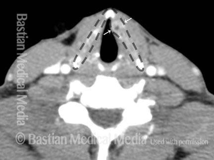
CT scan of the larynx (1 of 4)
CT scan of the larynx, showing the thyroid cartilage (outlined by gray dotted lines) and an abnormality deforming the thyroid cartilage on one side (between the white arrows). Note how the thyroid cartilage bulges on that side, as compared with the opposite side, and the black speck which indicates varying densities in the cartilage.
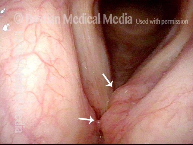
Endoscopic View of the Larynx (2 of 4)
Same patient, endoscopic view of the larynx, again showing the abnormality (at arrows). Here the abnormality looks similar to a saccular cyst, but the scan (and subsequent biopsy) shows that it is cartilaginous and a chondroma, not chondrosarcoma.
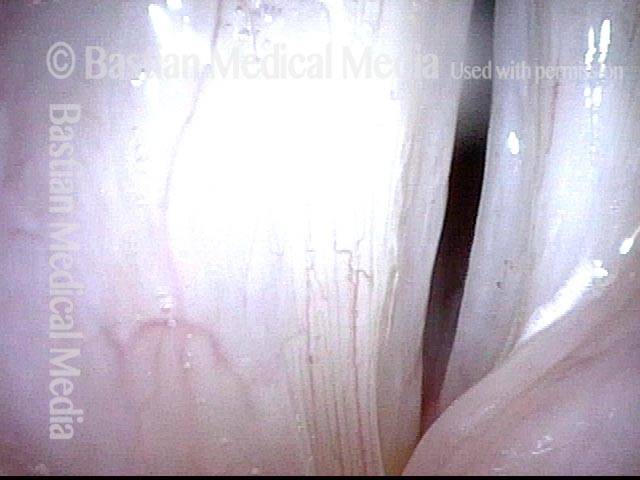
Chondroma of thyroid cartilage (3 of 4)
Closer view of the chondroma, showing an almost bi-lobed appearance.
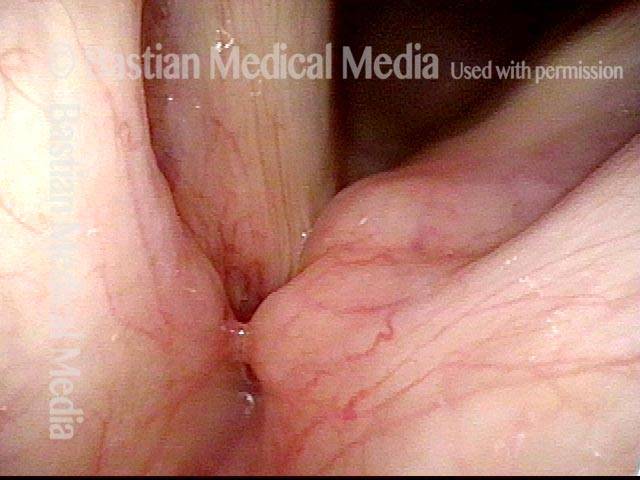
Left Vocal Cord Sits Lower Then the Right (4 of 4)
Under strobe lighting, which shows that the left vocal cord (right of photo) is apparently at a lower level than the opposite cord.