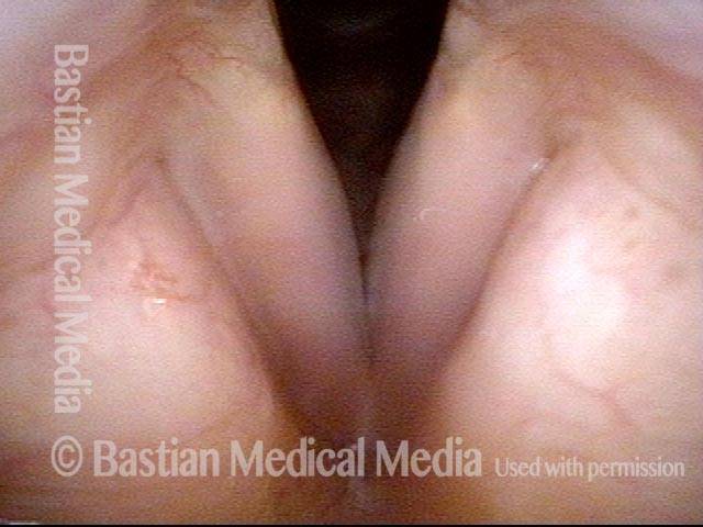
Polipi del fumatore, prima e dopo l’intervento chirurgico (audio con foto)
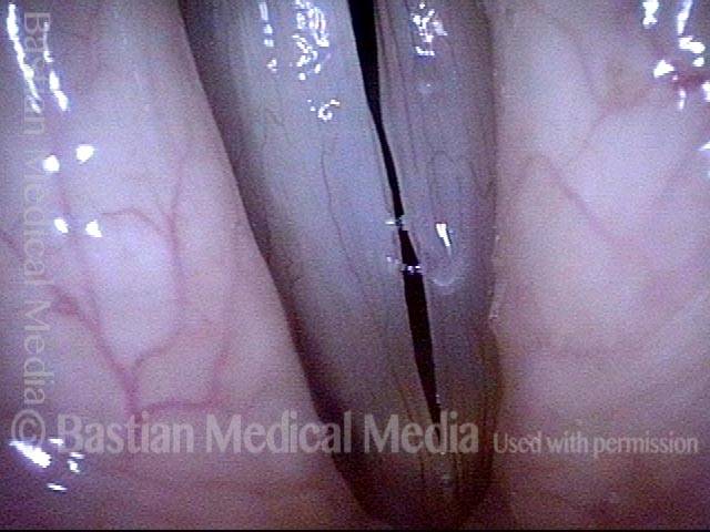
Campione vocale di un paziente con polipi del fumatore, PRIMA dell’intervento chirurgico (vedere le foto di questo paziente appena sotto): Stesso paziente, due mesi DOPO l’intervento chirurgico (le occasionali interruzioni di sillabe sono dovute alla recente chirurgia):Smoker’s polyps, BEFORE surgery (1 of 4)
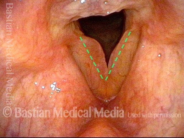
Even during quiet breathing, the convexity of the vocal cord margins (dotted lines show where normal margins would be) reveal the presence of smoker’s polyps.
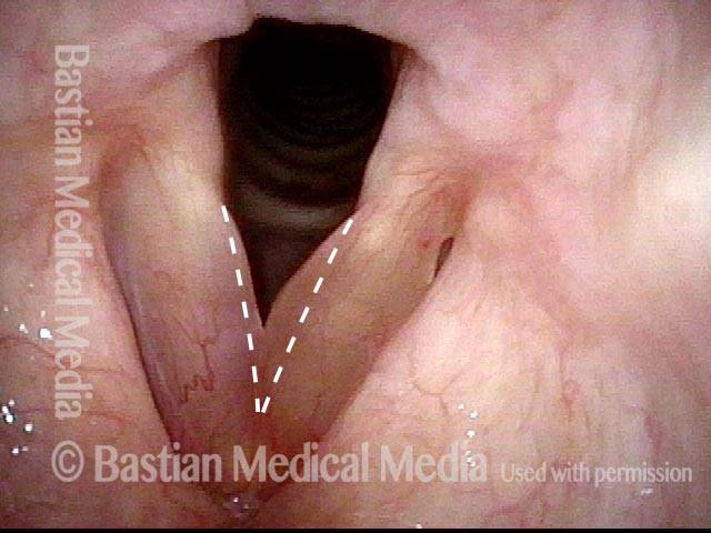
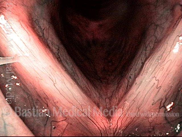
Smoker’s polyps, BEFORE surgery (2 of 4)
During inspiratory phonation: the polyps are drawn inward and are easier to see.
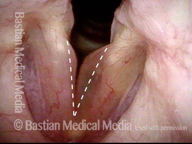
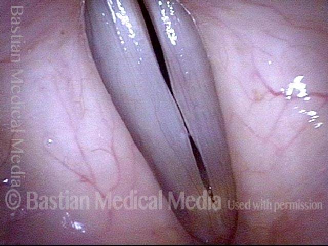
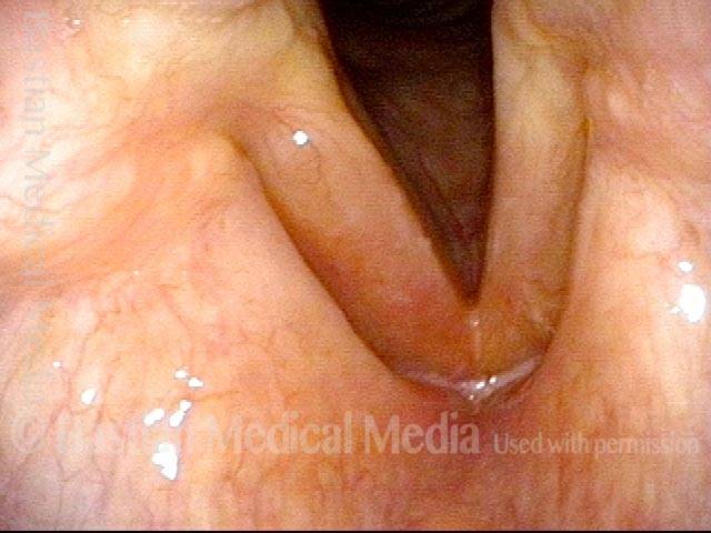
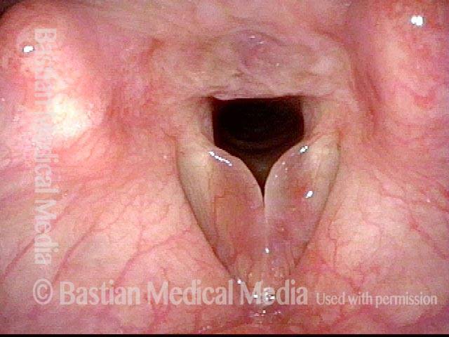
Smoker’s polyps, AFTER surgery (3 of 4)
Two months after surgery, during quiet breathing. The vocal cord margins are now straight.
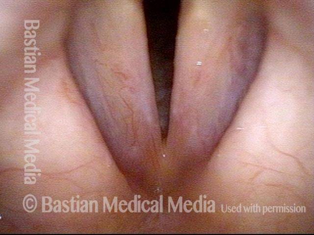
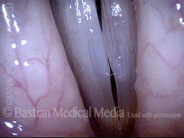
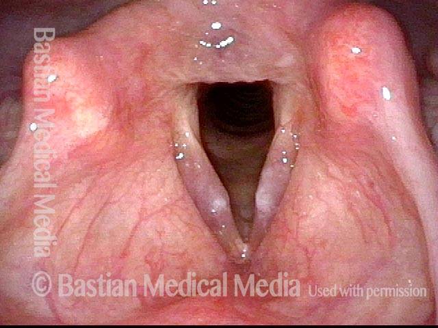
Smoker’s polyps, AFTER surgery (4 of 4)
During inspiratory phonation: the margins are drawn into a mildly convex contour, but far less than preoperatively. The patient’s voice is also much improved, albeit the occasional syllable dropouts due to recentness of surgery (listen to this patient’s voice samples in the audio section of the encyclopedia entry).
Noduli vocali polipoidi
Polypoid vocal nodules (1 of 4)
Polypoid vocal nodules in a “vocal overdoer” with phenomenology typical for a mucosal injury. Narrow band illumination (blue-green light) makes vasculature more prominent. Note also the fusiform (long, low-profile) swelling, best seen on the left cord (right of image).
Incomplete closure (2 of 4)
Phonation, strobe light, at the beginning of the closed phase of vibration; one can see that closure will be incomplete due to early contact of the polypoid nodules.
Polypoid vocal nodules (3 of 4)
Phonation, strobe light, closed phase of vibration, with persistent gaps anterior and posterior to the polypoid nodules.
Polypoid vocal nodules (4 of 4)
Phonation, strobe light, open phase of vibration, continues to show the mid-cord swellings.
Polipi del fumatore/Edema di Reinke
Smoker’s polyp / Reinke’s edema (1 of 2)
Quiet breathing, under standard light. The edematous mucosa is not yet evident.
Smoker’s polyp / Reinke’s edema (2 of 2)
Elicited inspiratory phonation in-draws and thereby reveals the edematous mucosa, greater on the right (left of image) than the left. The dashed lines indicate the normal location and contour of the vocal cords’ free margins.
Esempio 2
Smoker’s polyps / Reinke’s edema (1 of 3)
This patient is a long-term smoker, and also is talkative. Her voice has been gradually deepening for years. Here, with the vocal cords in abducted breathing position, one can only see somewhat underwhelming, broad-based, low-profile swelling, along with some hazy leukoplakia in the mid-cord.
Smoker’s polyps / Reinke’s edema (2 of 3)
Phonation. Again, there is only very low-profile, broad-based convexity of the margins, and again, the hazy leukoplakia in the mid-cords.
Smoker’s polyps / Reinke’s edema (3 of 3)
Elicited inspiratory phonation. Now, one can see that, contrary to the appearance in the prior two views, this patient in fact has moderate-sized “smoker’s-type” polyps, aka Reinke’s edema. The increased mass explains the virilization of the sound of this woman’s voice.
Polipi del fumatore in varie “pose”
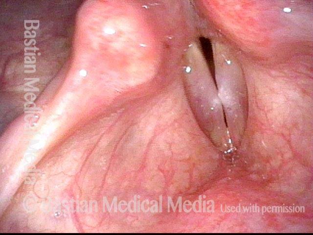
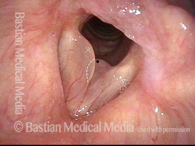
Smoker’s polyps in various “poses” (1 of 4)
Vocal cord abduction for breathing, during expiratory phase. Left polyp (right of photo) appears to be the only finding. This is in a middle aged smoker with several years of gradually deepening / masculinized and now rough voice. The black dot and white “X” are reference points, facilitating comparisons with the other photos.
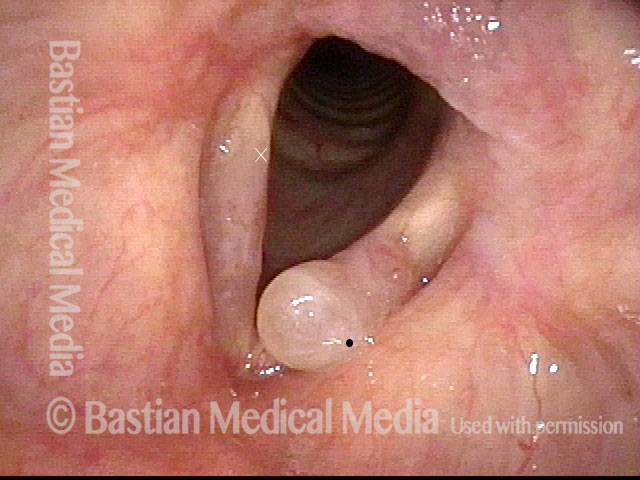
Polyp begins to fall off (2 of 4)
At the beginning of elicited rapid inspiration, showing the polyp beginning to be displaced from upper surface to the margin. That is, previously-unseen polypoid tissue (at ” X”) is now indrawing from upper surface of the right cord (left of photo) as well, and margin has become convex rather than straight as it was in photo 1.
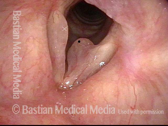
Polyps displace (3 of 4)
The left-sided polyp (right of photo) is now displaced below the margin of that cord. The right polyp (left of photo) is now fully displaced/ indrawn to the margin of the right cord (left of photo).
Edematous tissue causes a rough voice (4 of 4)
During voice-making, most of the edematous tissue relocates back to the upper surface of the cords where it vibrates chaotically to add not only masculine but also rough voice quality.