Papillomatosi respiratoria ricorrente (RRP) e altre lesioni indotte da HPV

Un disturbo in cui tumori simili a verruche o altre lesioni crescono in modo ricorrente all’interno delle vie aeree di una persona. Queste escrescenze sono causate dal papillomavirus umano (HPV) e possono verificarsi ovunque nelle vie aeree di una persona, come sulle corde vocali (di gran lunga il sito più comune), nella laringe sopraglottica o nella trachea. Se queste escrescenze vengono rimosse, ricresceranno quasi sempre o si ripresenteranno; quindi, “papillomatosi respiratoria ricorrente”.

Sintomi e rischi di papillomatosi respiratoria ricorrente

La RRP può essere pericolosa per la vita nei bambini piccoli, se non seguita e trattata attentamente, poiché le vie aeree di un bambino sono relativamente strette e possono potenzialmente essere completamente ostruite dalle escrescenze proliferative della malattia; inoltre, la RRP nei bambini tende a crescere ea ripresentarsi in modo più aggressivo.

Negli adulti, la RRP di solito compromette solo la funzione vocale (quando le escrescenze si verificano sulle corde vocali), sebbene possa anche compromettere la respirazione nei casi più gravi. Occasionalmente, la RRP può anche progredire verso il cancro, e quindi i pazienti che risultano essere ad alto rischio per questo (vedi sotto) devono essere monitorati attentamente.

Caratteristiche delle crescite

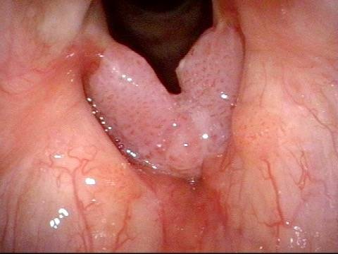
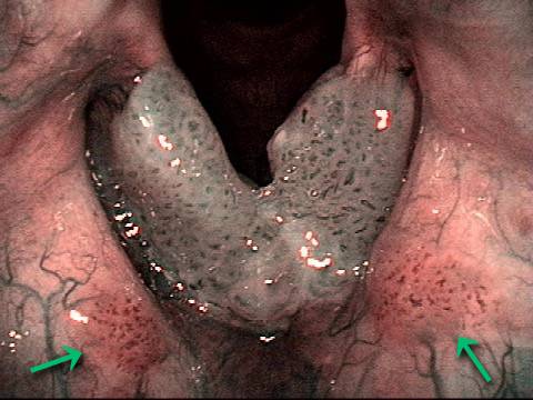
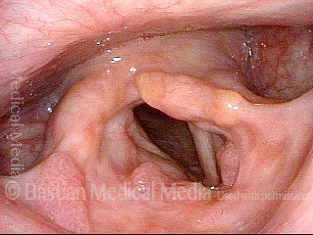
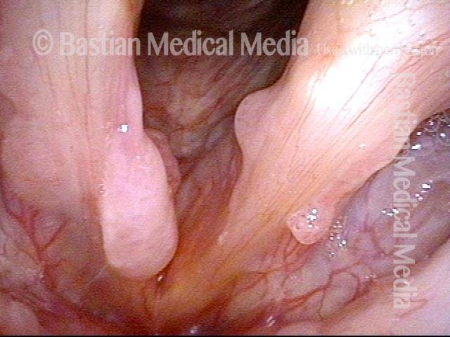
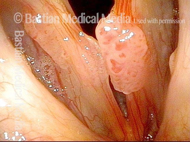
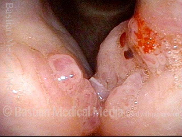
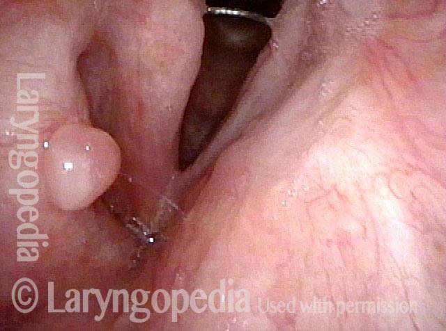
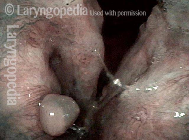
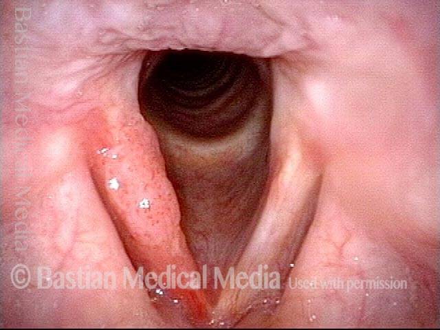
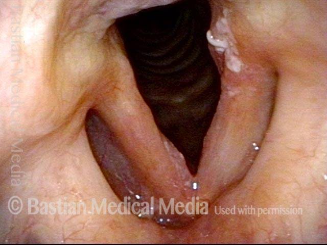
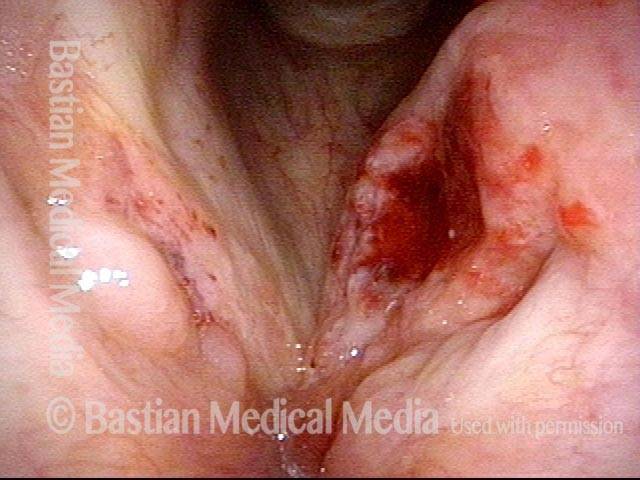
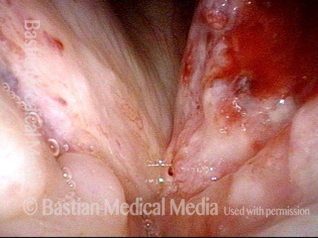
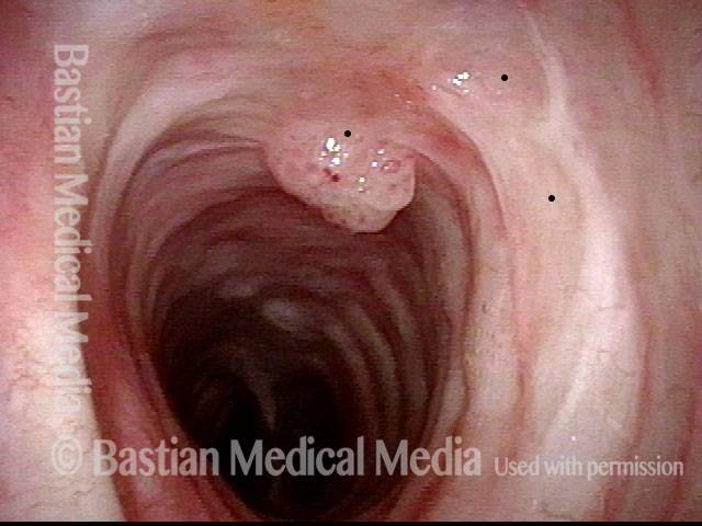
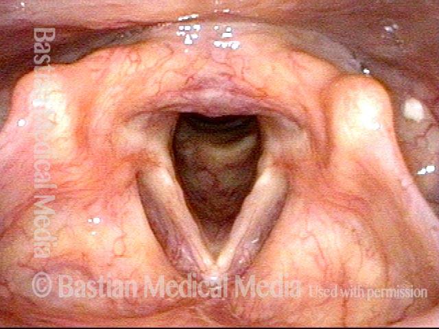
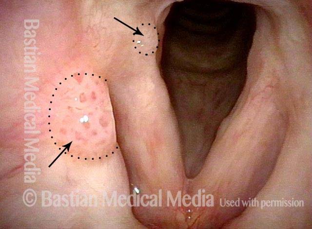
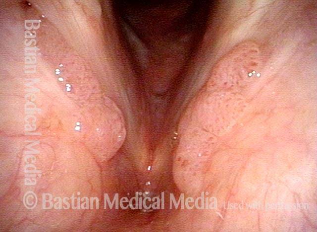
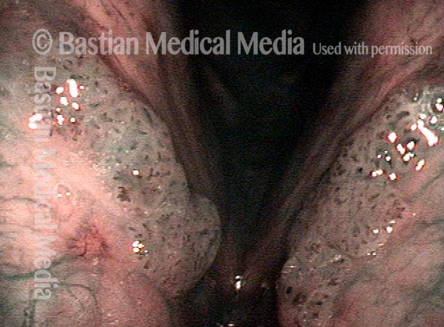
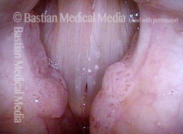
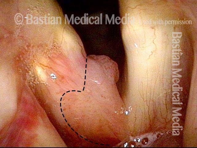
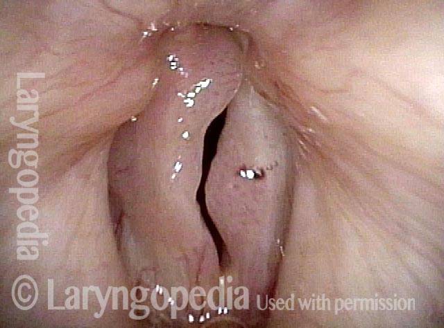
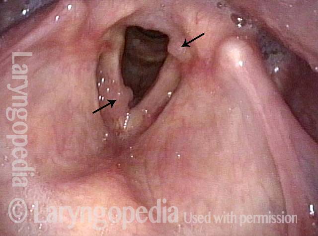
Le escrescenze solitamente associate a RRP sono tumori simili a verruche, o papillomi, che sporgono in modo evidente dalla superficie su cui crescono, spesso in grappoli simili a grappoli. Questi tipi di papillomi si osservano solitamente in pazienti con sottotipi di HPV 6 o 11, che sono entrambi sottotipi a basso rischio di contrarre il cancro.

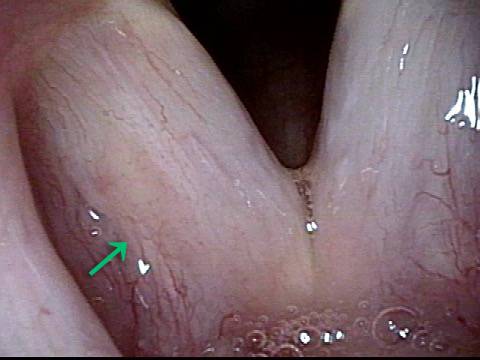
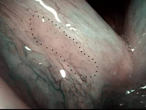
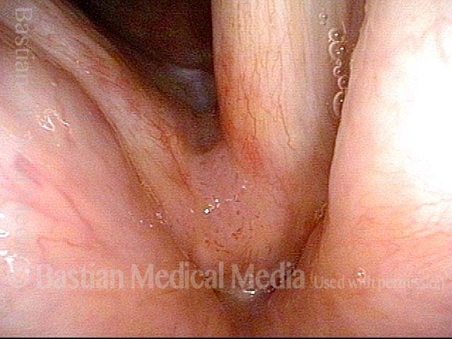
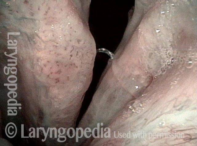
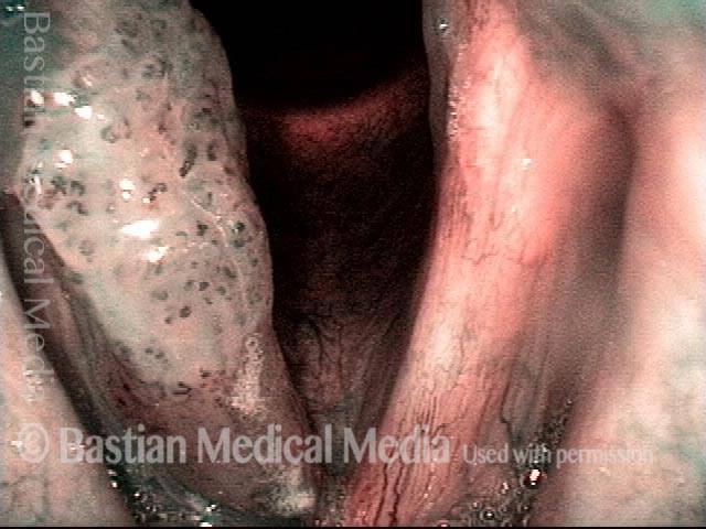
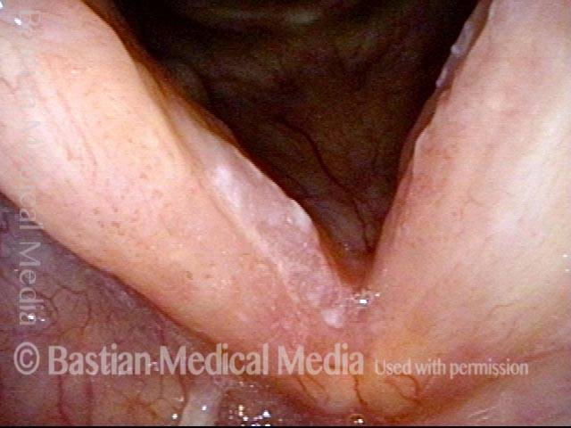
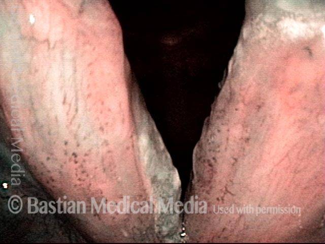
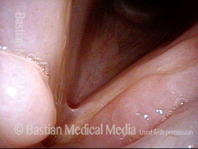
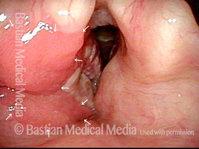
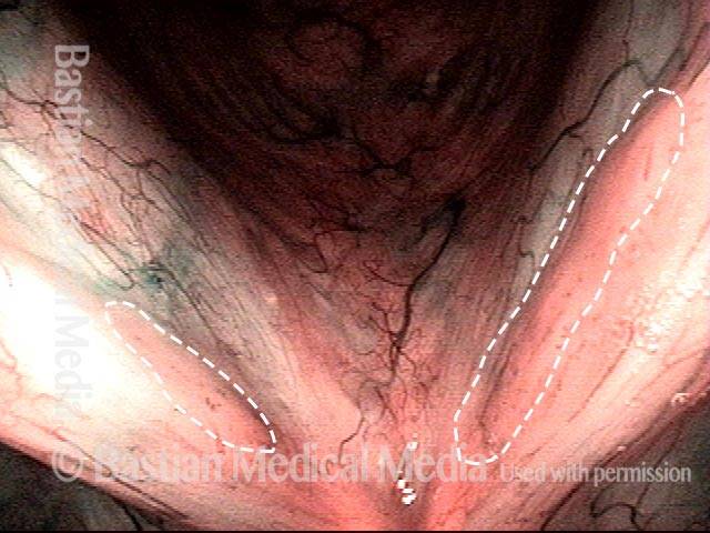
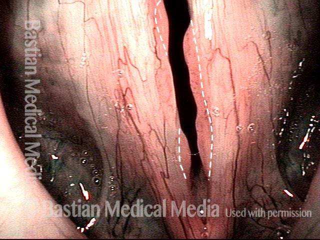
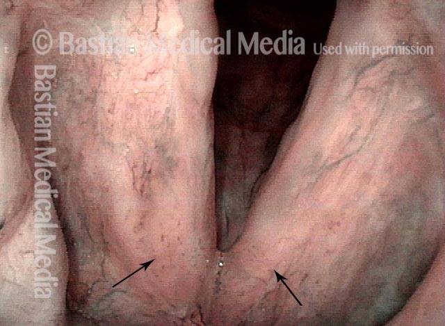
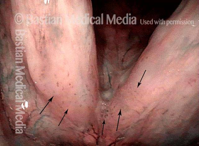
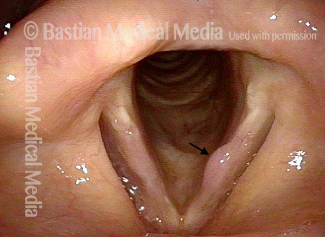
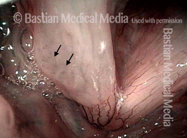
Ci sono alcuni pazienti HPV, tuttavia, che manifestano la loro infezione da HPV con escrescenze più sottili e vellutate all’interno delle vie aeree, escrescenze “varianti a tappeto”, per così dire. Sebbene queste escrescenze “varianti a tappeto” non abbiano l’aspetto simile a una verruca dei papillomi tipicamente associati a RRP, ci sono almeno alcuni punti chiave di somiglianza:

  1. Both the “carpet-variant” and wart-like growths are lesions that sometimes appear, either independently or together, in patients who have HPV;
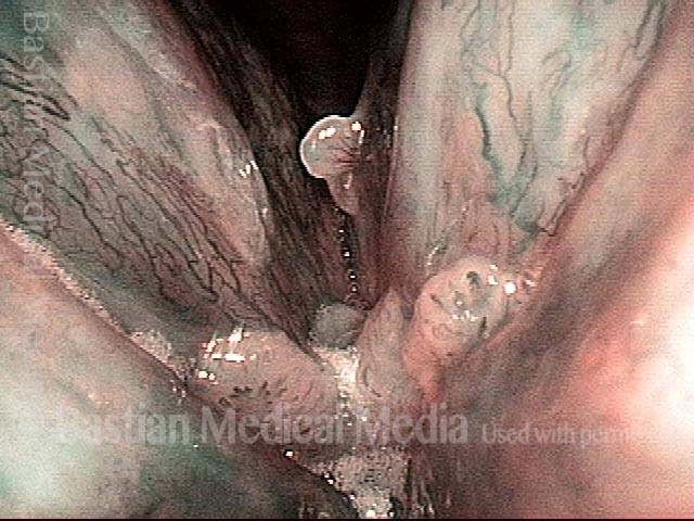
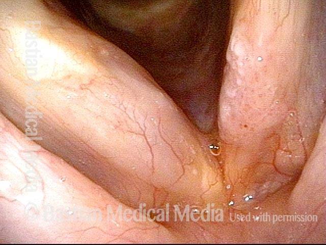
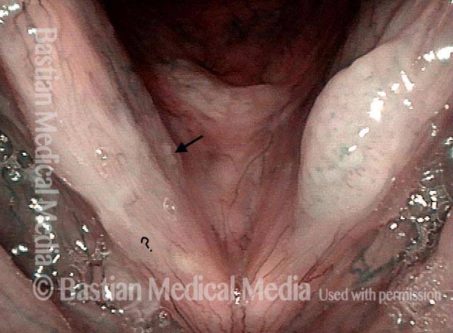
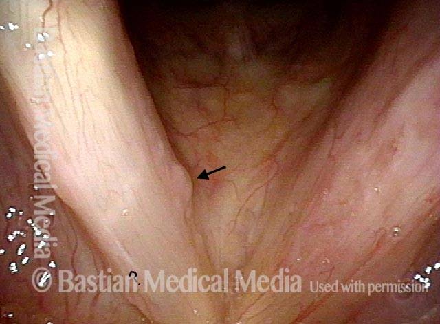
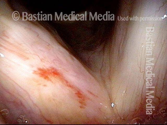
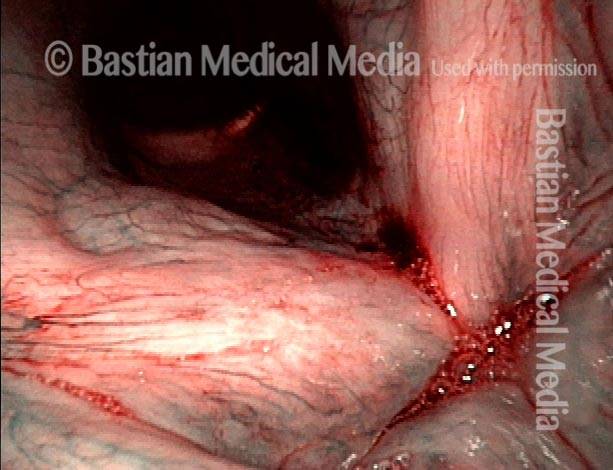
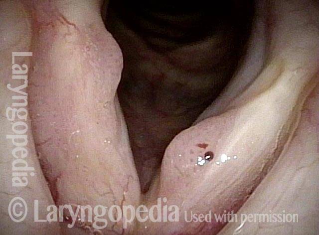
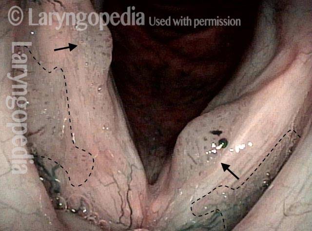
  2. Both the “carpet-variant” and wart-like growths are stippled with polka-dot vascular markings, because each “loop” in the “carpet” or each “grape” in the wart-like cluster has its own fibrovascular core, seen as a red dot;
  3. Both the “carpet-variant” and wart-like growths can disrupt voice function;
  4. Both the “carpet-variant” and wart-like growths usually recur if they are removed.

Because of these similarities, we consider these “carpet-variant” growths, even when the sole expression of the infection, to be at least a cousin to RRP, within the family of HPV-induced lesions. Many patients with this “carpet-variant” condition have HPV subtypes such as 16 or 18 that are higher-risk for cancer; such patients need to be monitored with particular care.

Trattamento della papillomatosi respiratoria ricorrente

Il trattamento primario per RRP e altre lesioni indotte da HPV è la rimozione chirurgica attenta e conservativa delle escrescenze. Poiché queste escrescenze si ripresentano quasi sempre, l’intervento chirurgico di solito deve essere eseguito su base ripetuta, ogni poche settimane nei bambini, ma in media molto meno spesso negli adulti. Un intervallo comune tra gli interventi chirurgici per i pazienti adulti è tra ogni sei mesi e ogni due anni, a seconda della velocità con cui la RRP o altra lesione correlata all’HPV si ripresenta e compromette nuovamente la funzione vocale del paziente.

Ci sono anche alcuni trattamenti medici che sono stati utilizzati in aggiunta alla chirurgia, tra cui, tra gli altri, interferone, indolo-3-carbinolo, vaccino intralesionale contro la parotite o MPR (morbillo-parotite-rosolia), cidofovir e bevacizumab.

Umiltà davanti al virus HPV: una recidiva di papillomi a dieci anni

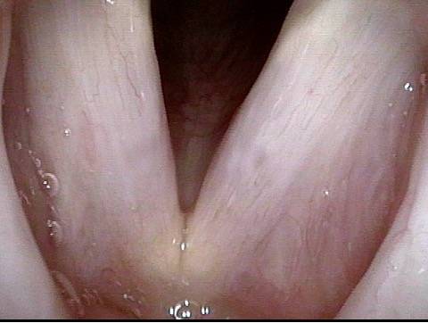
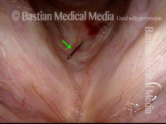
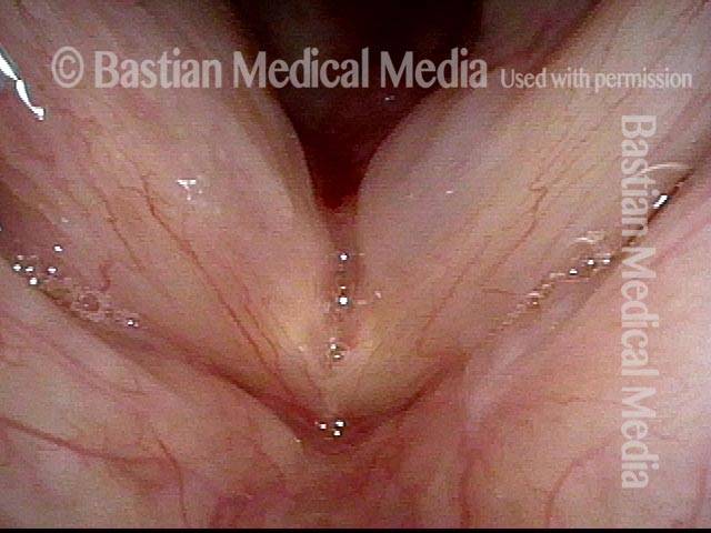
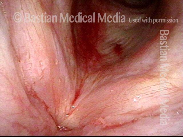
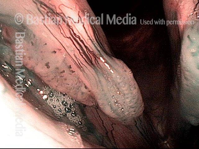
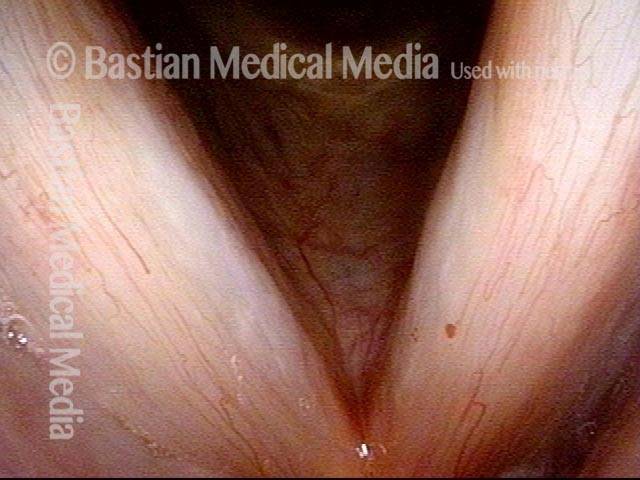
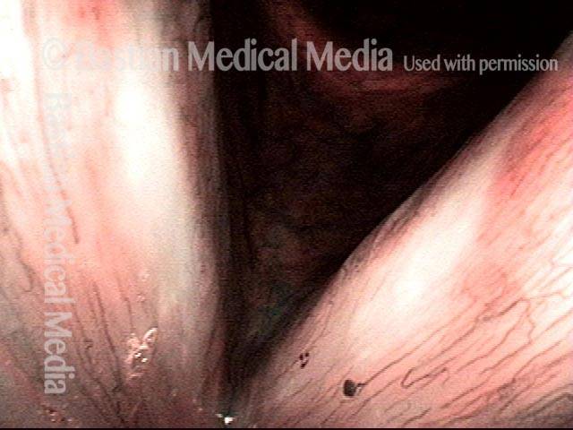
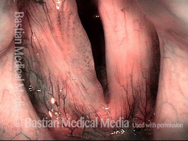
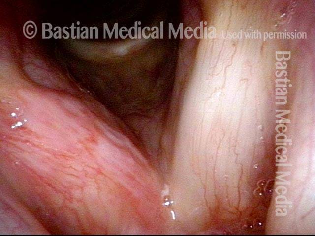
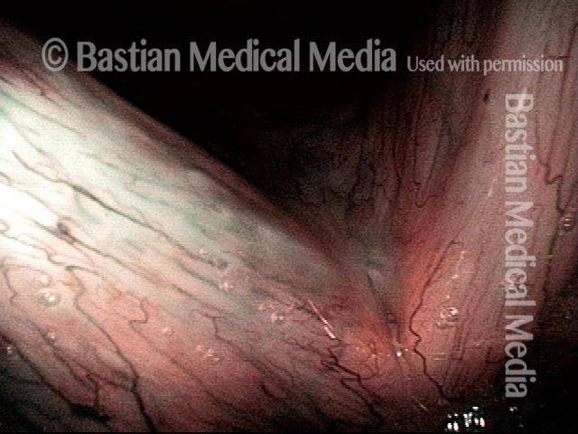
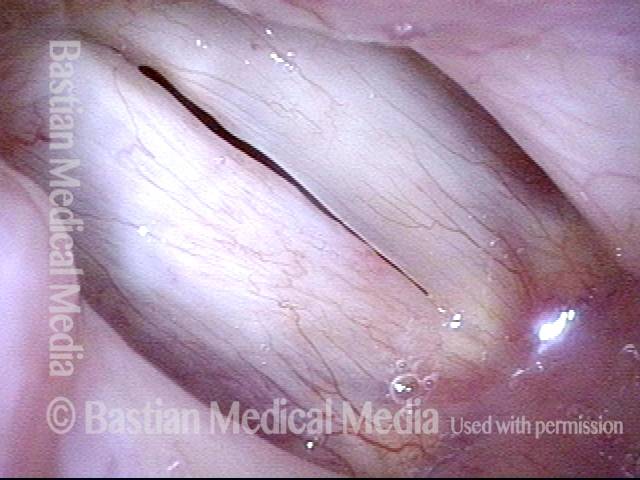
L’infezione da HPV è considerata cronica e provoca una crescita ricorrente di papillomi nella laringe. Tuttavia, a volte vediamo quelle che sembrano cure, o almeno remissioni a lungo termine. Questo sembra essere il caso qui. Dopo un intervallo di 8 anni di voce perfetta, il paziente ha avuto un improvviso aumento della raucedine verificatosi nelle poche settimane precedenti l’ultimo esame di seguito. Questo è un esempio del motivo per cui spesso diciamo a un paziente che sembra essere guarito: “Potresti essere guarito, ma di solito diciamo” remissione a lungo termine “. Lo scenario di questo paziente non è raro. La sua remissione a lungo termine di 8 anni è stata dovuta a un meticoloso intervento chirurgico? Cidofovir? La sua risposta immunitaria? È impossibile dire se sia stato uno o tutti questi fattori.

Visual Portfolio, Posts & Image Gallery for WordPress

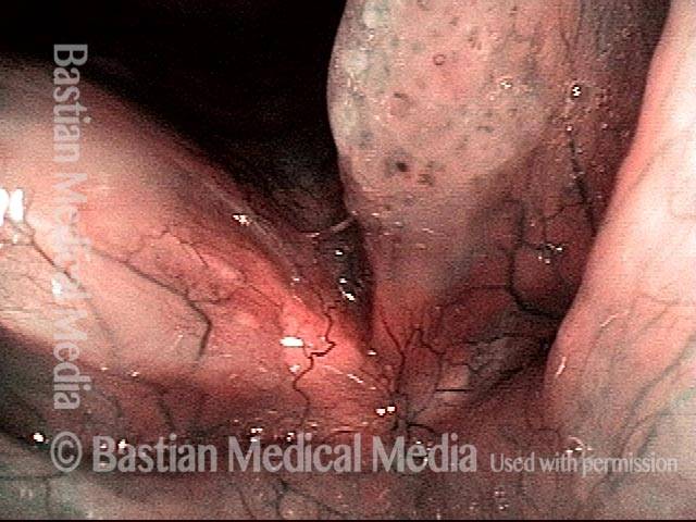
Stippled Vascularity (1 of 8)

In a somewhat distant view from 10 years earlier, under standard light, both vocal cords appear to be covered with stippled rather than (normal) linear or curvilinear vascularity. The diagnosis (biopsy confirmed): laryngeal papillomatosis aka RRP (recurrent respiratory papillomatosis), type 6.

HPV infection (2 of 8)

A somewhat closer view under blue-green narrow band light accentuates the stippled vascularity so typical of HPV infection. Both true cords are covered and there are patches on the false cords (arrows).

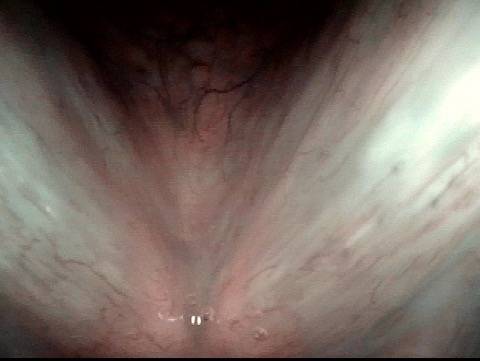
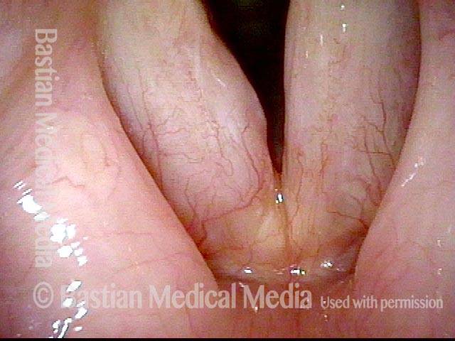
Stippled vascularity (3 of 8)

After surgery and cidofovir injection a year earlier, voice remains normal to the patient. Sharp eyes can pick up a tiny focus of stippled vascularity of the right cord (left of photo).

HPV vascular effect (4 of 8)

At closer range with narrow band light, the HPV vascular effect on the right cord is made more obvious (arrows). The patient requested a “curative mode” additional surgery to clear up this tiny residue and inject cidofovir.

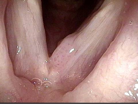
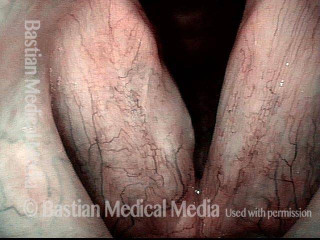
16 months later (5 of 8)

16 months later, voice remains normal to patient, and there is no sign of recurrent HPV vascularity or lesion.

Is it long-term remission? (6 of 8)

A slightly blurry narrow band view, again showing no sign of stippling. Is she “cured?” Or in long-term remission?

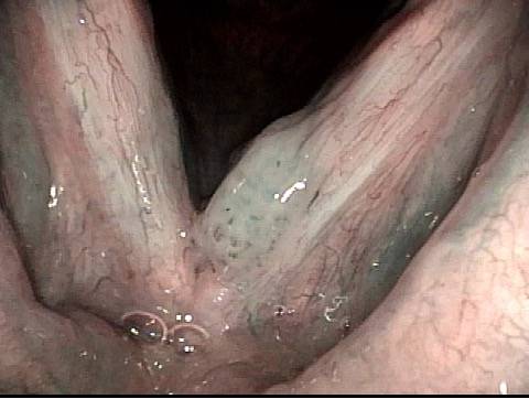
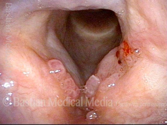
Recurrent Papilloma (7 of 8)

After 8 years of normal voice, the patient re-presented due to hoarseness, saying that her voice had been “perfect” up until a few weeks earlier. She was not sick, but noticed increased “drainage” and voice change. The explanation is a recurrent papilloma.

Stippled vascularity (8 of 8)

Narrow band light again accentuates the stippled vascularity. The patient is embarking on another “curative mode” series of surgical procedures, hoping to again put her into remission.

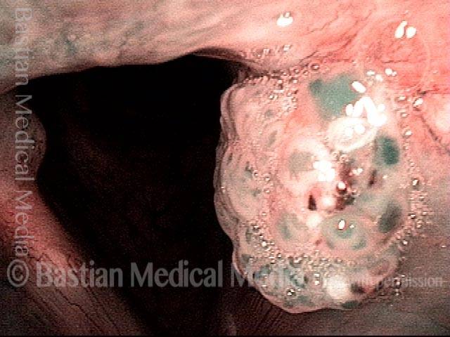
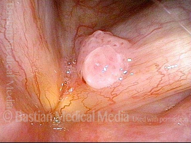
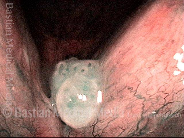
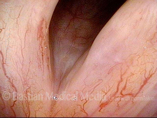
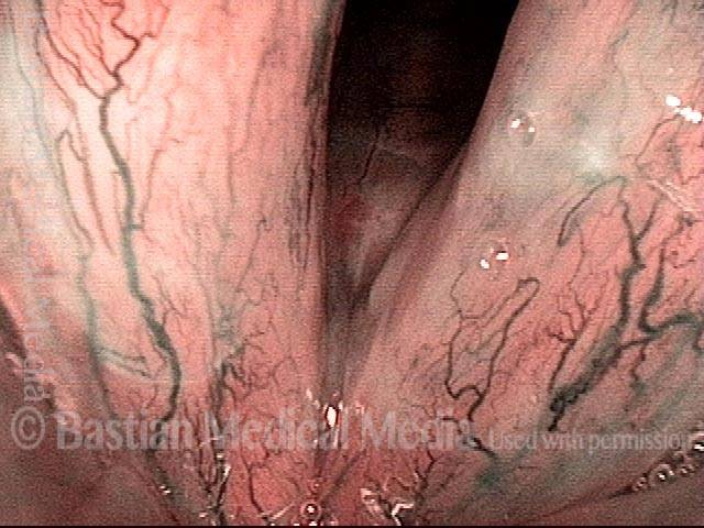
Papillomi, sottotipo HPV 11, prima e dopo la rimozione

Visual Portfolio, Posts & Image Gallery for WordPress

Papillomas: HPV Subtype 11 (1 of 4)

Papillomas at posterior vocal cords, with left side (right of image) much larger than right. This patient has HPV subtype 11.

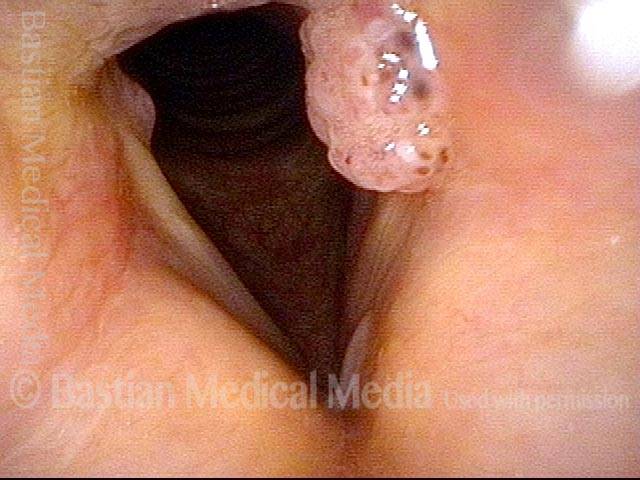
Papillomas: HPV Subtype 11 (2 of 4)

Closer view, under narrow band illumination, which accentuates the vascular pattern.

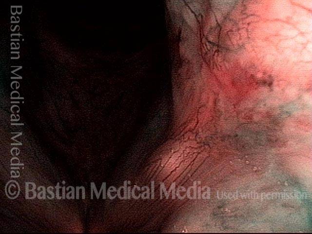
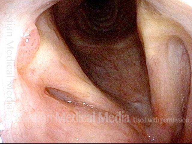
Papillomas, removed: HPV Subtype 11 (3 of 4)

Two weeks after microsurgical removal, cidofovir injection, and return of normal voice.

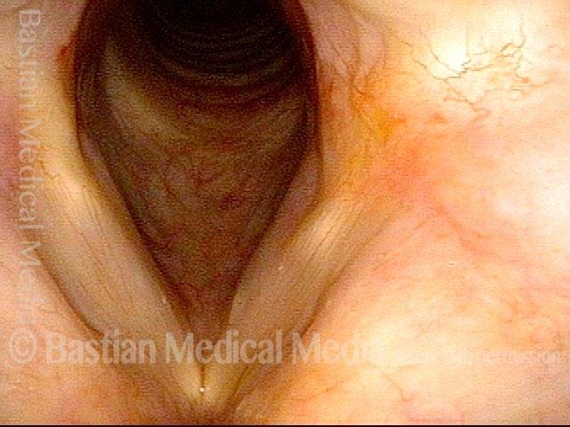
Papillomas, removed: HPV Subtype 11 (4 of 4)

Closer view of left posterior vocal cord, narrow band illumination. Notice that there are a few dot-like vascular marks. These are typical of HPV effect, and may presage recurrence.

Papillomi, HPV sottotipo 6, prima e dopo la rimozione

Visual Portfolio, Posts & Image Gallery for WordPress

Papillomas: HPV Subtype 6 (1 of 4)

Papilloma, left vocal cord (right of image), standard light. Voice is grossly hoarse. This patient has HPV subtype 6.

Papillomas: HPV Subtype 6 (2 of 4)

Same lesion, under narrow band illumination.

Papillomas, removed: HPV Subtype 6 (3 of 4)

After removal and cidofovir injection, normalized larynx. Voice is normal.

Papillomas, removed: HPV Subtype 6 (4 of 4)

Same view, under narrow band illumination.

Papillomi, HPV sottotipo 11

Visual Portfolio, Posts & Image Gallery for WordPress

Papillomas: HPV Subtype 11 (1 of 3)

Panoramic view, standard light, shows papillomas on the aryepiglottic cord, false cords, anterior face of arytenoid, and at anterior commissure. This patient has HPV subtype 11.

Papillomas: HPV Subtype 11 (2 of 3)

Closer view, standard light, shows more clearly the papillomas on the anterior face of the right arytenoid and at the anterior commissure.

Papillomas: HPV Subtype 11 (3 of 3)

Still closer view, to see more clearly the anterior commissure papilloma.

Esempio 2

Visual Portfolio, Posts & Image Gallery for WordPress

Papillomas: HPV Subtype 11 (1 of 2)

Vocal cords, narrow band light, showing papillomas on the upper surface of the anterior vocal cords. This patient has HPV subtype 11.

Papillomas: HPV Subtype 11 (2 of 2)

Tracheal view, standard light, showing scattered papillomas (arrows). The carina is in the distance.

Lesioni da HPV

Visual Portfolio, Posts & Image Gallery for WordPress

Lesions of HPV Subtype? (1 of 2)

Under standard light, faint stippled vascularity is seen, along with a general mild inflammatory response (pinkness). Patients like this are often misdiagnosed with acid reflux.

Lesions of HPV Subtype? (2 of 2)

Same patient, narrow band light. Now seen is the stippled, HPV-effect kind of vascularity of “carpet-variant” lesions.

Lesioni e papillomi da HPV, prima e dopo la rimozione e l’iniezione adiuvante

Visual Portfolio, Posts & Image Gallery for WordPress

Lesions and papillomas of HPV subtype? (1 of 8)

At initial diagnosis, as yet untyped for HPV. Multi-focal lesions on both vocal cords.

Subtle lesion (2 of 8)

Narrow-band illumination and a different viewing angle better reveal the more subtle lesion on the upper surface of the right cord (dotted circle).

Open phase (3 of 8)

Strobe light, open phase of vibration, showing mismatch.

1 week after removal (4 of 8)

One week after removal of papillomas, voice is dramatically restored. Strobe light, open phase of vibration. Compare with photo 3.

1 week after removal (5 of 8)

Strobe illumination, closed phase. Even in falsetto, oscillatory ability is preserved due to the precise and superficial removal of the papillomas.

Injecting adjuvant (6 of 8)

At three weeks after removal, the patient regards his voice as normal. The patient has neither lesion nor vascular change to suggest any residual or recurrent lesion. Needle in photo (arrow) positioned to inject adjuvant medication in attempt to prevent recurrence. This procedure is done in a voice lab under topical anesthesia, not the operating room.

After injecting adjuvant (7 of 8)

After both cords have been “inflated” with adjuvant medication. Note the convex, slightly blanched vocal cord margins, due to superficial infiltration of the medication.

After final adjuvant injection (8 of 8)

Nearly a month later, immediately after the third and final adjuvant injection (hence the blood below the vocal cords). The patient again regarded his voice as completely normal. No sign at this early point of recurrence of papillomas or other HPV lesions. Patients with focal disease as seen in photo 1 of this series not infrequently go into long-term remission or “cure,” though it may be impossible to discern the relative roles of surgery, adjuvants, and the patient’s immune system.

Papillomi, sottotipo HPV 55, in fase di remissione

Visual Portfolio, Posts & Image Gallery for WordPress

Papillomas: HPV Subtype 55 (1 of 4)

Papillomas of the vocal cords, in a patient with HPV subtype 55, which is intermediate-risk for progressing to cancer.

Papillomas: HPV Subtype 55 (2 of 4)

Same exam, with narrow-band lighting, which accentuates the vascular pattern of the papillomas.

Papillomas, in remission: HPV Subtype 55 (3 of 4)

Same patient, years later, in remission, and with normal voice. No sign of papillomas here or anytime during the prior three and a half years, after meticulous removal and Cidofovir treatment.

Papillomas, in remission: HPV Subtype 55 (4 of 4)

Same exam as photo 3, with narrow-band lighting. The vascular dots on the vocal cords are not HPV-related.

Papillomi, HPV sottotipo 31, andando in remissione

Visual Portfolio, Posts & Image Gallery for WordPress

Papillomas: HPV Subtype 31 (1 of 4)

Standard light, showing lesions on the vocal cords, in particular the stippled vascular pattern we call “HPV effect.” The patient’s voice was nearly gone, with numerous syllable drop- outs and a very effortful quality. Compare with photo 3.

Papillomas: HPV Subtype 31 (2 of 4)

Closer view, using narrow-band light to accentuate the vascular pattern of “HPV effect.” Biopsy and additional testing of these lesions showed squamous papilloma with moderate dysplasia, and HPV subtype 31 was confirmed, which is high risk for eventually causing cancer. After the patient underwent several injections of cidofovir, the lesions persisted but seemed to become more indolent. On compassionate grounds, this fairly young person was then prescribed celecoxib for six months.

Papillomas, in remission: HPV Subtype 31 (3 of 4)

Three years after photos 1 and 2, standard light view. Within two months of the start of celecoxib, voice improved very noticeably, and the “HPV effect” vascularity resolved. Still, it is unknown what roles in this recovery were played by the patient’s immune system, the cidofovir, and the celecoxib, respectively.

Papillomas, in remission: HPV Subtype 31 (4 of 4)

Closer view than photo 3, under narrow-band light.

Papillomi, HPV

Visual Portfolio, Posts & Image Gallery for WordPress

Papillomas: HPV Subtype? (1 of 2)

Papillomas involving both the true and false vocal cords.

Papillomas: HPV Subtype (2 of 2)

At higher magnification, the stippled vascular markings become more evident.

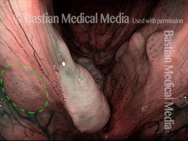
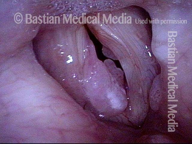
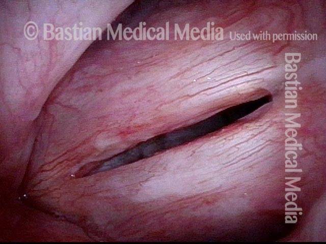
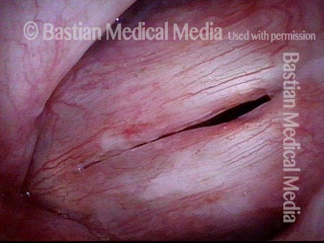
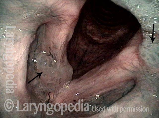
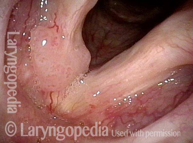
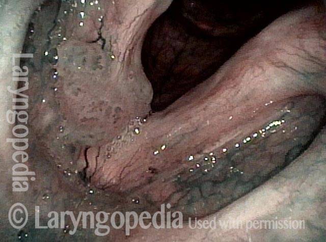
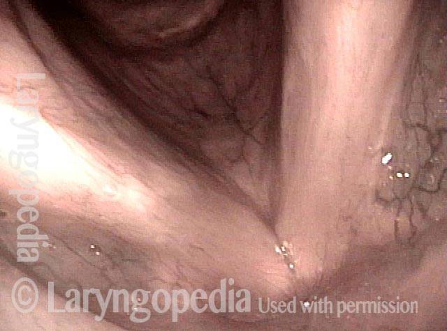
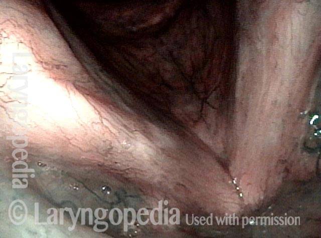
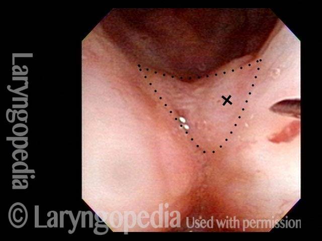
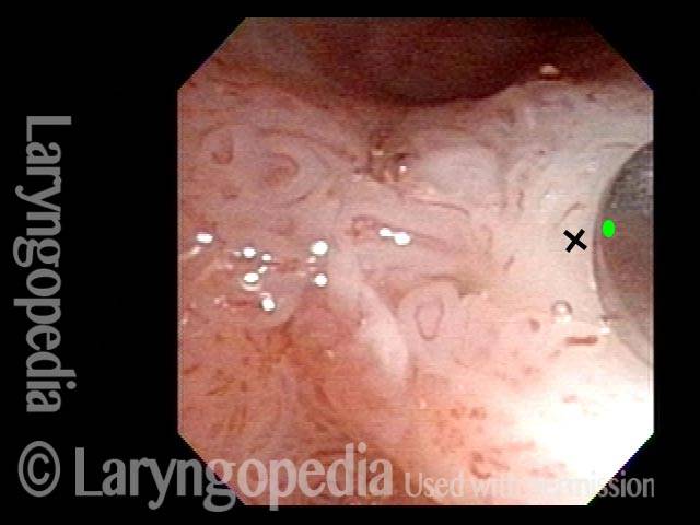
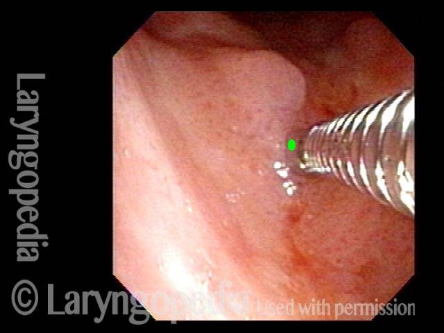
Superare le false piste per trovare il vero problema

Visual Portfolio, Posts & Image Gallery for WordPress

Obvious lesion not important (1 of 3)

Several months after removal of exuberant papillomas, voice remains quite good, but is becoming a little deeper. The obvious lesion here is not important; the subtle one is the key.

Granuloma (2 of 3)

Narrow band light reveals the spherical lesion to be a granuloma, not papilloma (which would have stippled vascular markings).

Carpet-varient papilloma (3 of 3)

At closer range, still under narrow band light, carpet-variant papilloma can be seen on the posterior right vocal cord (left of photo). This is the important finding.

Papillomi, sottotipo HPV 18 o 45

Visual Portfolio, Posts & Image Gallery for WordPress

Papillomas: HPV Subtype 18 or 45 (1 of 2)

Papilloma growths on the right vocal cord (left of image), standard light. This patient’s papillomatosis is caused by HPV, narrowed down to either subtype 18 or 45.

Papillomas: HPV Subtype 18 or 45 (2 of 2)

Slightly magnified view of the same papilloma growths under narrow-band illumination, which accentuates the vascular pattern.

Lesioni di HPV sottotipo 16

Visual Portfolio, Posts & Image Gallery for WordPress

Lesions of HPV Subtype 16 (1 of 3)

Recurring inflammatory and leukoplakic lesions caused by HPV subtype 16. A left vocal cord cancer (right of image) was removed several years earlier, and the patient developed a right vocal cord cancer almost a year later.

Lesions of HPV Subtype 16 (2 of 3)

Slightly magnified view, focusing on the anterior (frontward) ends of the vocal cords. The cords’ stippled vascularity, which often accompanies HPV infection, is more apparent here.

Lesions of HPV Subtype 16 (3 of 3)

A similar view to image two, but with narrow-band illumination, which accentuates the vascular pattern. Biopsy/removal of these lesions revealed high-grade dysplasia; re-biopsy almost a year after this examination returned a diagnosis of cancer.

Cancro, HPV sottotipo 16, prima e dopo la radiazione

Visual Portfolio, Posts & Image Gallery for WordPress

Cancer: HPV Subtype 16 (1 of 5)

Cancer, in a patient with HPV subtype 16. The divot and blood seen on the left vocal cord (right of image) are the result of a biopsy performed elsewhere (not by BVI physician) earlier the same day as this examination.

Cancer: HPV Subtype 16, after radiation therapy (3 of 5)

Six weeks after the end of radiation therapy, the tumor is no longer seen. However, part of the left cord (right of image) is missing, due to sloughing of the tumor that had eaten away part of the cord’s normal tissue.

Cancer: HPV Subtype 16, after radiation therapy (4 of 5)

Phonation. Strobe light, open phase of vibration, shows that the margin of the left cord (right of image) is at a lower level than the right’s, due to loss of some of the bulk of the cord where the tumor died and sloughed away.

Cancer: HPV Subtype 16, after radiation therapy (5 of 5)

Strobe light, closed phase of vibration. The more normal right cord (left of image) unsuccessfully attempts to reach the left cord’s residual upper surface mucosa. Voice is functional but hoarse.

Papillomi, HPV sottotipo 45

Visual Portfolio, Posts & Image Gallery for WordPress

Papillomas: HPV Subtype 45 (1 of 2)

Papillomas in the supraglottis, left of image. The pink, velvety area of papillomas is outlined by small arrows.

Papillomas: HPV Subtype 45 (2 of 2)

Closer view of the vocal cords, showing leukoplakia. This is presumably a second expression of the HPV infection, though the typical dotted or pointed vascular marks of HPV are not seen in the area of the leukoplakia.

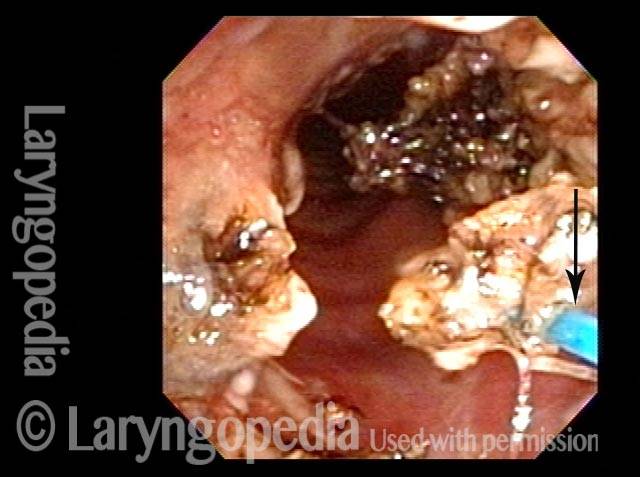
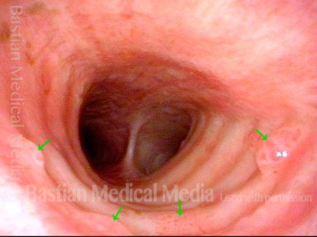
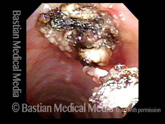
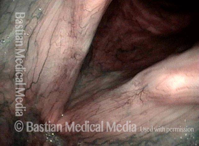
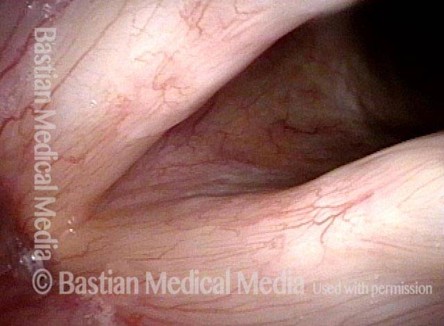
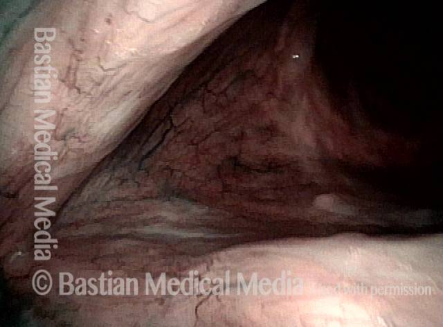
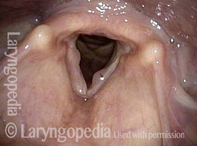
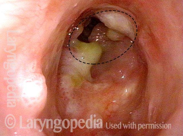
Papilloma medio-tracheale, trattato con laser al tulio

Visual Portfolio, Posts & Image Gallery for WordPress

Mid-tracheal papilloma, being treated by thulium laser (1 of 5)

The papilloma is seen attached to the posterior tracheal wall, at the midpoint of the trachea. Note the areas of scarring from prior laser procedures. The dots seen indicate reference points for photo 5.

Months after treatment: no papilloma (5 of 5)

Durable resolution of papilloma, many months afterwards. Compare with photo 1.

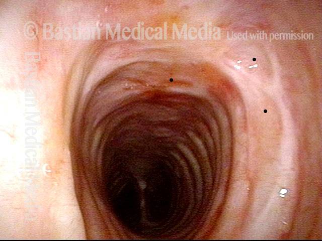
Mid-tracheal papilloma, being treated by thulium laser (2 of 5)

Using the channel scope, a blue glass fiber is extended from the tip of the scope.

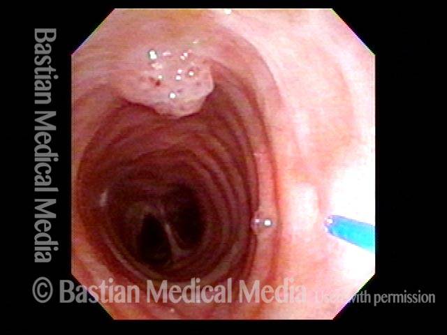
Mid-tracheal papilloma, being treated by thulium laser (3 of 5)

In a closer view, the papilloma has been mostly cauterized using near-contact (not touching) mode.

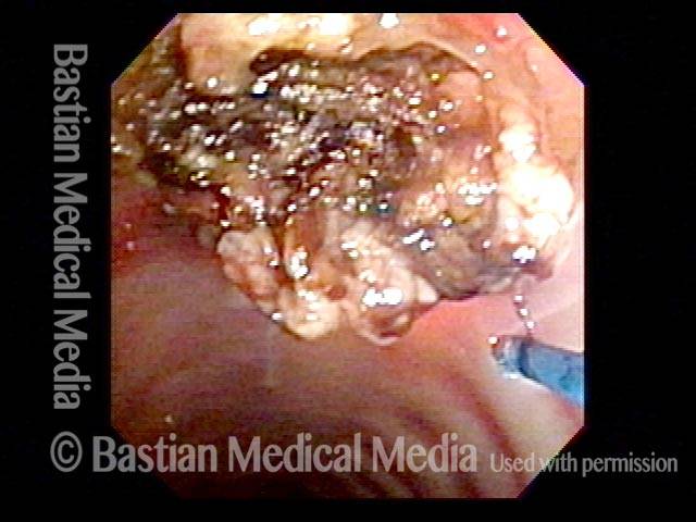
Mid-tracheal papilloma, being treated by thulium laser (4 of 5)

The papilloma is then penetrated multiple times to deliver laser energy to its base. Some of the papilloma is pulled off by attachment to the fiber, and the remainder will slough off and be swept upwards by the mucociliary blanket (thin layer of mucus being swept upward) within the trachea.

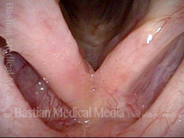
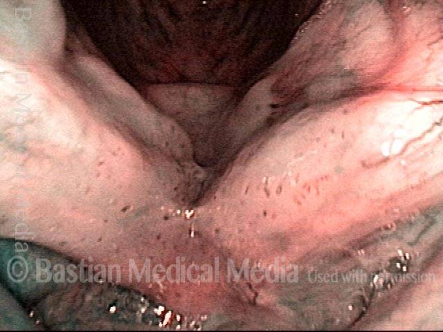
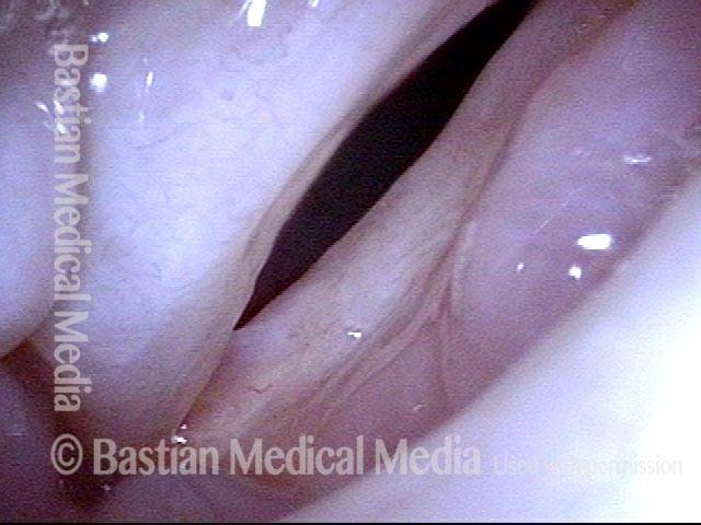
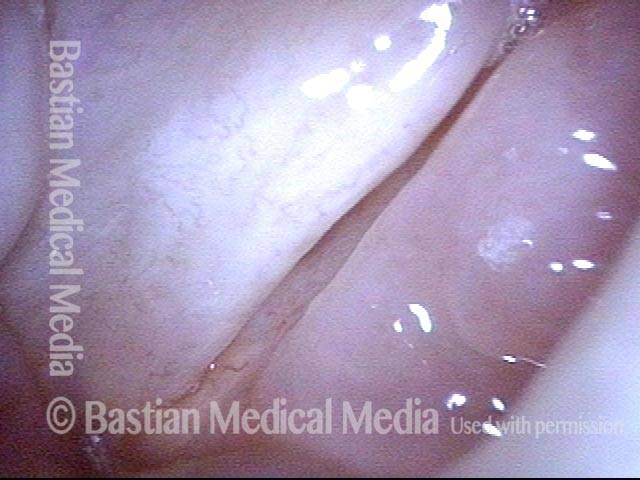
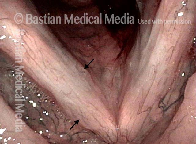
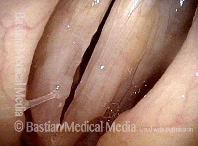
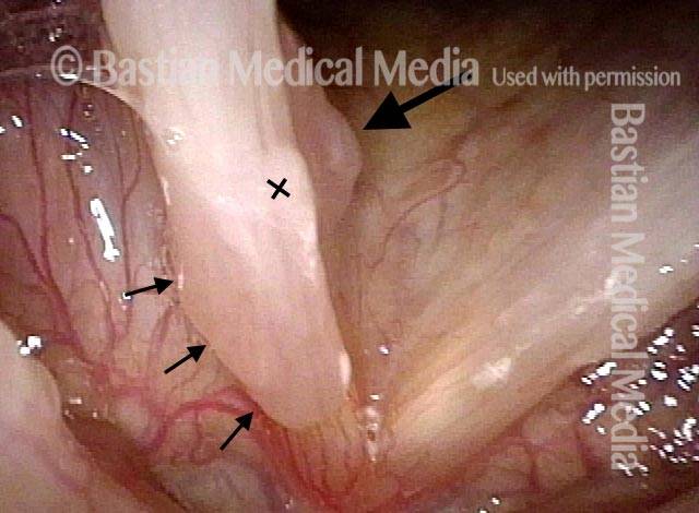
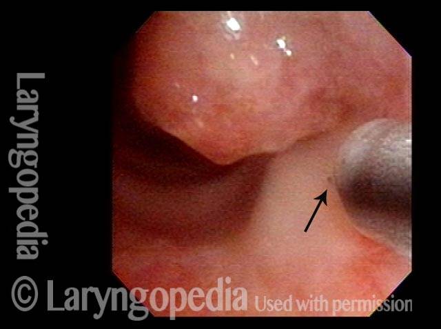
Papillomi sottili, HPV sottotipo 6

Visual Portfolio, Posts & Image Gallery for WordPress

Subtle papillomas, HPV subtype 6 (1 of 3)

After achieving a normal voice through several procedures, the patient came in for reexamination due to the return of mild huskiness. This distant panoramic view with standard illumination does not reveal any obvious papillomas.

Subtle papillomas, HPV subtype 6 (2 of 3)

At close range, using narrow band illumination, a subtle but definite HPV effect is seen. Notice the stippled vascular markings and the faintly increased pinkness at the margins of the cords, indicated by dotted lines.

Subtle papillomas, HPV subtype 6 (3 of 3)

With the vocal cords now at the pre-phonatory instant, these low-profile HPV-related papillomas are again seen, indicated by dotted lines.

Effetto vascolare dell’HPV

Visual Portfolio, Posts & Image Gallery for WordPress

Two papillomas (1 of 3)

Approximately one month after removal of papillomas and Avastin injection in a man who has battled aggressively-recurring disease caused by HPV, type 6. In this view using narrow band light, only two small papillomas are visible.

Stippled vascularity (2 of 3)

At closer range, careful inspection shows no papilloma (yet), but only the stippled vascularity typical of HPV infection.

HPV vascular effect (3 of 3)

HPV vascular effect is seen even more clearly.

Papillomi laringei Raramente possono essere trovati da … Accident

Visual Portfolio, Posts & Image Gallery for WordPress

Papilloma finding (1 of 4)

This young man had a tonsil problem and normal voice but during the initial head and neck examination was found to have a papilloma in his larynx. Rapid recurrence and spread triggered referral. Note stippled vascularity on masses along the edges of the false vocal cords.

Narrow band light (2 of 4)

Narrow band light makes the papillomas much more evident.

Closed phase (3 of 4)

Under strobe light, closed phase of vibration, the true cords are seen to be uninvolved, and this explains his normal voice.

Open phase (4 of 4)

Open phase of vibration, strobe light. The plan is definitive removal and HPV subtyping.

Vincere le battaglie contro il papilloma, ma non vincere la guerra… Ancora

Visual Portfolio, Posts & Image Gallery for WordPress

Left vocal cord lesion (1 of 8)

Middle aged woman with a 6-month history of hoarseness. Note the left vocal cord lesion (right of photo at arrow).

Narrow band light (2 of 8)

At closer range under narrow band light, the stippled vascular pattern suggests that this is HPV-related papilloma. Very tiny secondary lesions may be present at the arrow and (?)

One week after removal (3 of 8)

A week after removal (and proof of HPV subtype 6), the left cord (right of photo) shows expected pinkness. The tiny lesion under the right cord (left of photo) “escaped” and appears larger but is still not a verifiable papilloma, nor is the tiny lesion on the upper surface of the right cord (left of photo) at the (?)

2 months after removal (4 of 8)

Now 2 months after surgical removal of the original left cord lesion, that cord is healed and without evidence of papilloma. Voice is excellent—can pass for normal—but the tiny lesions previously seen are now verifiably flat papillomas (see stippled vascularity at arrows).

7 months after removal (5 of 8)

Now 7 months after original surgery, voice remains “almost” normal to the patient. Cord margins match well with voicing. Irregular margins are primarily due to overlying mucus.

Papilloma and mucus (6 of 8)

With abduction of the cords for breathing, a papilloma is seen below the margin of the right cord (left of photo at large arrow); the small arrows outline a peculiar “elevated” area that looks to be more than mucus the mucus seen at ‘X’.

Stippled vascularity (7 of 8)

At closer range, under narrow band light, the stippled vascular marks further define the papilloma. Note normalized vasculature on the left cord (right of view) where the original papilloma was found. The “battle” of the left cord (right of photo) may have been won…

HPV disease (8 of 8)

Still under narrow band light, stippled vascularity in the area of the arrows suggests that this elevated area also represents HPV disease.

Cosa ha “guarito” questo caso di RRP? Chirurgia? Cidofovir? Il sistema immunitario del paziente? Tutti e tre?

Visual Portfolio, Posts & Image Gallery for WordPress

Papilloma (1 of 8)

Papilloma on right vocal cord (left of photo), proven by biopsy elsewhere. The disease has both a projecting component along with ‘carpet-variant’ component seen only via stippled vascular marks (within dashed line).

Stippling (2 of 8)

Closer view under strobe light; stippling is seen more clearly. Compare the stippling with the linear capillaries of the opposite cord.

One week after surgical removal (3 of 8)

One week after removal and sub typing (HPV 6) and cidofovir injection. Under narrow band light there is residual bruising but no significant stippling.

Cidofovir injection (4 of 8)

At final office-based cidofovir injection. Blood from the injection is seen, but still no stippling.

Six months after surgical removal (5 of 8)

Six months after removal, papillomas have recurred at the margin of the vocal cord, but not on its upper surface where linear capillaries have replaced stippling.

One week after second removal (6 of 8)

A week after second removal of papillomas and cidofovir injection, with expected inflammation, but no visible remaining stippled vascularity.

4 months later, healed (7 of 8)

4 months later, the vocal cord has long since healed and narrow band light is used to accentuate capillaries. No HPV effect (stippling) is seen.

3 years later, no sign of papilloma (8 of 8)

3 years later, there is still no sign of papilloma or stippled vascularity (HPV effect).

Cura RRP? O solo remissione a lungo termine?

Visual Portfolio, Posts & Image Gallery for WordPress

Chronic hoarseness (1 of 4)

Chronic hoarseness, due to these papillomas, subsequently proven to be caused by subtype 6. Note HPV-effect vascularity.

4 months later (2 of 4)

A second surgery, 5 cidofovir injections (3 in office), and 4 months later, neither papilloma nor HPV vascular effect are seen here, under narrow band light.

8 months from start of treatment (3 of 4)

Now 8 months from the start of treatment, and 5 months since the final (office) cidofovir injection, there remains no evidence of abnormality. This view is under standard light.

Narrow band lighting (4 of 4)

An even closer view under narrow band light still shows no sign of HPV effect or papilloma. Voice is normal and vibratory flexibility is maintained when examined under strobe light.

I polipi hanno bisogno di uno sguardo da vicino: ecco uno dei motivi per cui

Visual Portfolio, Posts & Image Gallery for WordPress

“Polyps” diagnosis (1 of 4)

This patient is representative of persons initially diagnosed with “polyps,” based upon a view like this one.

Papillomas (2 of 4)

Now we can see that these lesions are papillomas by the powerful visual criterion of vascular stippling aka “HPV vascular effect.” Another clue of incorrect diagnosis, even with a distant view, would be the patient’s non-match with the vocal overdoer syndrome.

Vascular stippling (3 of 4)

Narrow band light at the same magnification accentuates the vascular stippling. Typical papillomas indicated by arrows, and faint lines online areas of “carpet variant” papillomas.

Prephonatory instant (4 of 4)

Prephonatory instant shows additionally that the right vocal cord lesion (left of photo) is in the wrong location for vibratory injury.

Remissione a lungo termine o addirittura “cura” di RRP/papilloma laringeo

Visual Portfolio, Posts & Image Gallery for WordPress

Chronic hoarseness (1 of 6)

In this distant view, the nature of the abnormality of the right vocal cord (left of photo) is not well seen, and the lesion of the left posterior cord (right of photo) is subtle.

Narrow band light (2 of 6)

Under narrow band light, the two discrete lesions are better identified as being papillomas due to the punctate (dotted) vascular markings.

Higher magnification (3 of 6)

Again under standard light but at higher magnification.

Higher magnification, narrow band lighting (4 of 6)

Back to narrow band light, to more carefully scrutinize the anterior right vocal cord lesion (left of photo).

Post-operation (5 of 6)

18 months after surgical removal and cidofovir injection, voice is excellent and there is no sign of recurrent papilloma.

Post-operation, narrow band lighting (6 of 6)

Under narrow band light, no stippled vascularity is seen. The question is: is remission due to surgery, cidofovir, or the patient’s immune system?

Anestetico locale iniettato provoca sbiancamento

Visual Portfolio, Posts & Image Gallery for WordPress

Infiltrating anesthetic (1 of 3)

A 27-gauge needle tip is poised to infiltrate local anesthetic lidocaine with epinephrine into the papillomas (within dotted line) located just below the anterior commissure. In a moment, the needle will enter the papillomas at the “X”.

Blanching (2 of 3)

The needle is buried and the tissue is blanching due to hydrostatic pressure of the injected fluid. The green dot is for reference with photo 3.

Subglottis being injected (3 of 3)

Farther below the vocal cords, the anterior subglottis is seen here being injected. The green dot is for reference with photo 2. The shank of the needle guide looks like a “Doctor Octopus” arm!

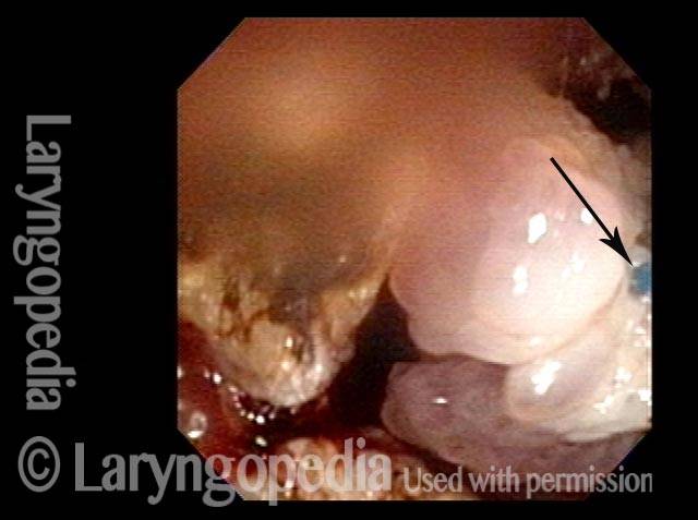
L’anestesia locale piuttosto che quella topica può consentire un intervento chirurgico tracheale abbastanza importante

Visual Portfolio, Posts & Image Gallery for WordPress

Obstructive papillomas (1 of 4)

This middle aged man has had lifelong RRP due to HPV 11. Primary focus of his recurrences for many years has been the trachea. Prior operative removals under general anesthesia have been challenging and recent work has used the thulium laser in an office setting. The papillomas seen here explain his mild pre-procedure stridor. His tracheal lumen should be approximately the size of the dotted circle.

Local injections (2 of 4)

A needle catheter passed through the channel of the scope is embedded at arrow, and is injecting 1% lidocaine with epinephrine (see blanched area). Numerous areas are similarly injected.

Removal of papilloma (3 of 4)

Here a large chunk of papilloma (stuck to the laser fiber at arrow) is being pulled away.

Improved breathing with papilloma removal (4 of 4)

An additional chunk is being detached. The patient’s breathing at the conclusion of this procedure is much improved. The tracheal lumen will be even larger when a lot of remaining coagulated papillomas slough away.
vocal cord wart-like growths YT Thumbnail
Riproduci video

Papillomi della laringe e della trachea

Questo video mostra escrescenze simili a verruche nella laringe e nella trachea (laringe e trachea) causate da un’infezione cronica con il papillomavirus umano (HPV).

pulsed-KTP laser coagulation YT Thumbnail
Riproduci video

Coagulazione laser pulsata-KTP dei papillomi delle corde vocali

Guarda una dimostrazione video della coagulazione laser dei papillomi delle corde vocali.

HPV in the Larynx YT Thumbnail
Riproduci video

Papillomatosi respiratoria ricorrente (RRP) | Che cos’è?

In questo video, il dottor Robert Bastian parla dell’infezione cronica da virus del papilloma umano (HPV) della laringe (soprattutto delle corde vocali), che causa raucedine.

Table of Contents

Subscribe
Notify of

0 Comments
Inline Feedbacks
View all comments
0
Click to see all comments.x