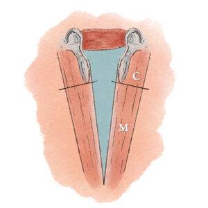
The glottis refers to the space between the vocal cords, and often at least informally includes at least the margin of the vocal cords and in some people’s minds, the true vocal cords themselves. The open (breathing) space between the anterior two-thirds of the vocal cord’s visible length is called the membranous glottis, or the musculomembranous glottis. Why this name? Because that part of the vocal cords is all soft tissue. The layers of the membranous glottis, from most superficial to deepest are:
- The mucosa, perhaps a millimeter thick, which comprises a layer of squamous epithelium and beneath that, a loose attachment zone called the lamina propria or Reinke’s space;
- The vocal ligament, another couple of millimeters thick, and made of elastin and collagen fibers;
- The thyroarytenoid muscle, which makes up the bulk of the cord;
- And then the unyielding thyroid cartilage.
 Disorders of the Membranous Glottis
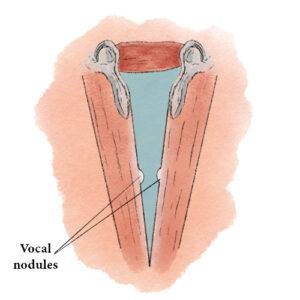
The mucosa of the membranous vocal cord is what vibrates during voicing; overuse can cause the hoarseness or even loss of voice from vibratory injury such as swelling, bruising, nodules, polyps, etc. Or voice can deteriorate due to infection (viral or bacterial) involving the membranous vocal cords or even tumors (mostly from tobacco use). The other one-third of each vocal cord’s visible length is called the cartilaginous glottis, inhabited by the arytenoid cartilages.