Normal vocal cords are muscular bands of tissue attached anteriorly inside the lower middle of the thyroid cartilage or “Adams Apple,” with the posterior ends inserted into the arytenoid cartilages which, in turn sit on top of the posterior ring of the cricoid cartilage.
Anatomy of the Vocal Cords
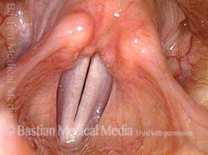
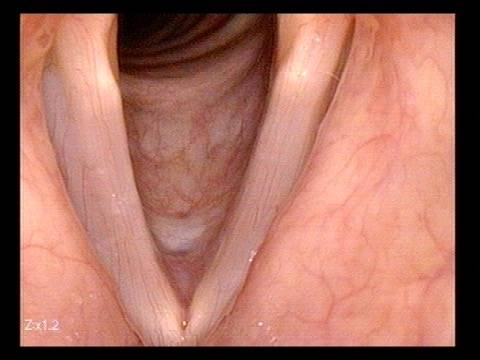
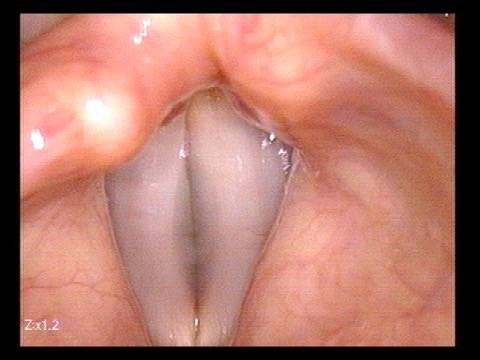
Vocal cords are also referred to as vocal folds, or vocal bands, or even, in some ways most appropriately, “laryngeal lips.” These lips take the shape of a “V” pointing straight anteriorly and the legs of the “V” proceeding horizontally in a posterior direction.
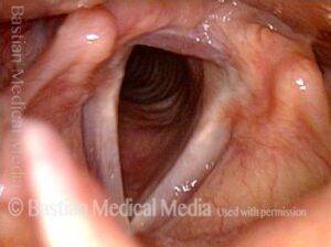
The vocal cords can assume an open (breathing) or closed (sound-making) posture. When the legs of the “V” are separated, a triangular glottic chink (opening) is created, and the cords become part of the airway. Inhaled air is drawn through the nose or mouth down into the throat and through the glottic chink into the trachea and then into the lungs.
When the legs of the “V” are forcefully pinched together, the result is breath-holding, with air unable to enter the windpipe or to leave the lungs.
If the cords are gently positioned together with air simultaneously emitted between them in a steady stream, then the vocal folds begin to vibrate, and one hears the sound of the voice. Of course, the vocal cords are also clenched together every time a person swallows (to protect from aspiration), coughs, or clears the throat (to expel mucus).
How do the Vocal Cords Move?
The larynx has a well-defined motor nerve supply. The recurrent laryngeal nerve is a branch of cranial nerve 10, the vagus “wandering” nerve. It comes from the lower part of the brainstem (medulla oblongata), descends through a small opening (jugular foramen) in the bone at the base of the skull.
From there it travels deep within neck downwards along the groove between the esophagus (swallowing passage) and trachea (windpipe). It then travels further down into the chest. It loops around great vessels in the chest (innominate artery on the right, and aorta on the left).
Only then does it turn upwards, return (recur) to the level of the larynx, and supply the seven paired muscles and one unpaired muscle. The superior laryngeal nerve is another branch of the vagus nerve, but it separates from the main trunk higher in neck and finds its way to the cricothyroid muscle.
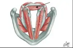
The posterior cricoarytenoid muscles open the larynx, and the (lateral cricoarytenoid, interarytenoid, and thryoarytenoid (the latter via tensing) bring the cords together.
The superior laryngeal nerve supplies what we sometimes call “the singer’s muscles.” Those paired muscles support in a special but not exclusive way, both high pitch phonation, and voice projection.
What Are Vocal Cords Made of?
The vocal cords are comprised of layers. Most of the bulk of the cords is contributed by the thryoarytenoid muscle. That muscle’s name signifies that it attaches from the inner surface of the thyroid cartilage to the arytenoid cartilage. The muscle is covered by a sheath of more dense tissue called the vocal ligament. The ligament is comprised of collagen and elastin fibers. The most superficial layer is called mucosa.
Mucosa is to the vocal folds as skin is to the back of the hand. Just as skin covers the entire surface of the human body, mucosa lines the interior spaces of the human body.
For example, our nose, mouth, throat, esophagus, trachea and bronchial tree, stomach, intestines are all lined by mucosa. Vocal cord mucosa is at the same time white, glistening, and delicate, as well as robust and ready to accept the rigors of vibration.
The stresses on the mucosa during vibration can be simplified to include two main forces: One is collision of mucosa at the point of vibratory contact. There is also a shearing force.
Place your index finger in the middle of the back of your hand and slide skin towards and away from yourself to get the sense of shearing forces. The number of “collisions and shearings” is the same as the pitch (Hertz) being produced. At C4 (middle C), the vocal cords collide and separate 252 times per second! The C above that is 504 times per second! Etc.
Vocal cord mucosa is specially designed for vibration. While attached firmly to the folds, it at the same time has a lot of freedom or “stretch ability.” Again, pinch the skin on the back of a hand planted firmly on the table in front of you and stretch it up and away to get the sense of the freedom of the mucosa to vibrate.
Another analogy is the child’s paddle ball toy. That is also an oscillating system. Yet the paddle may move only slightly to keep the red rubber ball oscillating back and forth over a great distance. In this analogy, muscle and ligament are the paddle and the mucosa is the red rubber ball attached by an elastic band.
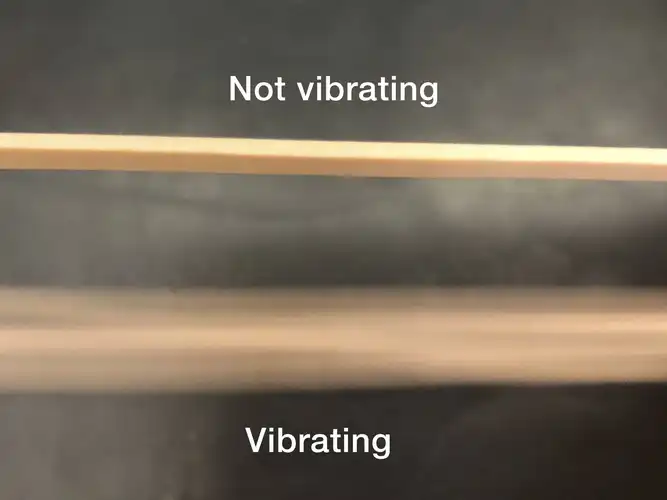
It is this flexibility of mucosa that allows it to oscillate or vibrate. So when voice is made, it is primarily mucosa, and not muscle or ligament, that is vibrating. Think by analogy of a trumpet player’s lips…. If you could watch them in slow-motion through a transparent mouthpiece, you wouldn’t so much see the lips as a whole vibrating as just the mucosa of lips. The exception might be a tuba player playing at very low pitch. Similarly, when the human voice vibrates at extremely low pitch there can be some participation of ligament in muscle in vibration. Still, in the general sense, it is the vocal fold mucosa that is the sound source for the human voice.
What Causes a Hoarse Voice?
When voice becomes hoarse, a variety of explanations may pertain. General explanation: Some process might stiffens the surface layer. By analogy, it would be harder to vibrate a thin sheet of leather than a thin sheet of silk.
Another might be process that thickens the mucosa. Silk cloth that is 2 mm thick would be harder to “vibrate” than if is only 1mm thick.
Yet a third source of hoarseness might be something that interferes with the straight line of the vocal cord margin so that when the person tries to bring the vocal cords together into a perfect match, something is in the way, allowing air leakage and turbulence. Think of an airplane trying to fly with a lump attached to both wings. The aerodynamic ability to slip through the air would be compromised and the whole airplane might “shudder” and lose the ability to fly at very high altitude.
Disorders of the Vocal Cords
Here is a partial list of examples of things that make the voice hoarse, or even take it away.
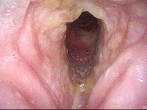
Viral or Bacterial Laryngitis
In this condition, the vocal fold mucosa becomes thicker and stiffer. Think of “pink eye of the vocal cords.” As the mucosa becomes thicker, its mass increases and the pitch of the voice drops. The tiny capillaries covering the vocal folds become more prominent. As the inflammatory process proceeds, Flexibility of the surface mucosa diminishes, sometimes to the point of complete laryngitis. At that point the vocal fold mucosa will not oscillate.
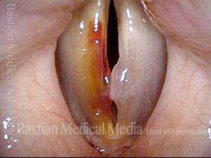
Mucosal Vibratory Injury
If a person uses the voice extremely aggressively for short period of time, or even mildly more than the mucosa is able to withstand over a long period of time, then the mucosa may thicken in order to protect itself.
Think, by analogy, gardener who shovels without gloves for many hours on the first day of spring. He or she may get blisters. This is simply an injury of the skin of the hands. If that same gardener were to continue shoveling day by day for several hours, then eventually the skin toughens and thickens in a more chronic kind of tissue reaction we call calluses.
The analogy with the vocal folds isn’t perfect, but it indicates to some extent what happens to vocal fold mucosa. Injury can be acute and fairly rapid to reverse, or can be chronic and more difficult to resolve. Whatever the injury, voice is altered: often a few notes are added to the low part of the range, and at the same time high notes are either lost or they become much more effortful to produce.
Other Lesions of the Cords
See also Other Benign Vocal Cord Disorders Not Caused by Vibration, Benign Laryngeal Tumors and Larynx Cancer.