Vocal Nodules
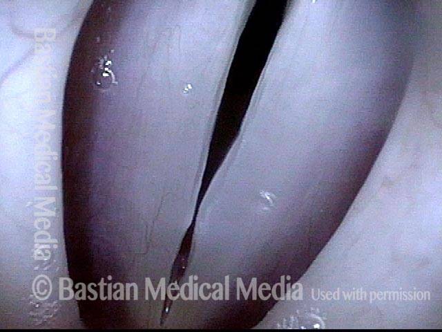
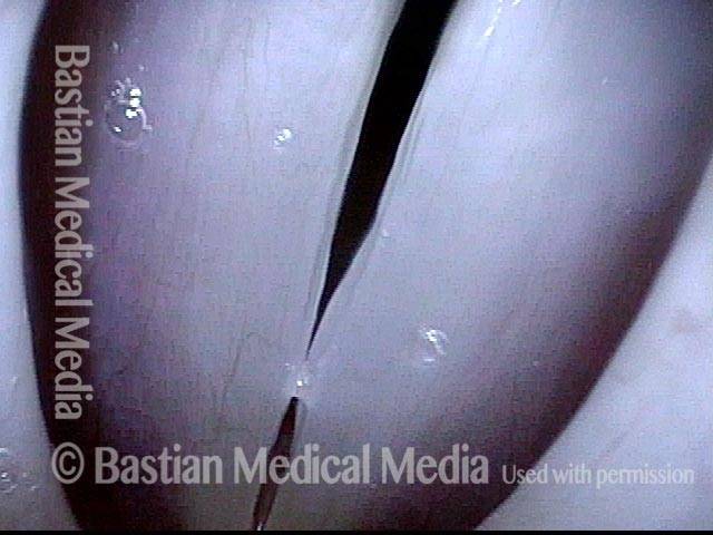
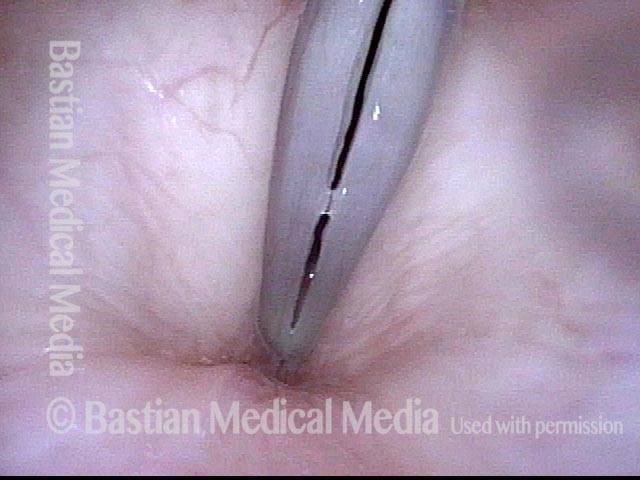
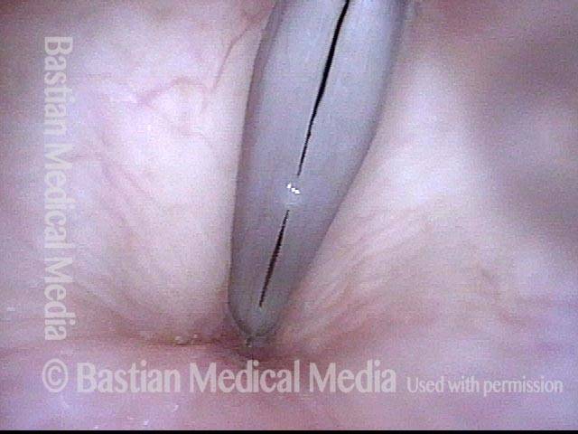
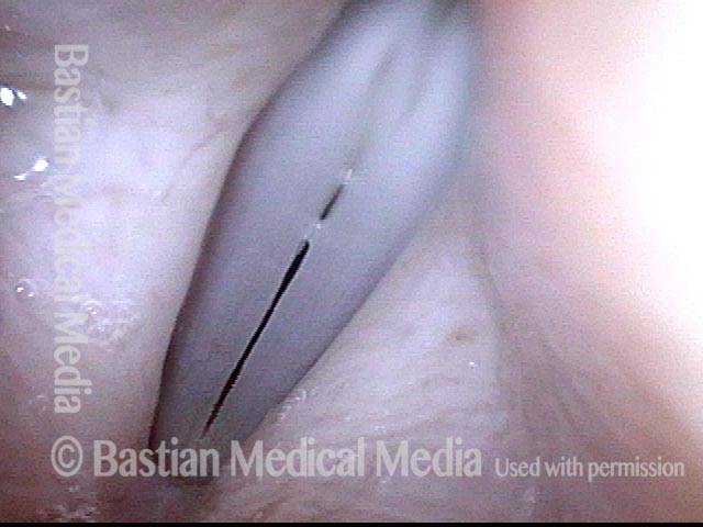
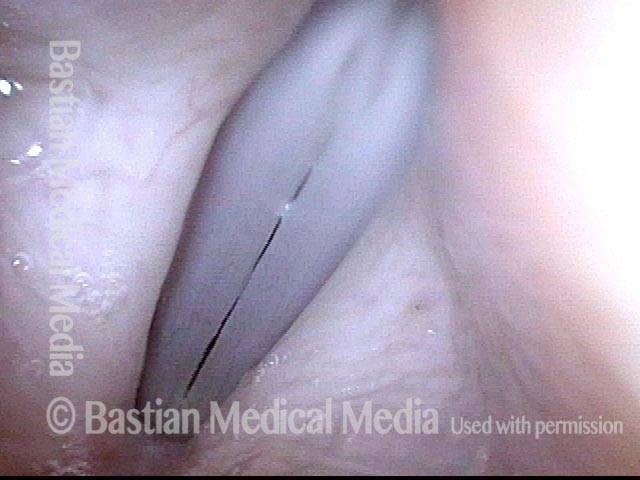
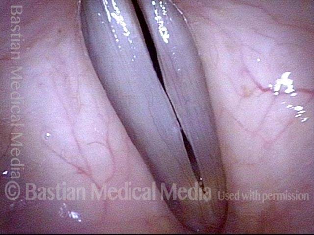
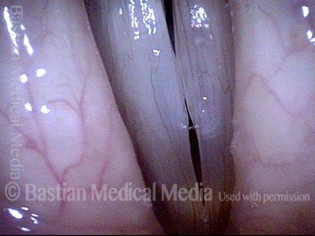
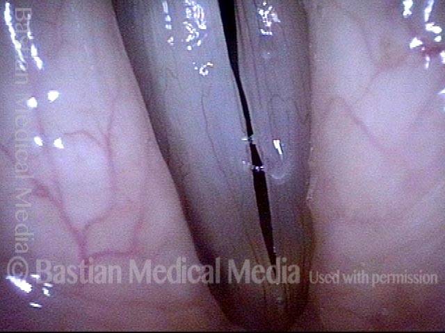
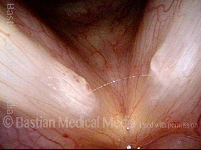
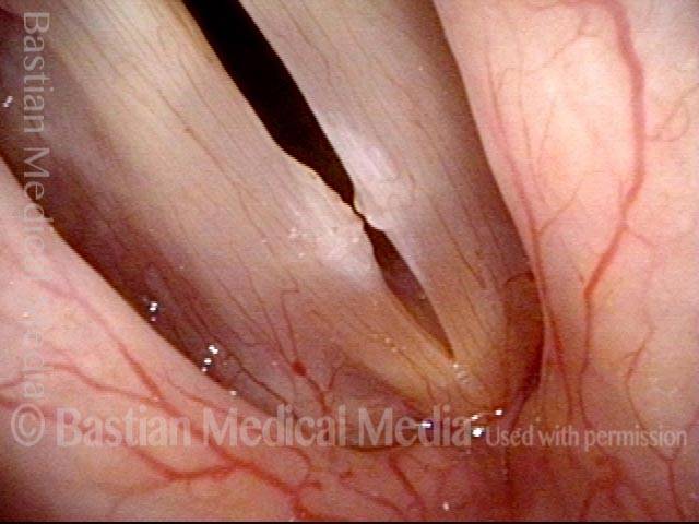
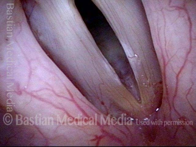
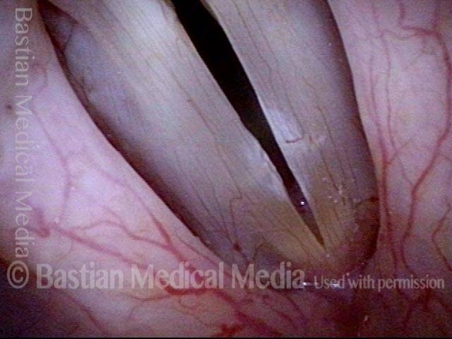
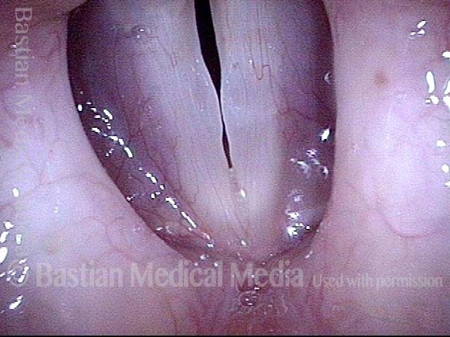
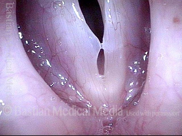
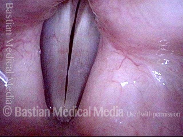
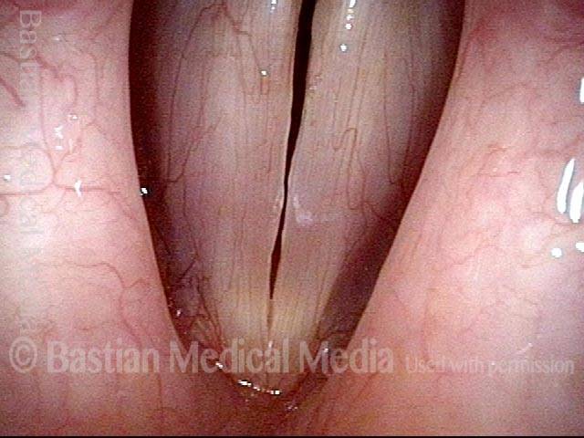
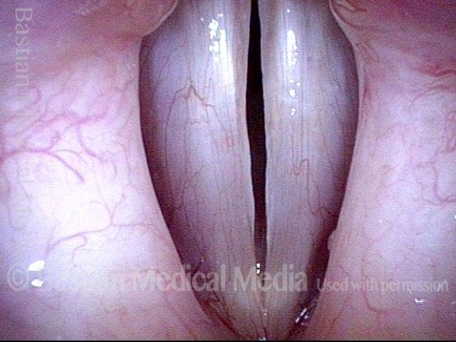
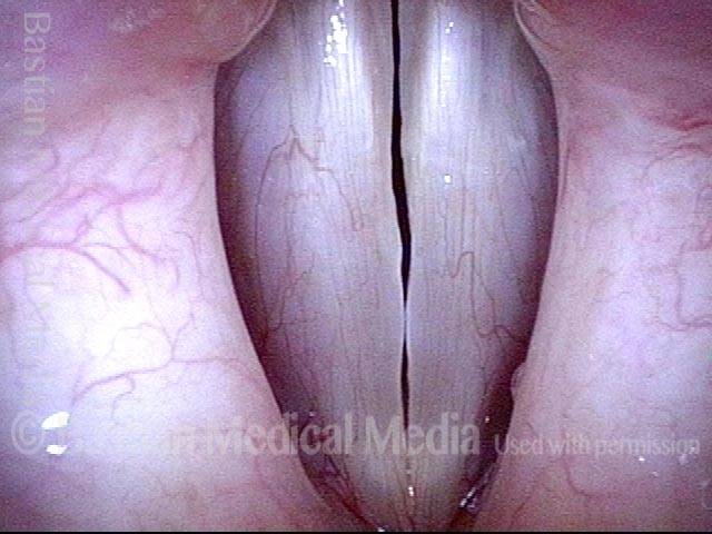
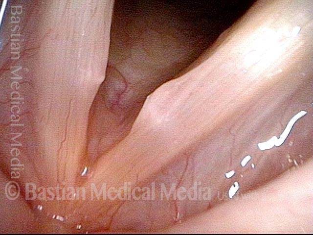
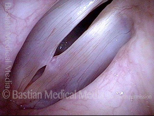
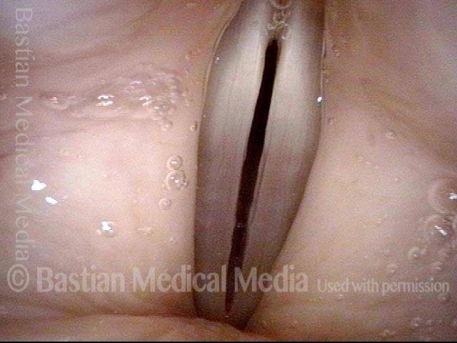
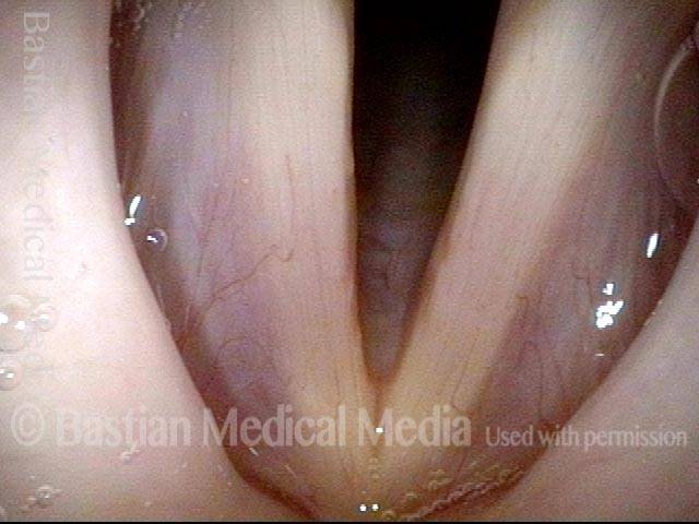
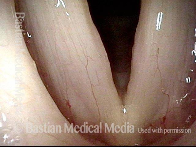
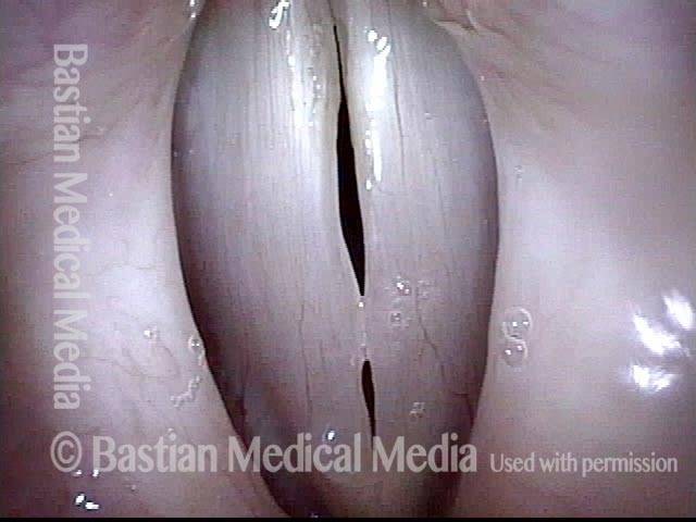
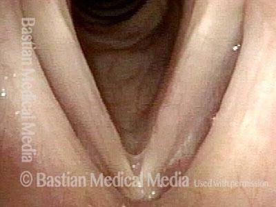
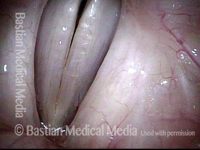
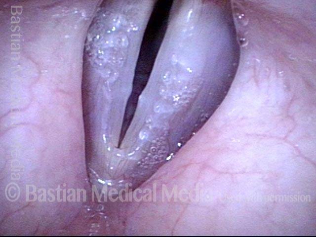
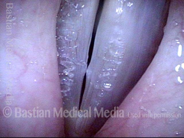
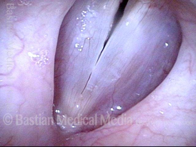
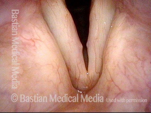
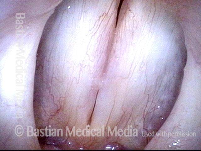
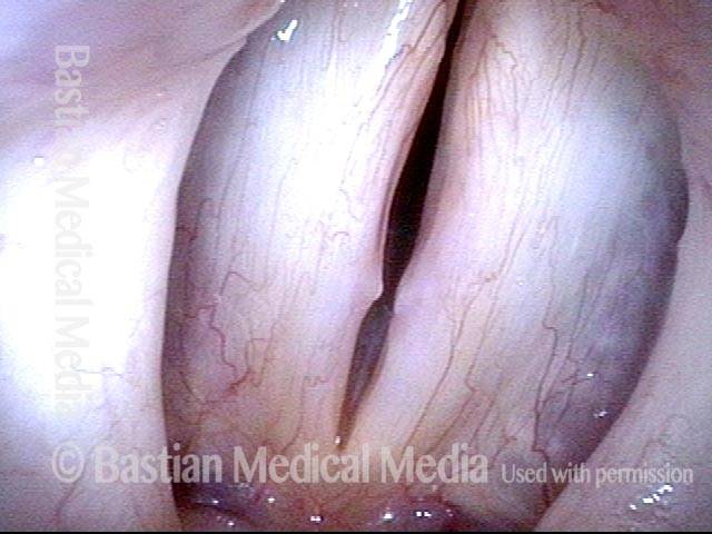
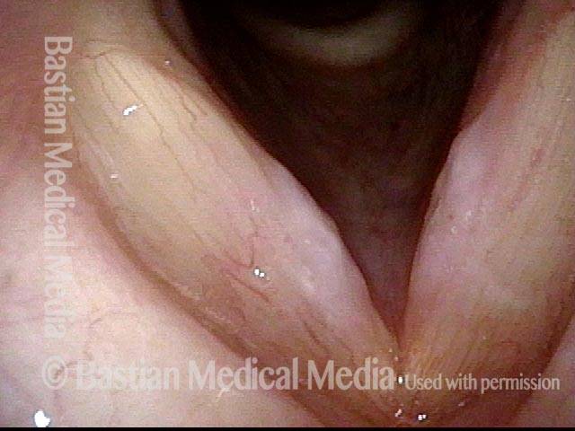
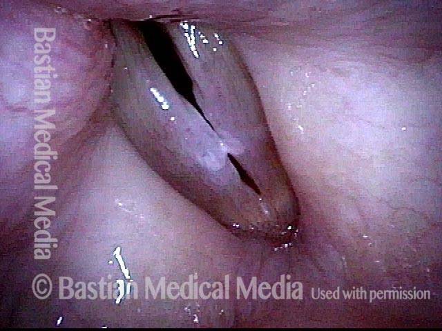
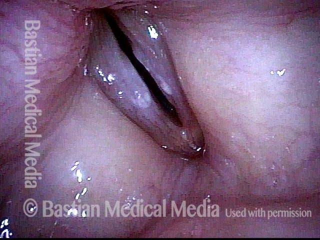
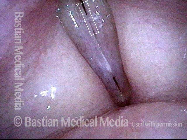
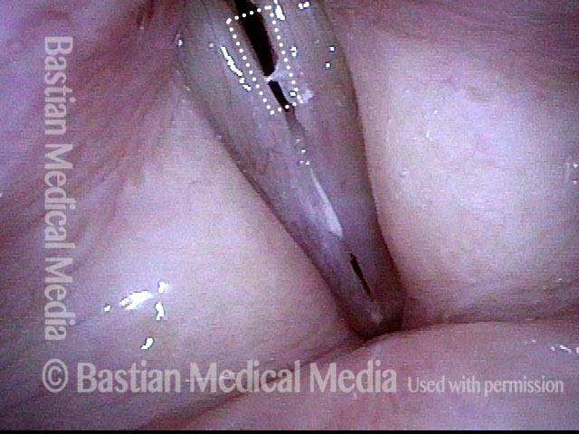
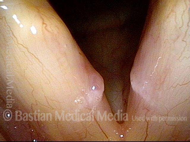
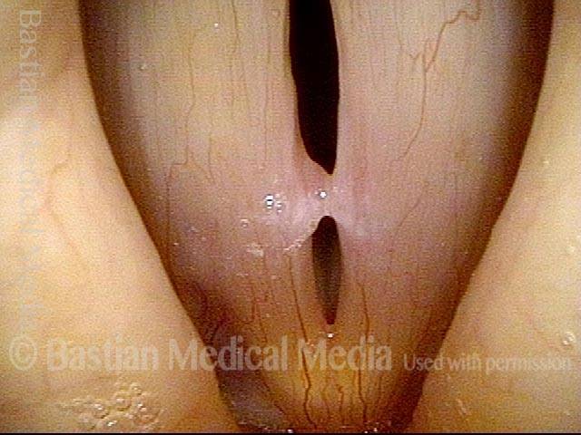
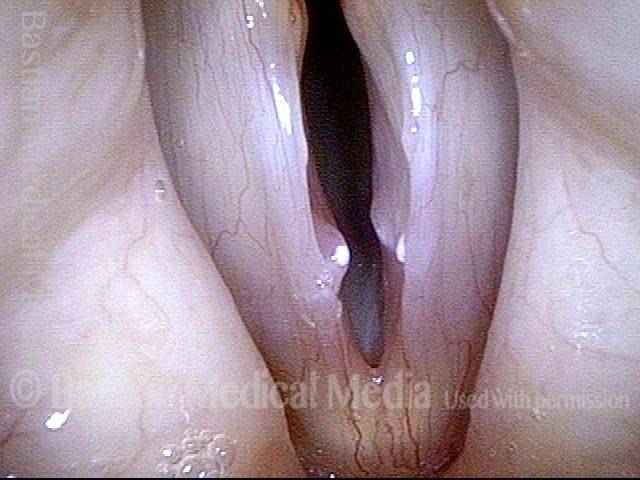
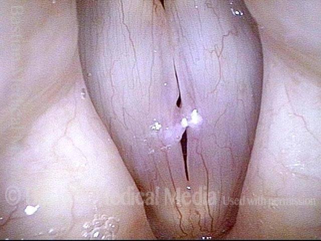
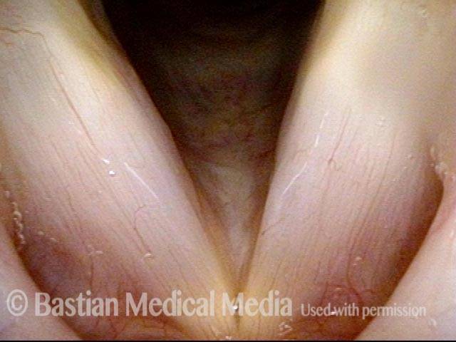
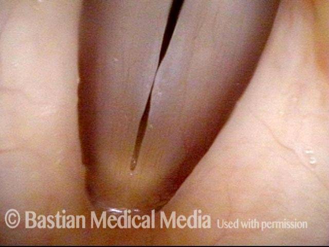
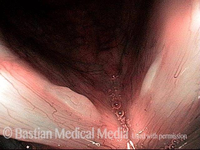
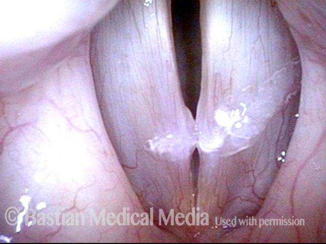
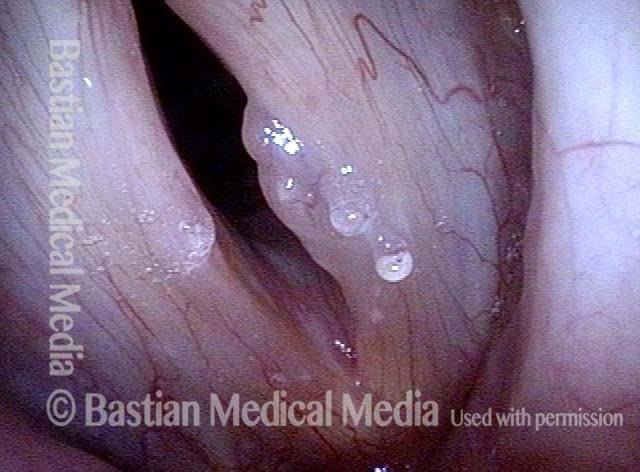
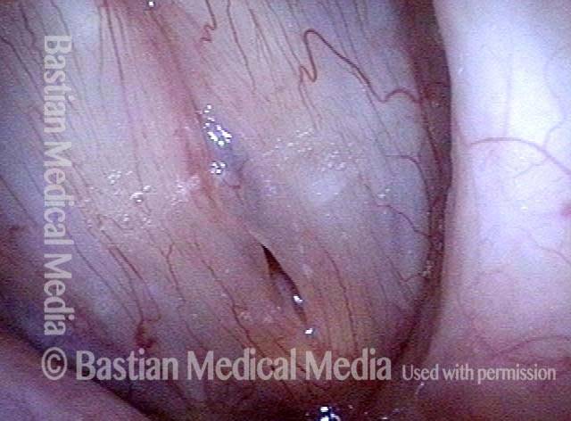
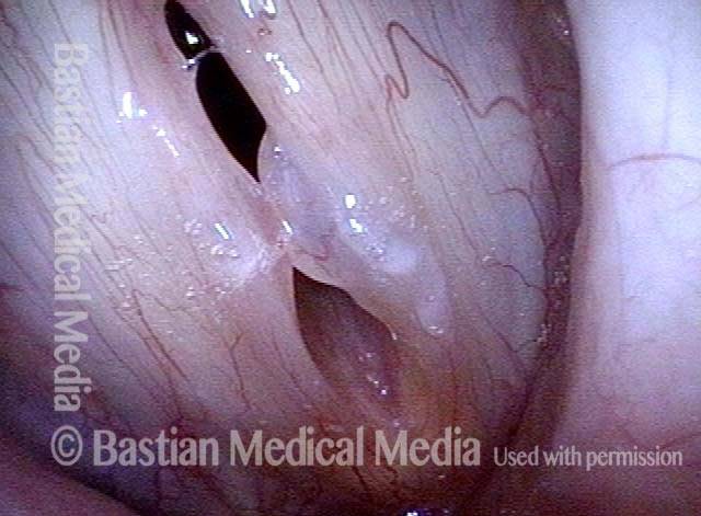
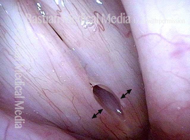
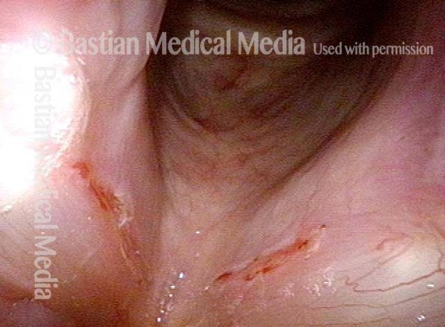
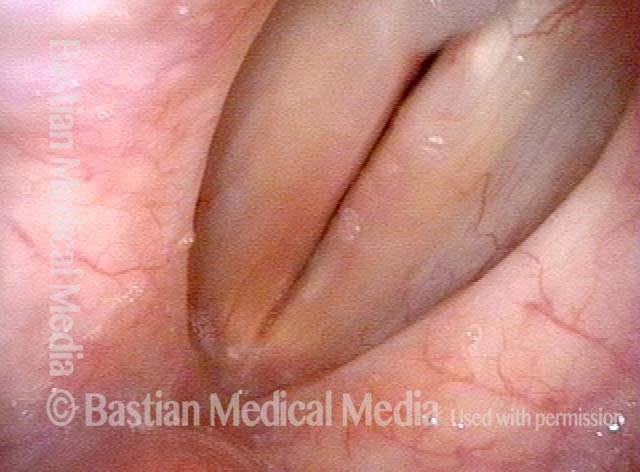
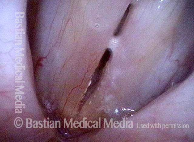
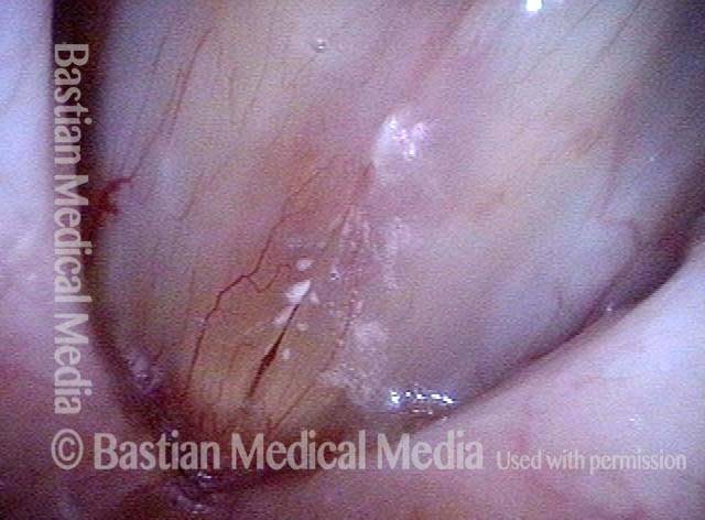
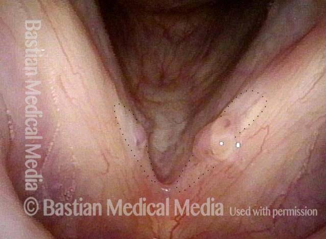
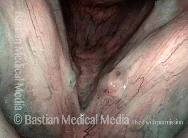
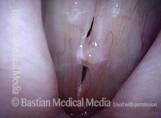
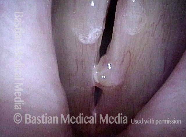
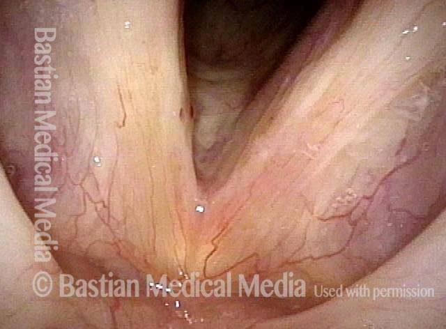
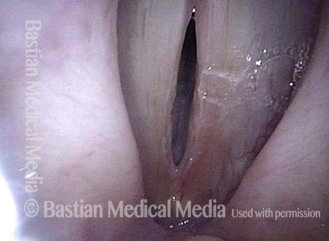
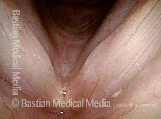
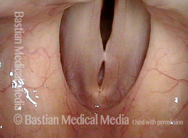
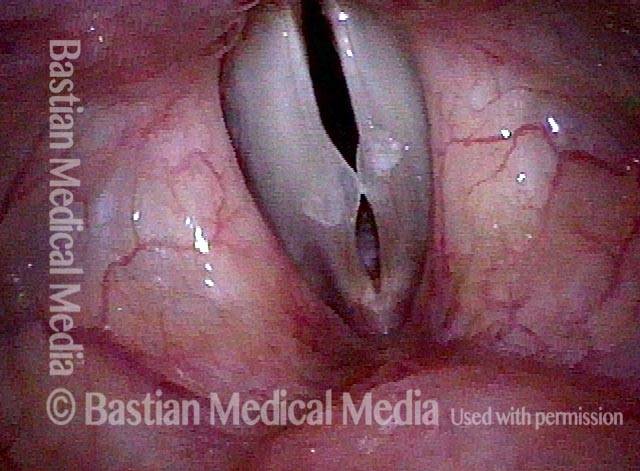
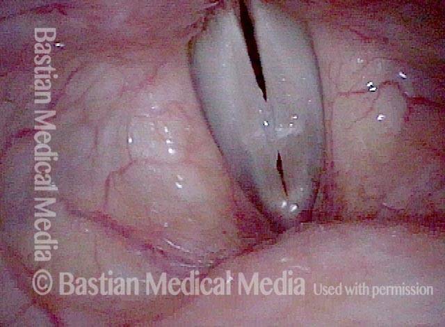
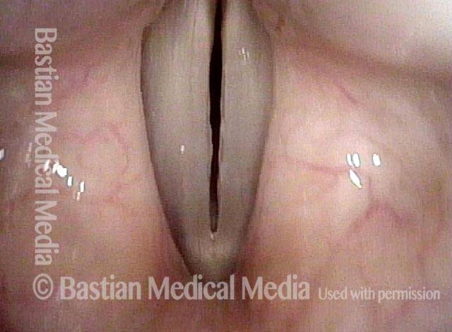
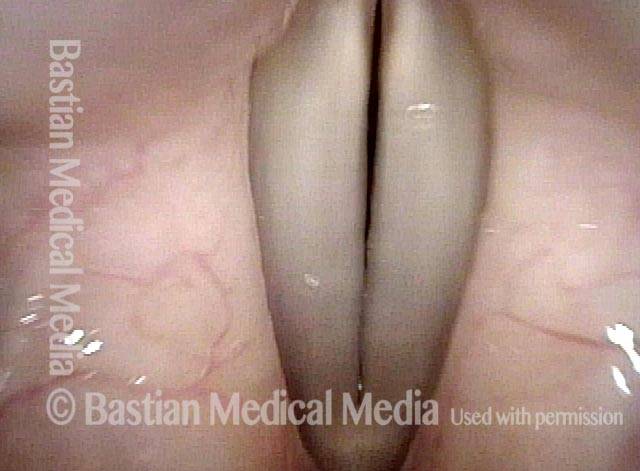
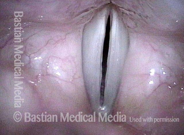
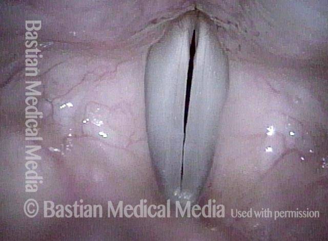
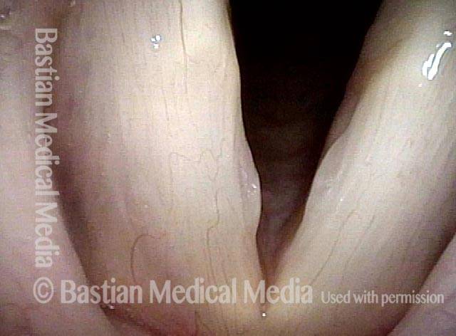
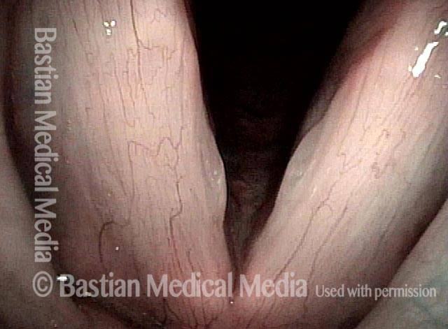
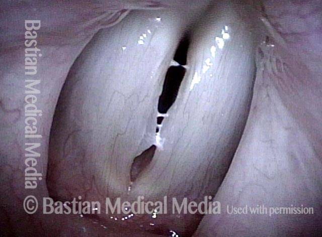
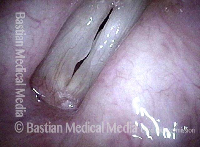
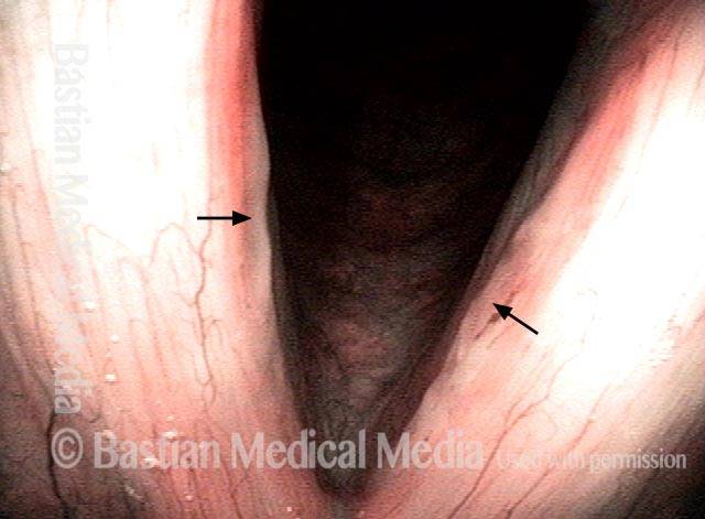
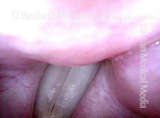
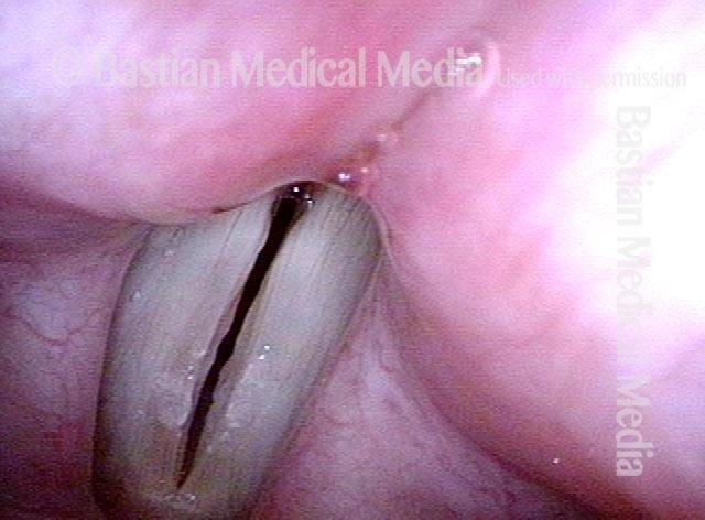
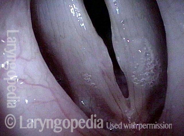
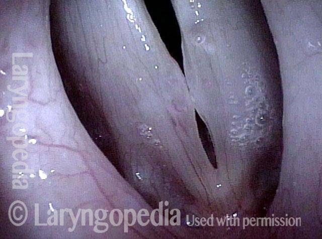
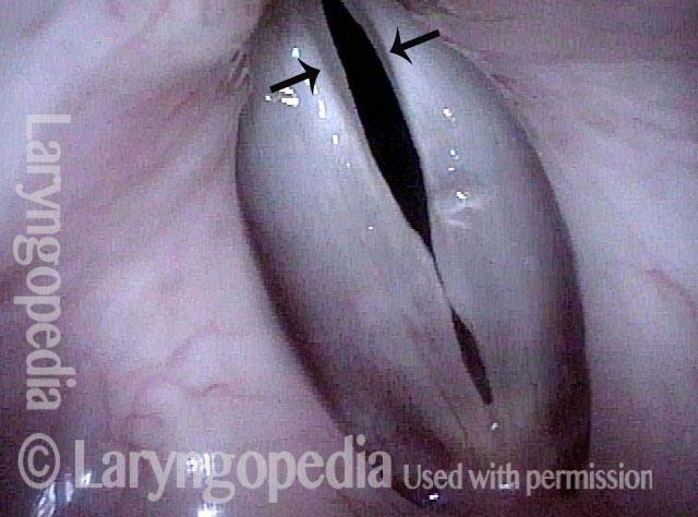
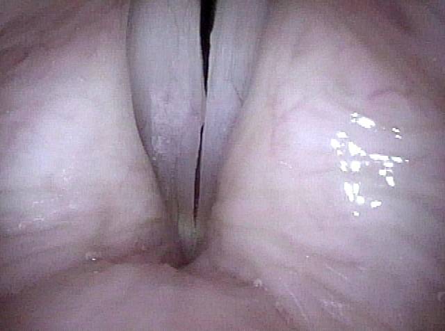
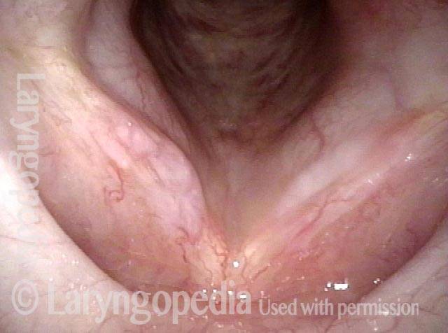
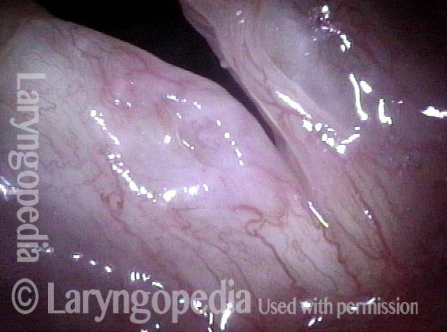
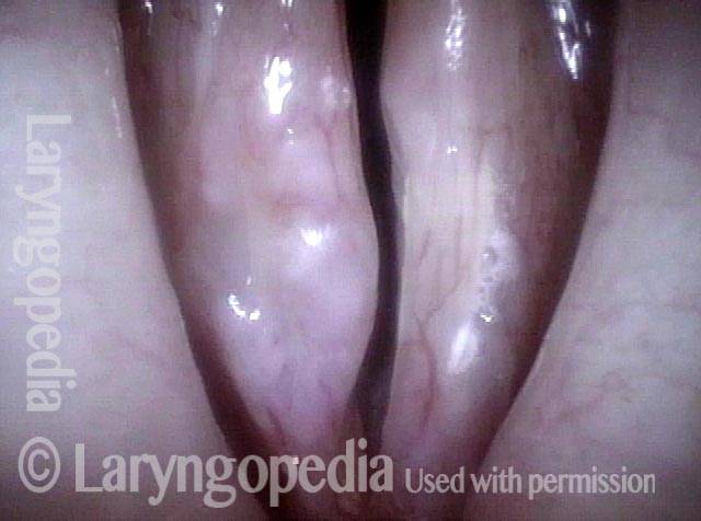
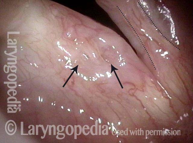
Small chronic swellings that appear in the junction of the middle and anterior thirds of the vocal fold. These swellings, or nodules (nodes), are vibratory injuries caused by vocal overuse. The most obvious symptom of medium-to-large nodules tends to be hoarseness. The top symptoms for nodules of any size may include:
- Difficulty with high, soft singing
- Day-to-day variability of vocal capability and clarity
- A sense of increased effort to produce voice, especially for singing;
- Reduced endurance, so that the voice becomes husky or “tired” after less voice use than formerly
- Phonatory onset delays, when there is a slight hiss of air before the voice “pops in.”
How nodules happen
When you overuse your voice, your body tries to cushion the vocal cords by pooling together edema (fluid) beneath the vocal cord mucosa (the surface layer of the cords); this pooled edema is like a small, low-profile blister on your finger. If after a few days you stop overusing your voice, the edema disperses readily, within a few days, and this “blister” on the vocal cords vanishes.
If, however, the amount or manner of voice use remains excessive for many weeks or months, then more chronic swelling materials (no longer just edema fluid) are laid down by the body, and the vocal cords develop true nodules.
Why nodes affect the voice
In either case (acute swellings or chronic nodules), this injury to the mucosa can impair the voice in two ways: it reduces the vibratory flexibility of the mucosa, and it interferes with the accurate match of the cords when they come together to produce voice. This impairment causes the voice to be hoarse or, more subtly, to suffer from onset delays, difficulty with high notes, and other similar problems.
Treatment for vocal nodules
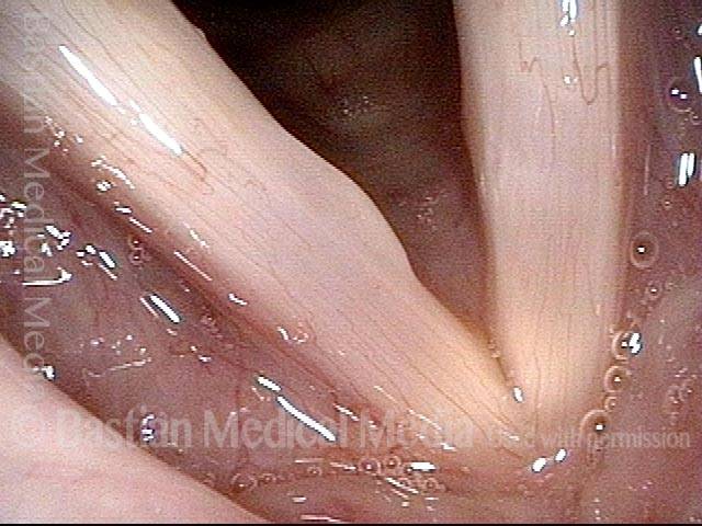
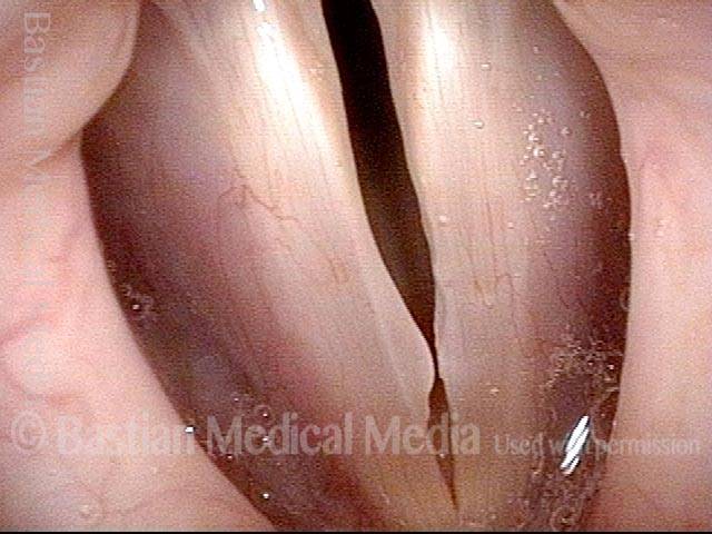
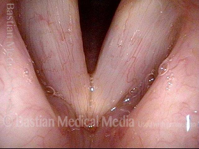
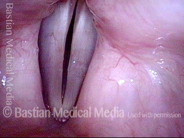
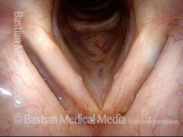
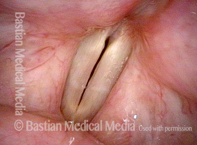
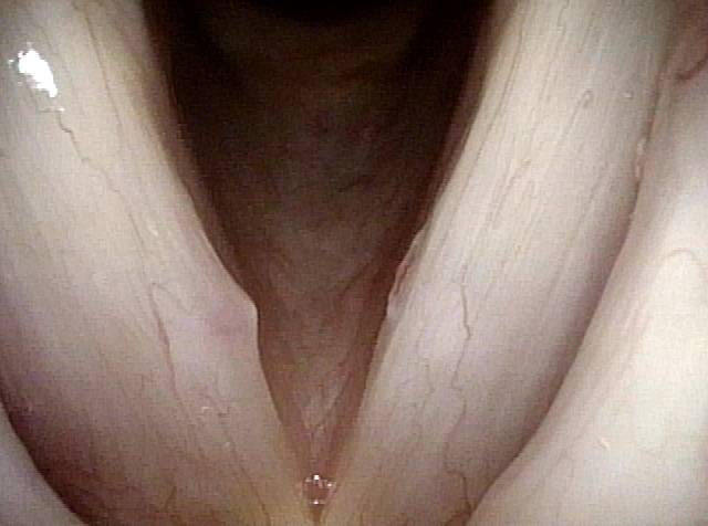
Nodules will often dissipate, with the help of rest and perhaps speech/voice therapy, over a period of weeks or months. Sometimes, the swellings are so stubborn that surgery is required.
How is the voice after treatment?
Vocal nodules’ effect on the voice, BEFORE surgical removal (see this patient’s photos just below): Same patient, seven weeks AFTER surgical removal of the vocal nodules:Vocal Nodules, Before and After Surgery
Vocal nodules (1 of 6)
Vocal nodules (1 of 6)
Breathy voice (2 of 6)
Breathy voice (2 of 6)
1 week after surgery (3 of 6)
1 week after surgery (3 of 6)
Closed phase (4 of 6)
Closed phase (4 of 6)
7 weeks after surgery (5 of 6)
7 weeks after surgery (5 of 6)
7 weeks after surgery (6 of 6)
7 weeks after surgery (6 of 6)
Polypoid Vocal Nodules
Polypoid vocal nodules (1 of 4)
Polypoid vocal nodules (1 of 4)
Incomplete closure (2 of 4)
Incomplete closure (2 of 4)
Polypoid vocal nodules (3 of 4)
Polypoid vocal nodules (3 of 4)
Polypoid vocal nodules (4 of 4)
Polypoid vocal nodules (4 of 4)
Vocal Nodules, Leukoplakia, and Capillary Ectasia
Vocal nodules, leukoplakia, and capillary ectasia (1 of 4)
Vocal nodules, leukoplakia, and capillary ectasia (1 of 4)
Vocal nodules, leukoplakia, and capillary ectasia (2 of 4)
Vocal nodules, leukoplakia, and capillary ectasia (2 of 4)
Vocal nodules, leukoplakia, and capillary ectasia: 6 months later (3 of 4)
Vocal nodules, leukoplakia, and capillary ectasia: 6 months later (3 of 4)
Vocal nodules, leukoplakia, and capillary ectasia: 6 months later (4 of 4)
Vocal nodules, leukoplakia, and capillary ectasia: 6 months later (4 of 4)
Vocal Nodules, Before and After Surgery
Vocal nodules (1 of 10)
Vocal nodules (1 of 10)
Prephonatory instant (2 of 10)
Prephonatory instant (2 of 10)
Translucency (3 of 10)
Translucency (3 of 10)
Open phase (4 of 10)
Open phase (4 of 10)
1 week after surgery (5 of 10)
1 week after surgery (5 of 10)
1 week after surgery (6 of 10)
1 week after surgery (6 of 10)
1 week after surgery (7 of 10)
1 week after surgery (7 of 10)
Vocal nodules: 10 weeks after surgery (8 of 10)
Vocal nodules: 10 weeks after surgery (8 of 10)
10 weeks after surgery (9 of 10)
10 weeks after surgery (9 of 10)
10 weeks after surgery (10 of 10)
10 weeks after surgery (10 of 10)
Example 2
Vocal nodules (1 of 4)
Vocal nodules (1 of 4)
Phonation (2 of 4)
Phonation (2 of 4)
After surgery (3 of 4)
After surgery (3 of 4)
After surgery (4 of 4)
After surgery (4 of 4)
Example 3
Vocal nodules, before surgery (1 of 4)
Vocal nodules, before surgery (1 of 4)
Before surgery (2 of 4)
Before surgery (2 of 4)
After surgery (3 of 4)
After surgery (3 of 4)
Vocal nodules, after surgery (4 of 4)
Vocal nodules, after surgery (4 of 4)
Vocal Nodules, Spicule-shaped
Vocal nodules (1 of 3)
Vocal nodules (1 of 3)
During phonation (2 of 3)
During phonation (2 of 3)
Closed phase (3 of 3)
Closed phase (3 of 3)
Example 2
Vocal nodules (1 of 4)
Vocal nodules (1 of 4)
Small nodules (2 of 4)
Small nodules (2 of 4)
Phonation (3 of 4)
Phonation (3 of 4)
Open phase (4 of 4)
Open phase (4 of 4)
Fibrotic Nodules
Fibrotic nodules (1 of 5)
Fibrotic nodules (1 of 5)
Fibrotic nodules: full-length vibration at low pitch (2 of 5)
Fibrotic nodules: full-length vibration at low pitch (2 of 5)
Fibrotic nodules: full-length vibration at low pitch (3 of 5)
Fibrotic nodules: full-length vibration at low pitch (3 of 5)
Fibrotic nodules: segmental vibration at high pitch (4 of 5)
Fibrotic nodules: segmental vibration at high pitch (4 of 5)
Fibrotic nodules: segmental vibration at high pitch (5 of 5)
Fibrotic nodules: segmental vibration at high pitch (5 of 5)
Vocal Nodules
Vocal nodules (1 of 4)
Vocal nodules (1 of 4)
Prephonatory instant (2 of 4)
Prephonatory instant (2 of 4)
Open phase of vibration (3 of 4)
Open phase of vibration (3 of 4)
Closed phase of vibration (4 of 4)
Closed phase of vibration (4 of 4)
Example 2
Vocal nodules (1 of 2)
Vocal nodules (1 of 2)
Vocal nodules (2 of 2)
Vocal nodules (2 of 2)
Example 3
Vocal nodules (1 of 1)
Vocal nodules (1 of 1)
Example 4
Vocal nodules (1 of 1)
Vocal nodules (1 of 1)
Vocal Cord ‘Wounds,’ 2 Hours after Surgery
Polypoid nodule, open phase (1 of 8)
Polypoid nodule, open phase (1 of 8)
Polypoid nodule, closed phase (2 of 8)
Polypoid nodule, closed phase (2 of 8)
Polypoid nodule, right and left cords (3 of 8)
Polypoid nodule, right and left cords (3 of 8)
Segmental vibration (4 of 8)
Segmental vibration (4 of 8)
Post-surgery wounds (5 of 8)
Post-surgery wounds (5 of 8)
Post-surgery, closed phase (6 of 8)
Post-surgery, closed phase (6 of 8)
Post-surgery, open phase (7 of 8)
Post-surgery, open phase (7 of 8)
Post-surgery, precise match (8 of 8)
Post-surgery, precise match (8 of 8)
Fibrosis as a Base to Nodules, Before and After Surgery
Bilateral polypoid nodules (1 of 8)
Bilateral polypoid nodules (1 of 8)
Narrow-band lighting (2 of 8)
Narrow-band lighting (2 of 8)
Closed phase (3 of 8)
Closed phase (3 of 8)
Open phase (4 of 8)
Open phase (4 of 8)
Two weeks after surgery (5 of 8)
Two weeks after surgery (5 of 8)
Phonation (6 of 8)
Phonation (6 of 8)
Margin fibrosis (7 of 8)
Margin fibrosis (7 of 8)
Open phase (8 of 8)
Open phase (8 of 8)
Pre-and 1 Week Post-removal Vocal Nodules
Vocal nodules (1 of 8)
Vocal nodules (1 of 8)
Muscular tension dysphonia (2 of 8)
Muscular tension dysphonia (2 of 8)
Open phase (3 of 8)
Open phase (3 of 8)
Closed phase (4 of 8)
Closed phase (4 of 8)
Post-op, one week (5 of 8)
Post-op, one week (5 of 8)
Phonation (6 of 8)
Phonation (6 of 8)
Open phase (7 of 8)
Open phase (7 of 8)
Closed phase (8 of 8)
Closed phase (8 of 8)
Vocal Nodule Postop Irregularity yet Match and Flexibility
Large vocal nodules (1 of 8)
Large vocal nodules (1 of 8)
Narrow band light (2 of 8)
Narrow band light (2 of 8)
One week after surgery (5 of 8)
One week after surgery (5 of 8)
Prephonatory instant (6 of 8)
Prephonatory instant (6 of 8)
Open phase (7 of 8)
Open phase (7 of 8)
Search not Only for Nodules, but Also for Segmental Vibration and Look at the Posterior Commissure for MTD
Open phase (1 of 4)
Open phase (1 of 4)
Closed phase (2 of 4)
Closed phase (2 of 4)
Segmental vibration (3 of 4)
Segmental vibration (3 of 4)
Posterior commissure (4 of 4)
Posterior commissure (4 of 4)
Ask Both Voice (via vocal phenomenology) and Larynx (via a motivated examination) to Give up their Secrets
Obvious mucosal injury (1 of 3)
Obvious mucosal injury (1 of 3)
Vocal nodules (2 of 3)
Vocal nodules (2 of 3)
Capillary ectasia (3 of 3)
Capillary ectasia (3 of 3)
Mottled Vocal Cord Mucosa May Hide Glottic Sulci
Vocal cord swelling and mucosa (1 of 4)
Vocal cord swelling and mucosa (1 of 4)
Same view under strobe light (2 of 4)
Same view under strobe light (2 of 4)
Closed phase (3 of 4)
Closed phase (3 of 4)
Glottic sulcus is visible (4 of 4)
Glottic sulcus is visible (4 of 4)
Nodules and Other Vocal Cord Injuries: How They Occur and Can Be Treated
This video explains how nodules and other vocal cord injuries occur: by excessive vibration of the vocal cords, which happens with vocal overuse. Having laid that foundational understanding, the video goes on to explore the roles of treatment options like voice therapy and vocal cord microsurgery.
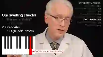
Vocal Fold Swelling Checks
In this video, Dr. Bastian introduces and demonstrates two swelling checks: two simple vocal exercises that can be used to help detect the early signs of vocal fold injury. He also discusses the physiology of vocal fold injury and explains how to make the best use of these swelling checks in your daily routine.
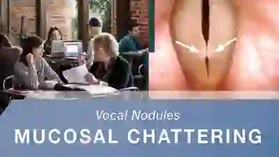
Mucosal Chattering
In this video, Dr. Bastian makes extensive use of elicitation with his own voice in order to hear the “vocal phenomenology” of a voice under evaluation. He has named one phenomenon he has not heard described elsewhere as mucosal chatter.
