An encyclopedia about voice, swallowing, airway, coughing, & other head + neck disorders.

Post-Radiation Telangiectasia

Post-radiation telangiectasia (teh-LAN-jee-ek-TAY-zhuh) refers to profuse neo-formation or atypical dilation of capillaries, most often observed in the laryngopharynx, as a delayed tissue response to radiation therapy. These vascular changes are benign, though they can appear dramatic on endoscopic examination.

Appearance

  • Bright red vascular markings scattered over irradiated mucosa, typically especially on the true cords, but can also appear on false cords, low aryepiglottic folds, petiole of epiglottis, etc.
  • May present as arborizing patterns of dilated capillaries lacking mass effect
  • They lack any corkscrew / bizarre pattern and are unassociated with leukoplakia, roughness, etc.

Timing

  • Rarely seen in the immediate or early post-treatment phase
  • More commonly appears as a mid- to long-term change, often 12 months or more after the conclusion of radiation therapy
  • They persist chronically once they are established

Clinical Significance

They are benign and without functional or symptomatic consequence. While by contrast, capillary ectasia can make the cords vulnerable to injury (bruising, hemorrhagic polyp), telangiectasias do not seem to manifest the same potential vibratory fragility.

It is important to inform patients prior to radiation therapy not only about logistics, early side effects, etc., but also that post-radiation telangiectasias may eventually occur. It can be a good practice to remind patients of this potential during early post-treatment visits, before they have appeared.  That is because these vascular changes can appear fairly suddenly between routine follow-up visits, and their striking appearance may cause anxiety when patients view their videoendoscopic examinations.

If so, the clinician can allay anxiety by reminding the patient: “Do you remember that I told you to expect that impressive, dilated capillaries may eventually appear some time after radiation is completed?” This reassurance helps normalize the finding and prevents confusion with disease recurrence.

Progression of Post-Radiation Treatment for T1-B Glottic Cancer

This 48-year-old man increasing hoarseness and a constant sore throat. Elsewhere, partly knowing of his loud workplace and boisterous personality, vocal nodules were diagnosed. Marked retrognathism necessitated office flexible endoscopic biopsy, returning a diagnosis of vocal early vocal cord cancer.

Due to bilateral abnormality and “impossible” surgical anatomy due to retrognathism, he was sent for radiotherapy rather than attempting laser resection.  The sequence of ulceration and eventual telangiectasia formation is found in this series.   

Visual Portfolio, Posts & Image Gallery for WordPress

Cancer or HPV? (1 of 6)

Under narrow band (blue-green) light, the right vocal cord (left of photo) has exuberant cake-icing leukoplakia. The left vocal cord (right of photo) has a lot of stippled vascular markings suggesting HPV effect. Some are marked by arrows. As mentioned, the biopsy returns a diagnosis of early cancer leading to radiotherapy.

Two months post radiation treatment (2 of 6)

In the initial weeks after radiotherapy is completed, mucosa has typically sloughed along with tumor. Mucosa has not yet regenerated. We do not have an examination from that time. At this interval, healing is complete, and the mucosal capillary pattern is fine-grained. Voice is good, and tumor has fully resolved on both sides.

Seven months post radiation treatment (3 of 6)

In this narrow band (blue-green) light view, post-radiation telangiectasia have begun to appear.

One year post radiation treatment (4 of 6)

Narrow band light, again showing telangiectasias. This more distant view reveals telangiectasias on the false vocal cords too.

Two years post radiation treatment (5 of 6)

Two years after radiation, the examination is stable. Voice remains very functional.

Four years post radiation treatment (6 of 6)

Under standard illumination to remind the viewer of the natural color. The slight protrusions at the blue arrows are “capillary knuckles” that may appear with telangiectasias long after radiotherapy.

Vocal Cord Cancer, before, during, and after Radiation

Visual Portfolio, Posts & Image Gallery for WordPress

Vocal cord cancer (1 of 12)

A 66-year-old man who complains of hoarseness. He smoked a pack a day for 50 years but quit five years ago. Note here the fullness and irregular contour especially of the left vocal cord ( right of photo). A biopsy confirmed this was cancer.

Vocal cord cancer (2 of 12)

At closer range, scattered leukoplakia and stippled vascular markings (suggestive of HPV effect, but HPV tested negative).

Vocal cord cancer, 3 weeks after radiotherapy (4 of 12)

Same patient, three weeks after the end of full-course radiotherapy. Distant view shows radiation-induced mucositis on the false and true cords, seen as areas of white, superficial ulceration. General redness is also a radiation effect.

Vocal cord cancer, 3 weeks after radiotherapy (5 of 12)

Closer view, showing that the main tumor of the left vocal cord (again, right of photo) has melted away. Note that the mucositis is generalized, and not necessarily focal to the area of tumor sloughing.

Vocal cord cancer, 2 months after radiotherapy (6 of 12)

Same patient, now two months after the end of radiotherapy. All of the visible tumor is gone, and voice is very good. Small anterior web. Compare with photo 1 of this series.

Vocal cord cancer, 2 months after radiotherapy (7 of 12)

Phonation. Compare with photo 3 of this series.

4 Years after radiation therapy (8 of 12)

Distant view of post-radiation telangiectasia.

Telangiectasia (9 of 12)

At closer range, instead of just “redness” we can see vascularity.

Closer look (10 of 12)

On closer inspection exuberant post-radiation telangiectasias are seen especially on the right cord. Also, post-radiation anterior commissure web.

Anterior Web and Telangiectasia (11 of 12)

Up close inspection of impressive, yet harmless vascularity.

Narrow band lighting (12 of 12)

Under narrow-band lighting (green and blue light). Since red light is absent, the capillaries display in even higher contrast black.

Telangiectasia, Gradually Developing Post-radiation

Visual Portfolio, Posts & Image Gallery for WordPress

Soon after end of radiotherapy (1 of 4)

Early after conclusion of radiotherapy for a left anterior vocal cord cancer. There is a small amount of resolving radiation mucositis (white patches) in the interarytenoid area.

1 year later: slight post-radiation telangiectasia (2 of 4)

Nearly a year later, with mild vascular prominence, especially left vocal cord (right of image).

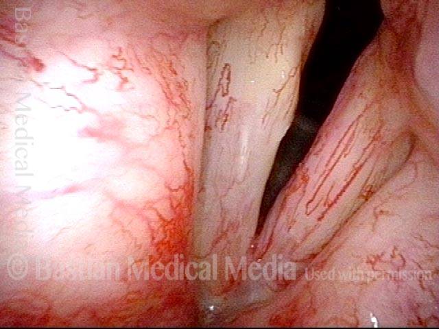
2 years later: post-radiation telangiectasia (3 of 4)

Now nearly two years after end of radiotherapy, much more prominent vascularity, called post-radiation telangiectasia. This side effect of radiation often begins to happen between one and two years after the end of radiotherapy. Notice that the post-radiation telangiectasias involve all areas that received radiation (including false vocal cords, etc.), and not just the area of original tumor. Voice remains very good.

3 years later: post-radiation telangiectasia (4 of 4)

Just over three years after the radiotherapy, telangiectasia now exuberant, but fairly stable. Voice remains very good.

Radiation: Telangiectasia Increases Slowly but is Maximal by 3 Years after End of Radiation

Visual Portfolio, Posts & Image Gallery for WordPress

Inflammation with moderate dysplasia (1 of 8)

Hoarseness began 2 years prior to this initial examination photo. This middle aged man has never smoked; has no reflux symptoms, nor has he had any response to empiric reflux therapy. Biopsy shows inflammation with moderate dysplasia. HPV testing was negative. No explanation for this chronic inflammation is ever found. A series of KTP laser treatments of stippled vascular areas and leukoplakia ensues.

Squamous cell carcinoma-in situ (2 of 8)

A year later, similar findings except appearance of a slight anterior commissure inflammatory web. Eventually, after an additional 2 years (5 years after onset of hoarseness) a second biopsy is triggered by aberrant, “corkscrew” capillaries. The diagnosis: squamous cell carcinoma-in situ. Laser excision is typically preferred for well-demarcated early vocal cord cancer, but the diffuse, superficial and bilateral abnormalities suggested radiotherapy instead.

Post radiation (3 of 8)

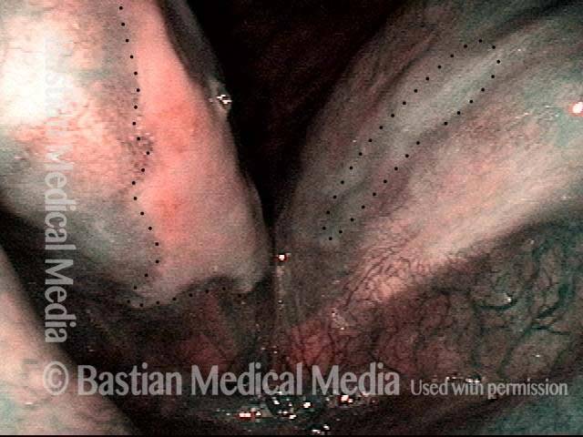
Six weeks after the end of his 30 radiation treatments, healing of the superficial ulceration (within dotted lines) is underway.

Narrow band light (4 of 8)

Closer view, now under narrow band light: A fine vascular pattern has returned except in the areas bounded by dotted lines.

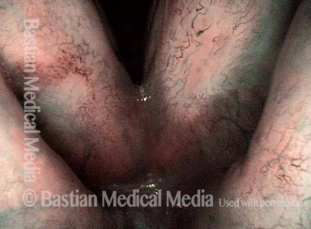
Regenerated vascular pattern (5 of 8)

Six weeks later (3 months after end of radiotherapy), the superficial ulceration has healed, and voice is very functional. Note the regenerated vascular pattern, and compare with photo 4.

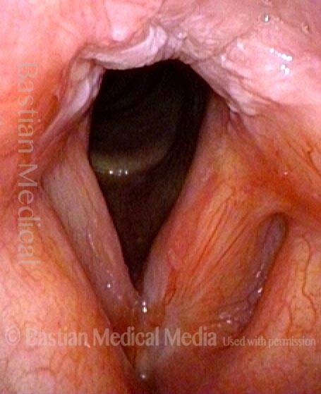
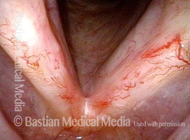
Post-radiation telangiectasias (6 of 8)

“On schedule” a year after the end of successful radiation therapy, post-radiation telangiectasias are becoming evident. Compare progression of these telangiectasias in photos 4, 5, 6, 7, and 8.

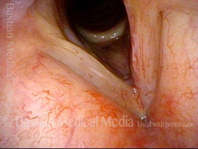
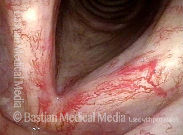
Standard light (7 of 8)

Now 3 years from the end of radiotherapy, radiation telangiectasia are “maximal” and stable. Voice remains very good.

Narrow band light (8 of 8)

Same view, under narrow band light.
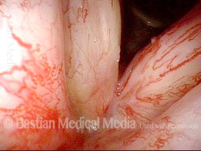
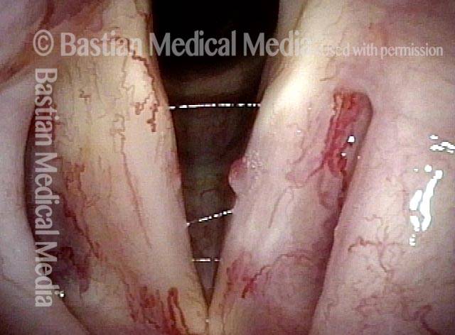
Office Laser of Post-Radiation Telangiectatic Polyp

Visual Portfolio, Posts & Image Gallery for WordPress

Post-radiation telangiectasias (1 of 4)

Years after radiotherapy for vocal cord cancer, the exaggerated capillaries are not typical capillary ectasia, but instead post-radiation telangiectasias. The “polyp” may be also radiation-related because there is no history of voice over-use.

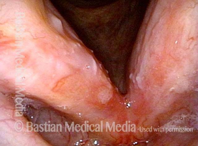
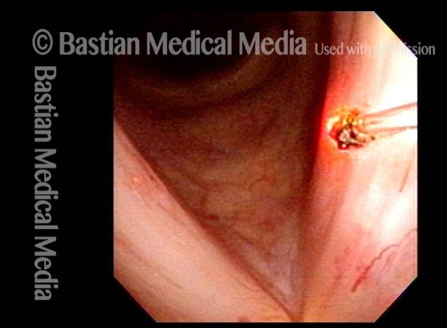
Pulsed-KTP coagulation (2 of 4)

At the conclusion of pulsed-KTP coagulation of the “polyp.”

“Polyp” pulled off (3 of 4)

The “polyp” has pulled off with the fiber.

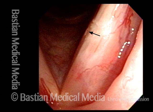
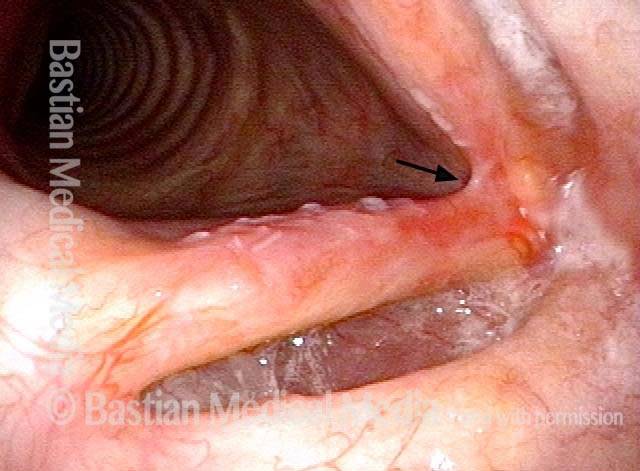
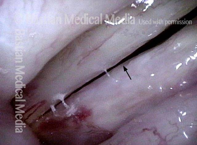
Three weeks later (4 of 4)

Three weeks later, the vocal cords now match, voice is improved, and the site of surgery (arrow) is healed.

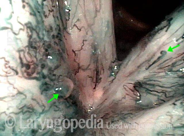
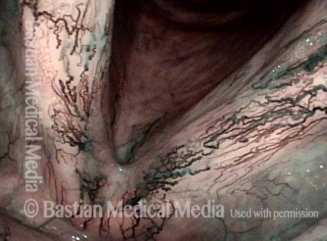
Telangiectasias and more after Radiotherapy

Visual Portfolio, Posts & Image Gallery for WordPress

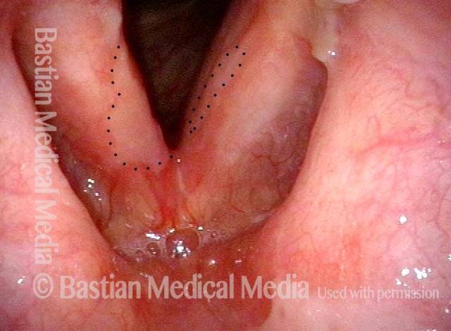
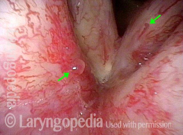
Post radiotherapy (1 of 2)

More than five years after radiotherapy for early vocal cord cancer, the post-radiation telangiectasias seen here are maximal and not expected to increase further. What can occasionally increase, are small polyp-like elevations (arrow to the left).

Narrow band lighting (2 of 2)

Narrow band light at closer range dramatically accentuates vascularity. Intense red becomes almost black; pink becomes blue-green.

Share this article

Support Laryngopedia
Are you a patient or doctor who loves Laryngopedia? If we've helped you, help us help others too!

Table of Contents