Common Usage
Suction drains are used routinely to evacuate blood and serum from under skin flaps in the early days after surgical procedures. When drainage diminishes to 30 ml or less in 24 hours, drains can be removed, most often within the first 3 or 4 postoperative days. At that point, they have served their purpose, and out they come.
Beyond Common Usage: Overview
There are several circumstances where the suction drain is used to treat a problem;
- Salivary fistula
- Air leak
- Esophageal perforation
- Necrosis
The origin of the concept and the details of “suction drainsmanship” can be found in the original article.
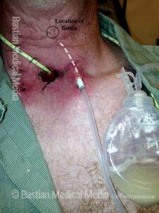
Suction Drain as a Treatment for Salivary Fistula
The first disorder for which suction drains are powerful treatment tools is salivary fistula. A “safety” suction drain can be placed strategically at the conclusion of laryngectomy, ready to capture the very first drop of saliva if the mucosal closure leaks. That “safety-monitoring” drain remains in position for 5 or more days before removal. If a fistula occurs, the drain is left in for as long as saliva or purulence continues to drain—sometimes for several weeks. When the fistula heals, the drain is backed out in steps.
Once surgeons master the concept and what to do for continuous vs intermittent leaks, etc., the patient can be taught and trusted to manage his or her fistula: that is, there are no IV’s; no dressing changes; no odor. The hospital stay is lengthened by only one or two days. Simple outpatient oversight occurs by phone and office visits.
Not only is patient management dramatically simplified by using suction as a treatment tool, but the surgical procedure proposed to the patient in the first place, might itself be of lesser magnitude. Suction drain management of fistulas is so safe and effective, that the need for protective flaps over carotid arteries, or to bolster mucosal closures is arguably diminished.
Suction Drains to Manage Air Leaks, Such as After Cricotracheal Resection
A second use is to manage air leaks after crico-tracheal resection for subglottic / tracheal stenosis. When a stenotic segment is entirely within the trachea, the repair is simple, because the repair is “trachea to trachea.” An airtight closure is therefore easy to accomplish.
The more difficult closure is trachea to cricoid, especially if part of the cricoid must be removed. The cricoid and trachea do not match in caliber. One can meticulously suture these two structures together with a dozen or more sutures. Yet, when airtightness is assessed, there is occasionally an air leak. One could spend a lot of time taking the repair down and “trying again.” Instead, suction drains can be placed in the tracheaoesophageal groove to manage this leak until healing closes it.
Suction Drains to Manage Hypopharyngeal or Cervical Esophageal Perforation
A third use might be to manage pyriform sinus or cervical esophageal perforation. Rather than attempting repair of the perforation, or widely draining the neck, a suction drain can be inserted through a tiny incision to actively “control” a perforation (or resulting abscess if there has been delay).
Suction Drain After Endoscopic Laser Cricopharyngeal Myotomy Antegrade Cricopharyngeus Dysfunction With or Without Zenker’s Diverticulm
A fourth use is after endoscopic laser cricopharyngeus myotomy. By definition, this procedure results in an esophageal perforation, because the muscular sphincter is divided “inside out,” first incising through hypopharyngeal / cervical esophagus mucosa.
A suction drain can be left in the area of the myotomy within the esophageal lumen and brought out through the nose to wall suction, to protect the patient from subcutaneous or mediastinal emphysema or even mediastinitis. There is a teaching video forthcoming to explain this technique.
Suction Drain for Non-Fistula Necrosis
Yet another use for suction drainage is under a transfer or free flap overlying a necrotic base. An example here might be the patient with widespread tissue breakdown due to a second course of radiotherapy or perhaps radioactive seed placement (brachytherapy).
In one actual scenario, the area of breakdown was at least 8 cm in diameter, with exposed scalene muscles and carotid artery. A pectoralis flap was placed over this necrotic bed. A drain between necrotic base and flap suctioned away liquifying necrotic material until the pectoralis flap could “bond with and feed” the base over which it was placed.