An encyclopedia about voice, swallowing, airway, coughing, & other head + neck disorders.

Torus Mandibularis

Torus mandibularis (also known as a mandibular torus) is a benign bony exostosis (growth of extra bone) arising from the medial (lingual) surface of the mandible. These growths are composed of dense cortical bone and most commonly occur bilaterally along the premolar region. Mandibular tori are typically asymptomatic and often discovered incidentally during dental or oral examinations. In most cases, they require no treatment unless they interfere with oral hygiene, speech, denture fitting, or surgical access.

Visual Portfolio, Posts & Image Gallery for WordPress

Torus mandibularis (1 of 2)

View inside the mouth, focused on the floor of the mouth, with tongue retracted. The “mounds” seen in the foreground are unusually large tori, which are touching anteriorly.

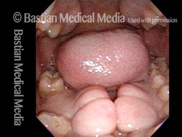
Torus mandibularis (2 of 2)

Looking more directly downward onto the tori, with the tongue now pointing upward at the roof of the mouth.

In laryngology, mandibular tori are clinically relevant because, when large, they can significantly impede exposure of the larynx during operative microlaryngoscopy. Adequate visualization of the vocal folds depends on compression of the floor of the mouth by the laryngoscope blade, which usually sits against the inner surface of the anterior mandible, allowing the distal end of the scope to angle anteriorly toward the anterior commissure of the glottis. While the soft tissues of the floor of mouth normally deform under this pressure, mandibular tori—being rigid bony structures—do not compress. As a result, large tori may prevent anterior angulation of the laryngoscope, making visualization of the vocal folds difficult or, in some cases, impossible.

Surgical Management

Surgical removal of mandibular tori is generally reserved for patients in whom the tori cause functional problems, including interference with denture placement or (rarely) repeated difficulty with laryngeal exposure during microlaryngoscopy. Resection is typically performed by an oral and maxillofacial surgeon under general anesthesia, although smaller tori may be removed under local anesthesia with sedation.

The procedure begins with a mucosal incision along the lingual gingiva overlying the torus, followed by careful elevation of a mucoperiosteal flap to expose the bony prominence. The torus is then reduced or removed using a combination of osteotomes, rotary burs, or powered saws, taking care to avoid injury to adjacent structures, particularly the lingual nerve and submandibular duct. The bony surface is smoothed to eliminate sharp edges, and the mucosal flap is repositioned and closed with absorbable sutures.

Postoperative care typically includes oral hygiene measures, a soft diet, and short-term analgesia.

Alternatives for the Laryngologist

In the modern era, torus mandibularis may turn attention away from O.R. management of recurrent laryngeal lesions like papillomas or leukoplakia, toward office laser (thulium, blue, pulsed-KTP) treatments.

How to Overcome Torus Mandibularis to Treat Glottic Carcinoma (Laryngeal Cancer)

This patient was suspected to have an early glottic carcinoma. The lesion was superficial, and the patient strongly preferred transoral laser excision over primary radiotherapy. During attempted microlaryngoscopic excision, however, a large anterior mandibular torus prevented adequate exposure of the anterior one-third of the lesion, making complete endoscopic resection impossible.

Rather than proceeding directly to radiotherapy to manage the residual disease, the patient requested an alternative approach. The residual anterior tumor was subsequently treated using an office-based thulium laser delivered via a glass fiber passed through a flexible endoscope. This allowed targeted coagulation of the remaining superficial lesion without the need for further operating room exposure or resorting to radiotherapy.

This approach has resulted in durable disease control, with nearly five years of cancer-free follow-up to date.

Torus Mandibularis

The inner (lingual) surface of the mandible is indicated by the dotted line. A prominent anterior mandibular torus prevents the laryngoscope from advancing anteriorly the full distance indicated by the white arrow. As a result, the distal tip of the laryngoscope (not shown, within the larynx) is unable to rotate anteriorly toward the anterior commissure of the vocal cords, explaining the inability to visualize the anterior extent of the tumor.

Share this article

Support Laryngopedia
Are you a patient or doctor who loves Laryngopedia? If we've helped you, help us help others too!