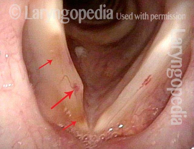
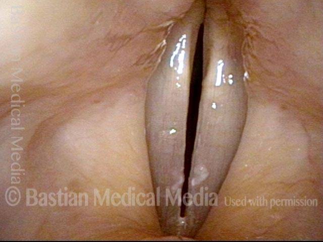
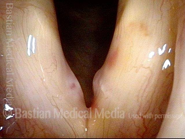
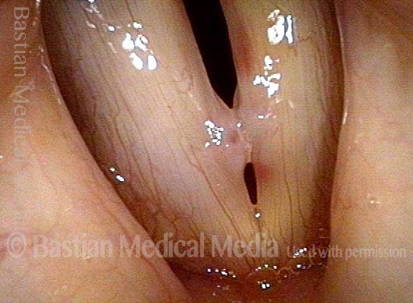
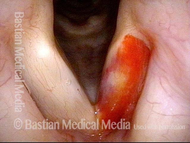
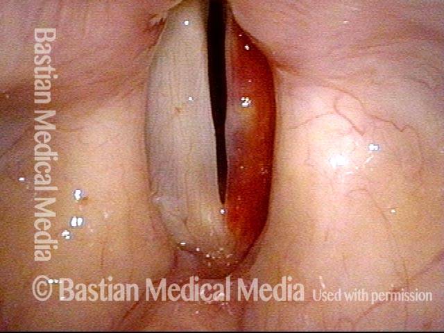
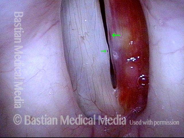
An encyclopedia about voice, swallowing, airway, coughing, & other head + neck disorders.

Vocal Cord Bruising (Hemorrhage)

Vocal cord bruising happens when tiny blood vessels (capillaries) in the vocal folds rupture, allowing blood to leak into the tissue. In addition to a visible bruise, this often causes sudden hoarseness and loss of upper voice.

Common triggers include aggressive or prolonged voice use such as yelling or extremely loud singing, forceful coughing, or a loud sneeze. Occasionally, there is no obvious cause in the patient’s memory, particularly when significant capillary ectasia is present.

There are two main types of vocal cord hemorrhage. To set up an analogy, think of a wet street after rain. If the pavement is new and smooth, with no low spots, the wet surface will dry quickly once the sun comes out. If, however, water collects in a pothole as a puddle, it may take much longer to evaporate—and this also indicates the need for street repair.

Returning to bruising, a superficial hemorrhage consists of a thin suffusion of blood very near the surface of the vocal fold (wet pavement). There is no change in the vocal fold margin, and vibration under strobe light can be normal, which explains why the voice is often only minimally affected. The bruise typically disappears rapidly.

If, on the other hand, there is ad “street puddle” of blood, like a micro–goose egg or “blood blister,” absorption occurs much more slowly. This type of hemorrhage may evolve into a polyp and can require removal of a residual lesion.

Initial Treatment

Once the type of bruising has been identified (thin suffusion versus blood blister or hemorrhagic polyp), the patient typically rests the voice for several days. Voice use should be minimal, though absolute voice rest is not usually necessary. Other general measures of voice care—such as good hydration and reflux management when present—are advisable.

A follow up visit can be useful within a short time if a superficial bruise, and the person can often resume voice use, including vocal performance, before all traces of the bruise have absorbed. A longer interval for reexamination makes sense for hemorrhagic polyps, since recovery is expected to take much longer.

After any vocal fold hemorrhage, follow-up examination is important, even if the voice becomes normal. The purpose is to look for an ectatic capillary, an abnormally enlarged surface blood vessel. These fragile vessels increase the risk of repeat hemorrhage and, if unidentified, may remain silent until another bruise occurs. For this reason, identification and prophylactic treatment, as described below, are strongly recommended.

If an ectatic capillary is found after resolution of the bruise, laser coagulation is often recommended, either in the office or the operating room. Commonly used lasers include CO₂, KTP, thulium, or blue laser systems. While the choice of laser is of some interest, the surgeon’s experience and judgment are more important; lasers tuned to the wavelength of hemoglobin (such as pulsed-KTP or blue laser) are theoretically ideal, but the CO₂ laser can also work very effectively.

If a blood blister becomes a persistent polyp, microsurgical removal may be needed. This is most often performed in the operating room, although in selected cases an office-based laser approach may be appropriate. With modern surgical techniques and an experienced surgeon, voice quality is almost always fully restored.

Prognosis Summary

Superficial bruises resolve rapidly without permanent damage. Blood blisters or hemorrhagic polyps recover more slowly and may require microsurgical repair, but in both cases, with expert care, outcomes are excellent, commonly returning the voice to its original capabilities.

A Vocal Cord Bruise that Could Happen to Anyone

While capillary ectasia (as seen in other photo series here) markedly increases vulnerability to vocal cord bruising, every human vocal cord has capillaries on its service, and even normal capillaries can leak and cause a bruise with sufficient vocal trauma.  In this person, with an aggressive cough, her normal capillaries are the source of the bruise.

Visual Portfolio, Posts & Image Gallery for WordPress

Bruised Vocal Cord (1 of 2)

View from a distance, showing a bruise of the right cord. But is there a vulnerable (ectatic) capillary that explain this bruise?

Bruised Vocal Cord (2 of 2)

At closer range, the normal capillaries are seen; there is no dilated capillary to explain the bruise. This bruise could happen to anyone, with sufficient vibratory (loud voice) or cough trauma.

Vocal Cord Bruises (Hemorrhage) often Initially Obscure the Ectatic Capillary “Culprit”

A person can experience sudden hoarseness at a time of sustained heavy voice use, or even immediately following a “scream,” or even a loud sneeze. The explanation might be a bruise of a vocal cord. This can happen to anyone but is far more likely if the person has underlying capillary ectasia.

When a bruised vocal cord is seen, therefore, the question is: “Is this a fluke bruise that can happen to anyone, or is it one explained by capillary ectasia?” In this instance, the answer is yes to capillary ectasia.

Visual Portfolio, Posts & Image Gallery for WordPress

Singer’s Bruised Vocal Cord (1 of 8)

In this singer, bilateral margin swellings are seen, but the right cord (left of photo) is bruised. Sometimes we must wait to see the culprit capillary or capillaries. Here, we can already suspect that this is the explanation (from following photos) since the bruise is a few weeks old and the blood in tissue partially resorbed.

Bruise under Strobe Light (2 of 8)

Strobe views (giving us apparent slow-motion vibration) help us see the margin elevations more clearly.

Abnormal capillary (3 of 8)

At very close range, the suspicion of an abnormal capillary is confirmed. But will it be there when the bruise is completely gone?

Bruise is gone (4 of 8)

Several weeks later, the bruise is indeed gone, and the vulnerability-inducing capillaries are seen clearly in this distant view.

Capillaries under narrow band light (5 of 8)

At close range under narrow band light, the capillaries come into full view. Notice one small capillary also on the left cord margin (right of photo).

Capillaries touch during phonation (6 of 8)

Phonatory view again reveals the margin swellings bilaterally, but with the ectatic capillaries in the point of maximum vibratory contact, where they increase the risk of another bruise.

Post-surgical repair (7 of 8)

After surgical repair of the ectasia and margin swellings (distant view), open for breathing.

Margins match (8 of 8)

Also after repair, voicing position showing again absence of the capillary ectasia and much better match of the margins. Upper voice is restored; vibratory flexibility is normal even at very high pitch.

Vocal Cord Bruise / Hemorrhage, Before and After Rest and Surgery

Visual Portfolio, Posts & Image Gallery for WordPress

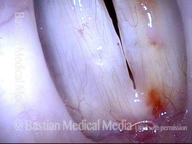
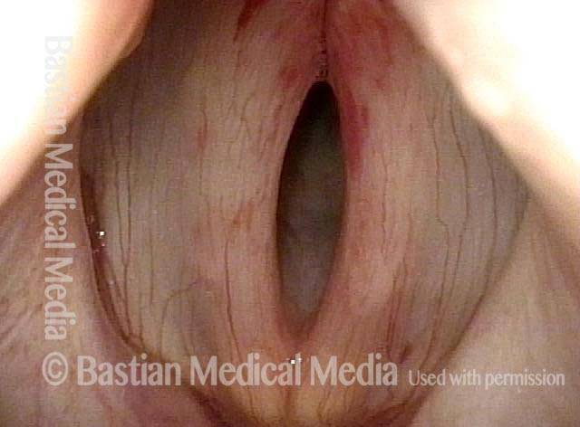
Vocal cord bruise / hemorrhage (1 of 4)

Bruise, right vocal cord (left of image), estimated one week old, in combination with large polypoid vocal nodules. Note the yellowish discoloration, indicating partial breakdown of the hemoglobin (source of red color of blood) into hemosiderin as bruise is beginning to be cleared away.

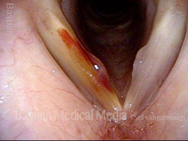
Vocal cord bruise / hemorrhage (2 of 4)

Same patient, during phonation.

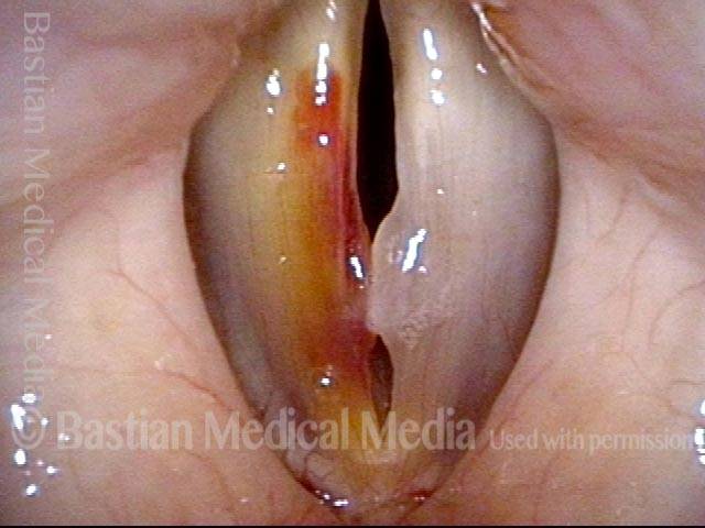
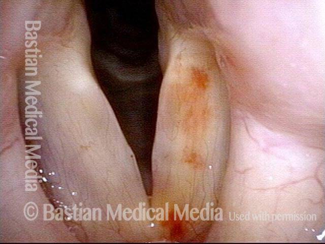
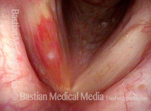
Vocal cord bruise / hemorrhage, after rest and surgery (3 of 4)

Bruise was allowed to resolve, and then patient underwent vocal cord microsurgery one week prior to this examination.

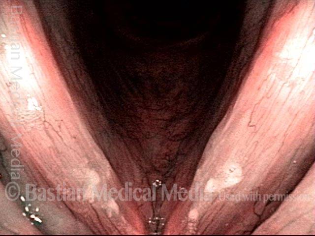
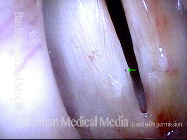
Vocal cord bruise / hemorrhage, after rest and surgery (4 of 4)

Same patient, during phonation. Note the patient’s tendency to phonate with a gap between the cords, as though the vocal cords “remember” the early contact that used to require separation during voicing. This gap can be lessened through expert speech (voice) therapy.

Vocal Cord Bruise / Hemorrhage

Visual Portfolio, Posts & Image Gallery for WordPress

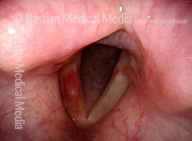
Vocal cord bruise / hemorrhage (1 of 2)

Bruise, left vocal cord (right of image), estimated three weeks old, in combination with vocal nodules, capillary ectasia. There is likely an ectatic capillary also on the same side, within the nodule, that is the source of the leaking of blood into the tissues. When the bruise first occurred, it would have been most evident in the area of the nodule. As time passes, the central part of the bruise typically resolves first, with the last area to disappear anterior and posterior, as shown here. Note also the faint yellowish discoloration of the left cord, indicating residual hemosiderin (breakdown products of blood in tissue).

Vocal cord bruise / hemorrhage (2 of 2)

Same patient during phonation under standard light.

Vocal Cord Bruise / Hemorrhage, Before and After Rest

Visual Portfolio, Posts & Image Gallery for WordPress

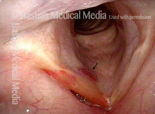
Vocal cord bruise / hemorrhage (1 of 6)

Breathing position, standard light. Notice a superficial bruise of the left cord (right of image), still very bright red. The source vessel cannot be identified with certainty here, but would be expected to be in the mid-cord, where the bruising is least evident. This is because mucosal oscillation tends to “massage” the bruise anteriorly and posteriorly, when the bruise is a thin-suffusion “wet pavement” type rather than a pocket or “puddle” of blood.

Vocal cord bruise / hemorrhage (2 of 6)

Prephonatory instant, standard light, shows that the margin of the left cord (right of image) is relatively straight. This suggests that the bruising is a very thin layer, and not a pocket of blood (as mentioned in photo 1).

Vocal cord bruise / hemorrhage (3 of 6)

Closer phonatory view, strobe light, also shows subtle elevation of the right cord (left of image), and a tiny ectatic capillary (small arrow), both of which can suggest that this person has been using the voice a lot. Left cord shows a darker linear bruise (larger arrow). After the bruising clears, perhaps it will become evident that this is in fact the ectatic capillary.

Vocal cord bruise / hemorrhage: after 2 weeks of rest (4 of 6)

After two weeks of relative voice rest, standard light. Notice the yellowish discoloration, which represents the breakdown products of hemoglobin. No obvious culprit capillary is seen. The last blood to resorb is always at the periphery from the point of origin.

After 2 weeks of rest (5 of 6)

Strobe light, open phase of vibration. No obvious ectatic vessel is seen, except for the vessel on the non-bruised side (at arrow) that was seen in photo 3.

After 2 weeks of rest (6 of 6)

Strobe light, closed phase, shows small margin swellings, greater on the left cord (right of image) than on the right cord. This swelling is being addressed by ongoing mild voice conservation, “on the fly” – i.e., while the person carries on with her work.

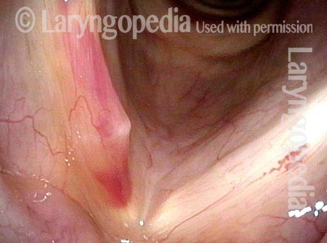
Bruise Caused by Cough

Visual Portfolio, Posts & Image Gallery for WordPress

Bruise caused by violent coughing (1 of 2)

Closer view of bruise, with small collection of white mucus in the middle.

Closer view of bruise (2 of 2)

A person with violent sensory neuropathic coughing may injure the vocal cords, as illustrated by this bruise, right vocal cord (left of photo).

Vocal Cord Bruising From Coughing

Visual Portfolio, Posts & Image Gallery for WordPress

Bruise from coughing (1 of 3)

This man had an episode of aggressive coughing a week earlier. Note the bruising over the vocal processes, which receive the major collisional force during coughing.

Pre-phonatory instant (2 of 3)

The vocal processes are approaching the point of touching (contact would occur gently with onset of talking and more aggressively with coughing).

Phonation (3 of 3)

Vocal cords are now in full contact. Note the unrelated moderately-severe vocal cord bowing.

The Evolution of Vocal Cord Bruising and Emergence of a Vulnerable Capillary

Visual Portfolio, Posts & Image Gallery for WordPress

Margin swelling and bruising (1 of 2)

This professional woman is extraordinarily dynamic and intense, and must talk all day to do her work. Here, the right vocal cord (left of photo) is bruised due to vibratory trauma. The margin swelling on the right causes her hoarseness more than the bruising, however.

Six weeks later (2 of 2)

Six weeks later, the bruise is mostly resolved. The capillary that “leaked” blood to form the bruise is now seen more clearly (long arrow). This ectatic capillary can be seen easily now when looking back at photo 1. The short arrows indicate the residual “smudges” of discoloration caused by breakdown products of the bruise. The last evidence of widespread vocal cord bruising is always in these two locations.

Resources for Further Reading

Bastian, R.W. (2015). Cummings Otolaryngology—Head & Neck Surgery, 899-927. Elsevier/Saunders. ISBN-13: 978-1455746965

Cordano VP, Alvarez ML, Cabrera JM, Napolitano CA. Acute vocal fold hemorrhage: retrospective analysis in a voice unit. J Voice. 2025;S0892-1997(25)00023-X.

Clark CM, Sulica L, et al. Current management of vocal fold hemorrhage: a survey of American Broncho-Esophagological Association members. J Voice. 2025;S0892-1997(25)00046-3.

Azadeh Ranjbar, P., Maxwell, P. J., Barna, A., Balouch, B., Hawkshaw, M., Al Omari, A. I., Lyons, K., Alnouri, G., & Sataloff, R. T. (2025). An Exploration of the Risk Factors, Severity, and Sequelae of Vocal Fold Hemorrhage in a Population of Voice Patients Before and After Diagnosis. Journal of voice : official journal of the Voice Foundation, 39(6), 1584–1593.

Lennon, C. J., Murry, T., & Sulica, L. (2014). Vocal fold hemorrhage: factors predicting recurrence. The Laryngoscope, 124(1), 227–232.

Klein, A. M., Lehmann, M., Hapner, E. R., & Johns, M. M., 3rd (2009). Spontaneous resolution of hemorrhagic polyps of the true vocal fold. J Voice. 23(1), 132–135.

Share this article

subglottic bruise
This individual occasionally coughs to the point of hoarseness. Particularly noteworthy is the subglottic bruise (arrow, dotted line) caused by profound Valsalva-retching kind of coughing. The rest of the right cord (left of photo) is also bruised.
Vocal nodules & other voice injuries YT Thumbnail

Nodules and Other Vocal Cord Injuries: How They Occur and Can Be Treated

This video explains how nodules and other vocal cord injuries occur: by excessive vibration of the vocal cords, which happens with vocal overuse. Having laid that foundational understanding, the video goes on to explore the roles of treatment options like voice therapy and vocal cord microsurgery.

Support Laryngopedia
Are you a patient or doctor who loves Laryngopedia? If we've helped you, help us help others too!

Table of Contents