An encyclopedia about voice, swallowing, airway, coughing, & other head + neck disorders.

Hemorrhagic Polyp

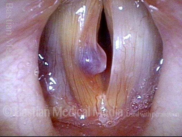
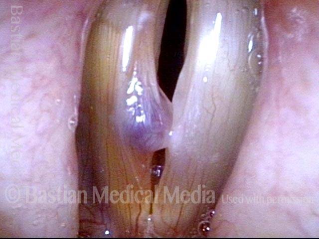
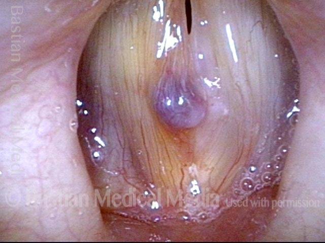
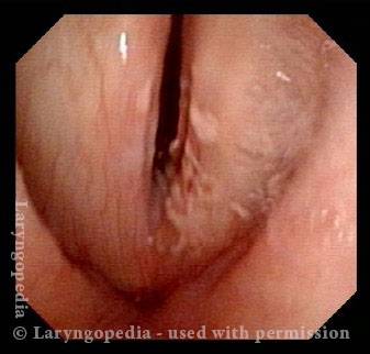
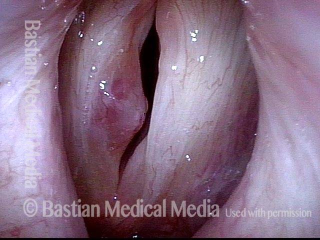
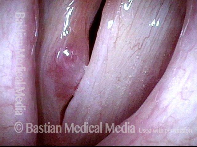
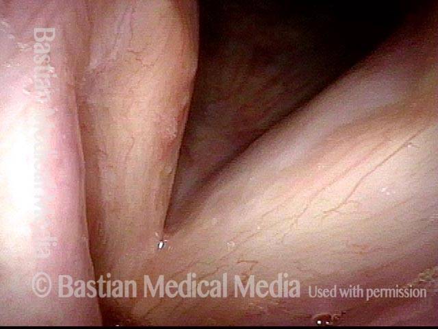
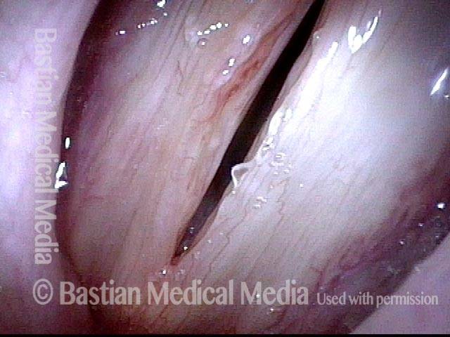
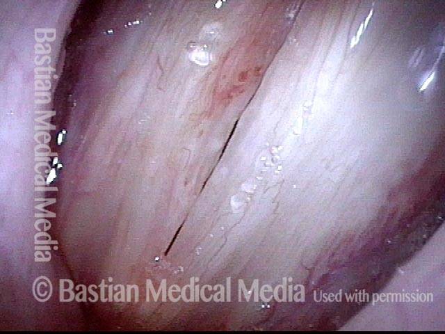
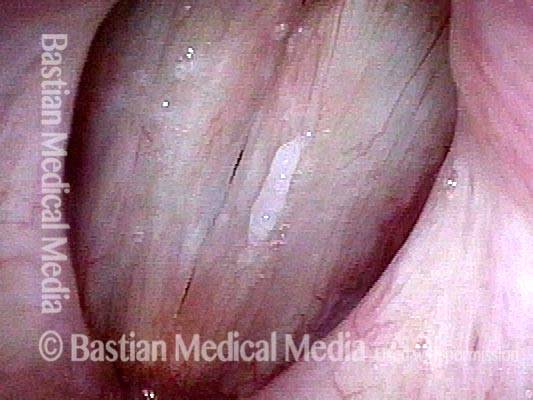
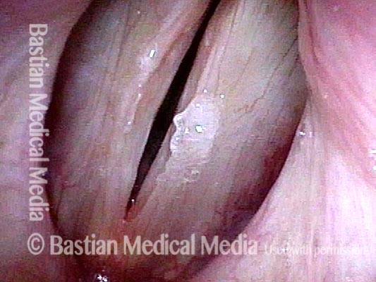
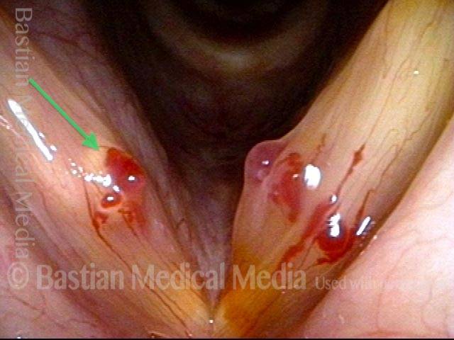
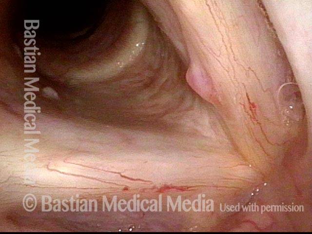
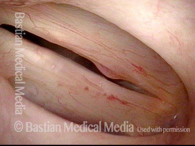
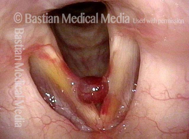
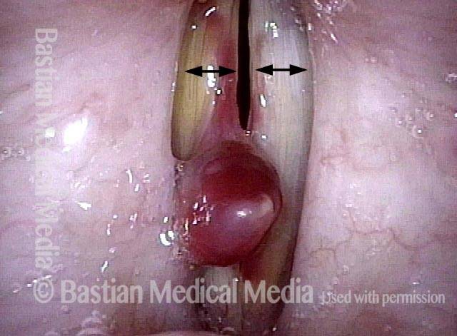
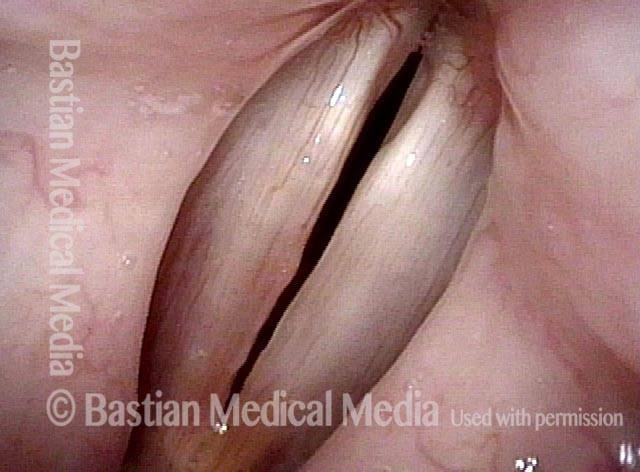
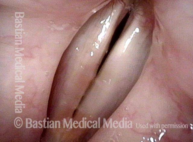
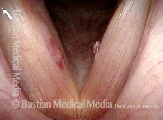
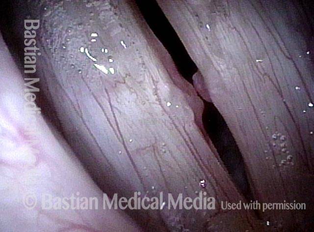
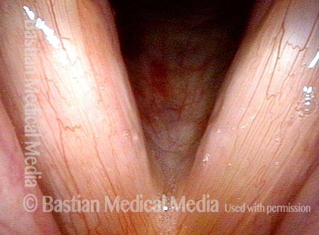
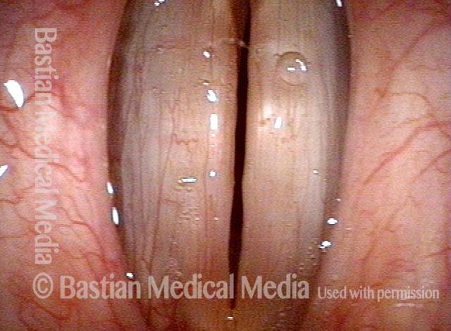
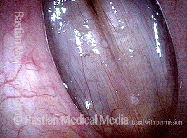
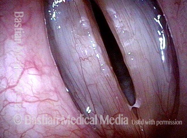
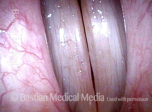
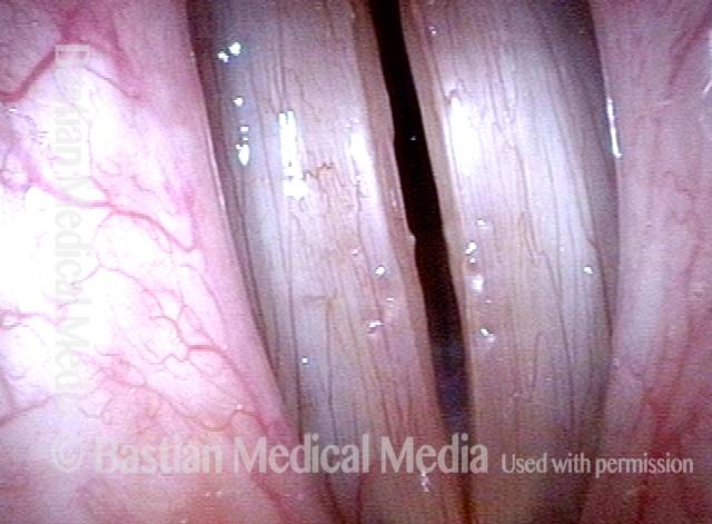
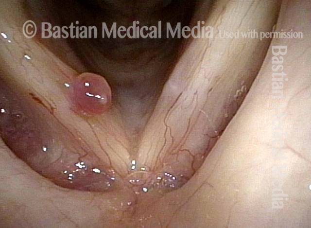
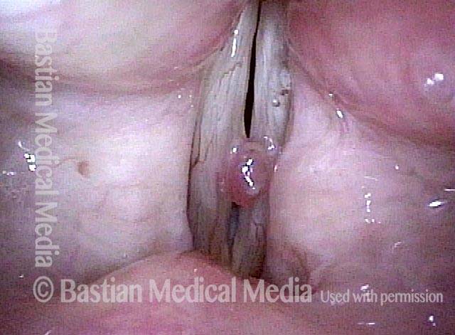
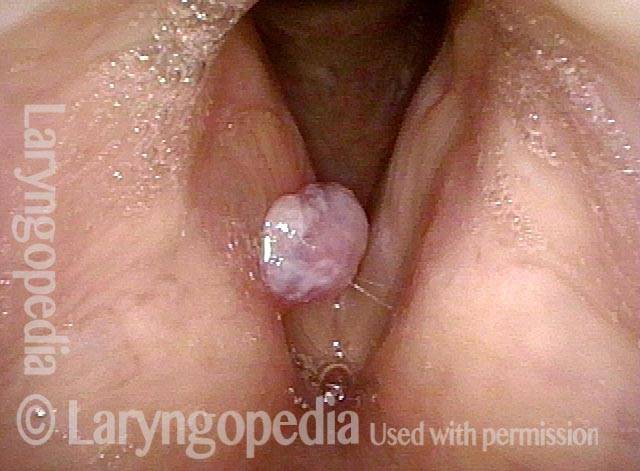
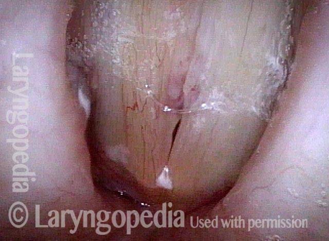
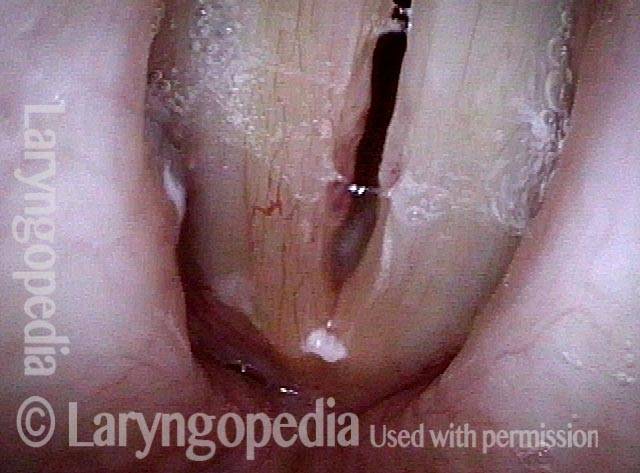
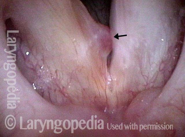
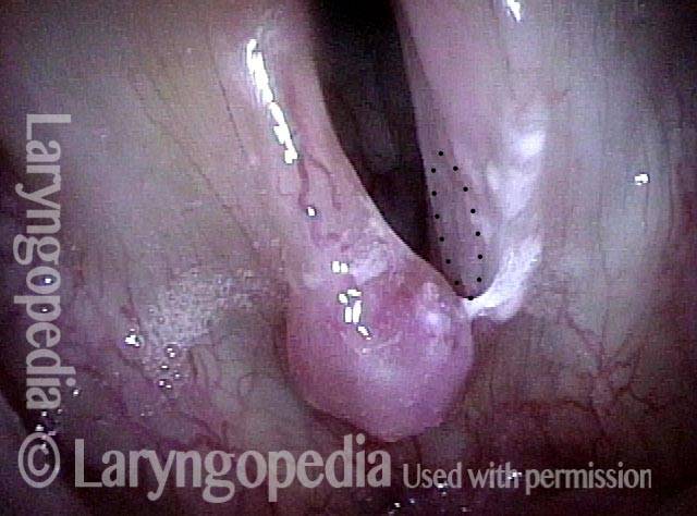
Hemorrhagic polyp is a vocal polyp that looks like a “blood blister” on the vocal cord. It may occur because of acute vocal trauma—sudden and extreme overuse of the voice—and may result in abrupt and fairly severe hoarseness that is persistent. In time, the blood may resorb and leave a translucent polyp; this kind of polyp may be prone to re-bruising intermittently.

Small hemorrhagic polyps may heal on their own, but usually require many months to do so. Larger ones should be surgically removed. Fortunately, the prognosis for full recovery after surgery is excellent.

Hemorrhagic polyp

Visual Portfolio, Posts & Image Gallery for WordPress

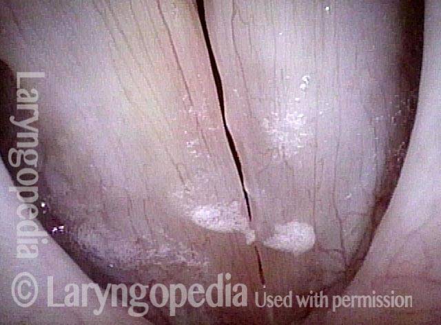
Hemorrhagic polyp (1 of 2)

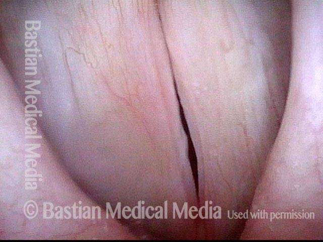
Large hemorrhagic polyp right fold, with large polypoid nodule and vascularity, left fold, during breathing (Lab).

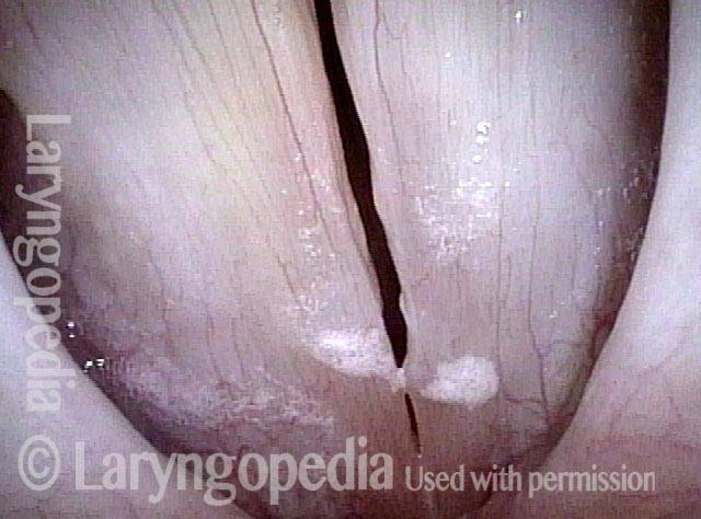
Hemorrhagic polyp (2 of 2)

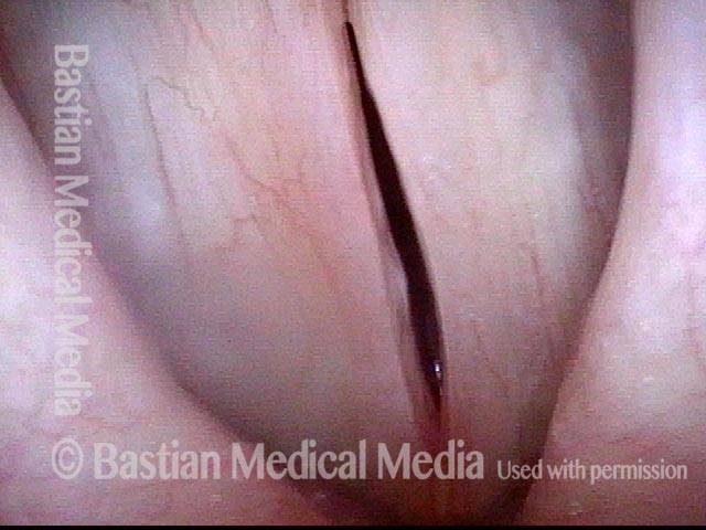
Same patient during phonation, showing now faint bruise anterior part of left-sided lesion (Lab).

Example 2

Visual Portfolio, Posts & Image Gallery for WordPress

Hemorrhagic polyp (1 of 4)

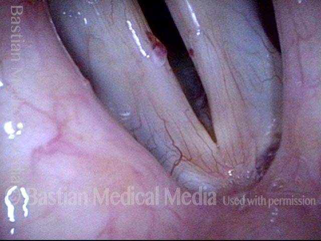
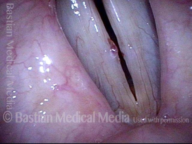
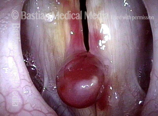
Hemorrhagic polyp, right cord. Notice the “blood blister” appearance. Recent further bleeding evident from yellowish discoloration of upper surface of the cord, due to breakdown products of a bruise, estimated two weeks earlier. Hemorrhagic polyps sometimes re-bruise intermittently.

Hemorrhagic polyp (2 of 4)

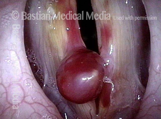
Open phase of vibration, strobe light, at low vocal pitch.

Hemorrhagic polyp (3 of 4)

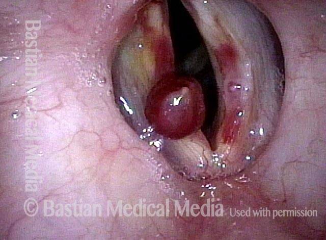
Vocal cords are coming into vibratory contact, beginning of closed phase.

Hemorrhagic polyp (4 of 4)

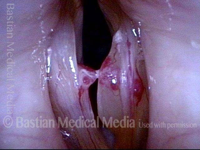
Closed phase of vibration, strobe light, showing that the polyp shifts to upper surface of the vocal cord.

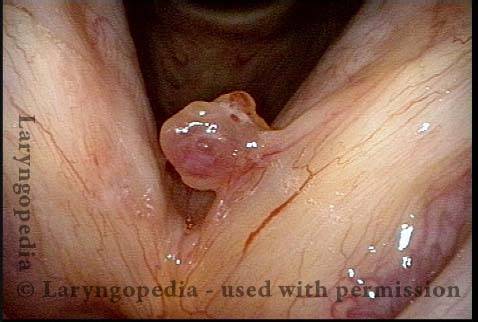
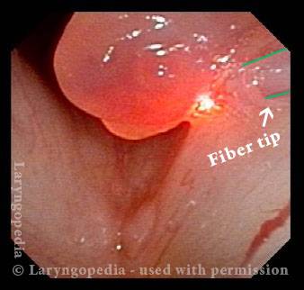
A Hemorrhagic Polyp that is Pedunculated (attached by a “stalk”)

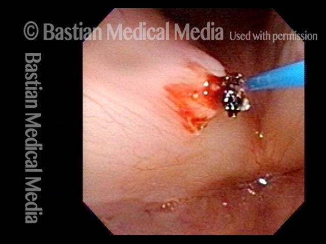
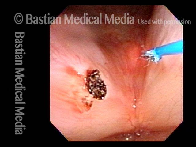
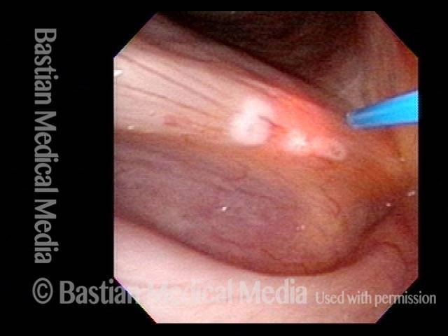
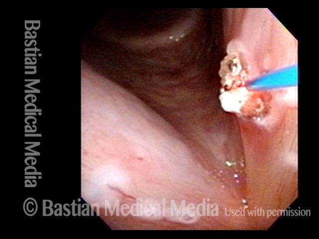
The thulium or other videoendoscopy lab (office-based) laser can be used for vocal cord injuries in persons who will not or cannot undergo general anesthesia. Reasons that patients might choose this options include severe medical issues such as:

  • Heart disease that makes general anesthesia risky
  • Morbid obesity
  • Severe phobia of general anesthesia
  • Anatomy that makes operative (O.R.) laryngoscopy difficult or impossible; enormous torus mandibularis, extreme retrognathism, kyphosis, and trismus are some examples.
Visual Portfolio, Posts & Image Gallery for WordPress

Hemorrhagic polyp (1 of 4)

This man is a major vocal overdoer with injury of both vocal cords, but the left cord polyp (right of photo) is the main cause of hoarseness.

Laser removal (2 of 4)

At very close range, the tip of the laser fiber is being used to coagulate the point of attachment.

Voice immediately improves (3 of 4)

After successful removal of the polyp with thulium laser—voice instantly improved.

5 months post-laser (4 of 4)

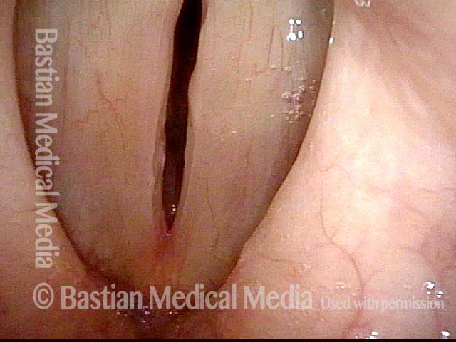
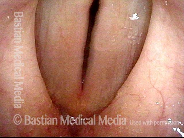
The patient continues to “overdo” with evidence of bilateral margin swelling and inflammation, but voice remains much better than it was with the polyp.

Hemorrhagic Polyp, Before and after surgery

Visual Portfolio, Posts & Image Gallery for WordPress

Hemorrhagic polyp (1 of 8)

Standard light, close-up view of a hemorrhagic polyp of the vocal cord.

Hemorrhagic polyp (2 of 8)

Strobe light, open phase of vibration.

Hemorrhagic polyp (3 of 8)

Strobe light, closed phase of vibration.

Hemorrhagic polyp: 1 week after surgery (4 of 8)

Same patient, one week after surgical removal of the polyp, standard light.

Hemorrhagic polyp: 1 week after surgery (5 of 8)

Strobe light, open phase of vibration. Compare with photo 2. Note here that the vibratory amplitude of both cords is the same, showing that the operated cord remains flexible.

Hemorrhagic polyp: 1 week after surgery (6 of 8)

Strobe light, closed phase of vibration. Compare with photo 3; the vocal cords now match much better during voicing, and the voice is completely normalized.

Hemorrhagic polyp: 7 months after surgery (7 of 8)

Seven months later. Strobe light, closed phase of vibration. The patient feels his voice is normal, and swelling checks don’t indicate any impairment.

Hemorrhagic polyp: 7 months after surgery (8 of 8)

Strobe light, open phase of vibration.

Hemorrhagic Polyp, Treated By Thulium Laser

Visual Portfolio, Posts & Image Gallery for WordPress

Hemorrhagic polyp, treated by thulium laser (1 of 8)

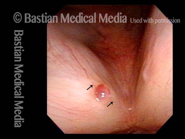
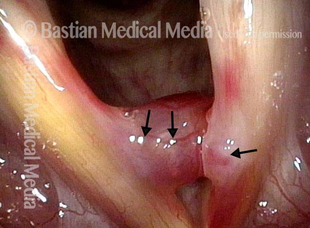
Hemorrhagic polyp, right vocal cord (left of image). This professional singer has struggled with severe limitations for six months. Note the feeding vessel, both anterior and posterior to the polyp, at arrows. These will be the first target of treatment.

Hemorrhagic polyp, treated by thulium laser (2 of 8)

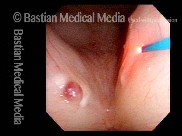
Using near-contact mode with a thulium laser, the feeding vessels have been coagulated, to reduce bleeding when the polyp itself is addressed. In contrast to what would be seen with a pulsed-KTP laser, one can see here hazy superficial coagulation affecting epithelium surrounding the vessels—so superficial that it will not affect vibratory flexibility.

Hemorrhagic polyp, treated by thulium laser (3 of 8)

The remaining laser energy is delivered to the polyp in contact mode, while stretching it away from the cord.

Hemorrhagic polyp, treated by thulium laser (4 of 8)

At the conclusion of the procedure, the polyp is released from the fiber. There is no damage to the vocal cord surrounding the polyp. A follow-up visit will be scheduled as “possible laser,” in case there is any residual polyp that did not slough off.

12 weeks after thulium laser treatment (5 of 8)

Twelve weeks after removing the hemorrhagic polyp via thulium laser. View under standard light, at the pre-phonatory instant. Both the patient and physician regard the patient’s voice as completely normal in quality and capabilities.

12 weeks after thulium laser treatment (6 of 8)

During phonation, with vibratory blur. Standard light.

12 weeks after thulium laser treatment (7 of 8)

View under strobe light. During phonation, at the closed phase of vibration, for the pitch B-flat 4 (~466 Hz).

12 weeks after thulium laser treatment (8 of 8)

Open phase of vibration, also at B-flat 4 (~466 Hz).

Capillary Ectasia and Hemorrhagic Polyp, Before and After Treatment

Visual Portfolio, Posts & Image Gallery for WordPress

Capillary ectasia and hemorrhagic polyp (1 of 4)

Open phase of vibration, strobe light.

Capillary ectasia and hemorrhagic polyp (2 of 4)

Partially closed phase of vibration, strobe light.

Capillary ectasia and hemorrhagic polyp, after treatment (3 of 4)

Abducted breathing position after vocal cord microsurgery, standard light. Note that the right cord is normalized, the capillary ectasia on the left is smaller, but persists in spite of spot-coagulation. A simple pulsed-KTP laser procedure in the videoendoscopy procedure room abolished this residual lesion.

Capillary ectasia and hemorrhagic polyp, after treatment (4 of 4)

Pre-phonatory instant, standard light, showing excellent match. Oscillatory ability entirely normal into extreme upper range in this professional singer.

Capillary Ectasia and Hemorrhagic Polyp

Visual Portfolio, Posts & Image Gallery for WordPress

Capillary ectasia and hemorrhagic polyp (1 of 2)

Note the differing patterns of capillary ectasia—fine “stream;” meandering “river,” and “pond.” Brilliant white areas are reflection of light; more indistinct submucosal white area right cord raises question of possible cyst (arrow).

Capillary ectasia and hemorrhagic polyp (2 of 2)

Strobe illumination better reveals the polypoid component of the swellings. Some mucus accumulation, especially posteriorly.

Capillary Ectasia and Hemorrhagic Polyp, Treated by Thulium Laser

Visual Portfolio, Posts & Image Gallery for WordPress

Capillary ectasia and hemorrhagic polyp (1 of 7)

Open position for breathing, standard light. There is capillary ectasia on both vocal cords, and there is also a hemorrhagic polyp of the left vocal cord margin (right of photo).

Capillary ectasia and hemorrhagic polyp (2 of 7)

During voicing, the polyp interferes with accurate approximation of the vocal cords, which explains this man’s chronic hoarseness.

Capillary ectasia and hemorrhagic polyp, thulium laser treatment (3 of 7)

Using the thulium laser to spot-coagulate and interrupt the flow in dilated capillaries.

Capillary ectasia and hemorrhagic polyp, thulium laser treatment (4 of 7)

Coagulation of the polyp, with fiber tangential to the vocal cord and sometimes lifting medially during contact mode. A second, similar procedure was needed a few weeks later, only for residual polyp.

Capillary ectasia and hemorrhagic polyp, after treatment (5 of 7)

Several weeks later, capillary areas are blanched, but the vocal cord mucosa is fully mobile.

Vocal cord margin (6 of 7)

Vocal cord margin match and mucosal flexibility are best tested in high voice. This is strobe light, closed phase of vibration, at F4 (~349 Hz).

Capillary ectasia and hemorrhagic polyp, after treatment (7 of 7)

Also at F4 (~349 Hz). The mucosa of both cords is completely supple. The patient considers his voice to be perfectly normal—”original equipment.”

Hemorrhagic Polyp with Added Rumble at Low Pitch and Segmental Vibration at High Pitch

Visual Portfolio, Posts & Image Gallery for WordPress

Young woman with hoarse voice (1 of 8)

Young woman who fits the “vocal overdoer” profile and who has been hoarse for many years. The hoarseness has become intolerable in the past 2 months.

Inspiratory phonation (2 of 8)

At much closer range, with the right polyp (left of photo) displaced with inspiratory phonation to show the considerable left cord (right of photo) injury. Note large vessel coursing along the right polyp (left of photo) and the capillary prominence within the left vocal cord (right of photo) injury as well. Note as well the bruising distant from the origin and yellowish discoloration of partially resolved bruising.

Closed phase (3 of 8)

Strobe light, closed phase at F4. (349 Hz)

Open phase (4 of 8)

Open phase of vibration, also at F4.

Open phase, rumble (5 of 8)

At F3 (175 Hz), open phase of vibration. At this pitch the polyp flutters and adds a second low rumbling sound to the main pitch she is singing.

Closed phase, segmental vibration (6 of 8)

The patient sings B-flat 5 (932 Hz) using only the posterior segment with vibration indicated by bi-directional arrows.

One week post-op, prephonatory instant (7 of 8)

One week after surgical removal, prephonatory instant, at C5 (523 Hz). Note the gap between the cords, much the same as preoperatively in prior photos.

One week post-op, phonation (8 of 8)

Phonation also at C5. Vibratory blur is equal bilaterally, and upper voice dramatically restored, even before “surgical laryngitis” has resolved.

Pre-op and Very Early Post-op Mucosal Match and Flexibility in Male Singer

Visual Portfolio, Posts & Image Gallery for WordPress

Bilateral chronic injuries (1 of 8)

Young music teacher and choral director with chronic hoarseness for more than a year. Note bilateral chronic injuries, and also recent bruise of the right cord (left of photo).

Closeup of injuries (2 of 8)

Closeup of injuries and their “refusal” to let the cords approximate, when attempting (unsuccessfully) to sing a high pitch.

Post microsurgery (3 of 8)

7 days after vocal cord microsurgery, the voice can already pass for normal quality and capability. Compare with photo 1.

Prephonatory instant (4 of 8)

Prephonatory instant at high pitch, showing that the match of the cords is already markedly restored. The tiny elevation will disappear with further healing.

Closed phase, A-flat 3 (5 of 8)

Closed phase of vibration at A-flat 3 (208 Hz), seen under strobe light.

Open phase (6 of 8)

This open phase shows what is equally important: that both cords display equal vibratory flexibility; that is, there is no stiffness or scarring.

Dramatically improved match (7 of 8)

This man also has clear, normal falsetto voice: closed phase of vibration at A-flat 4 (415 Hz), again showing dramatically improved match.

Equal mucosal flexibility (8 of 8)

Again more important is evidence of equal mucosal flexibility between the two sides, as seen in this open phase of vibration, at the same pitch.

Before and After Repair of Dilated Capillaries and Hemorrhagic Polyp

Visual Portfolio, Posts & Image Gallery for WordPress

Hemorrhagic polyp (1 of 4)

Kindergarten teacher with severe double pitch and hoarseness. Right vocal cord hemorrhagic polyp and scattered ectatic (dilated) capillaries.

Attempted voice production (2 of 4)

Attempting to produce voice with interference and “rattling” of the polyp.

Post surgery (3 of 4)

A few weeks after surgical removal of the polyp, and spot-coagulation of the dilated capillaries.

Closed phase (4 of 4)

Closed phase of vibration at high pitch. Much improved match and normal high voice. Broad-based residual swellings are likely chronic from many years of teaching kindergartners.

Pedunculation Defined in Pictures

Visual Portfolio, Posts & Image Gallery for WordPress

Polyp (1 of 7)

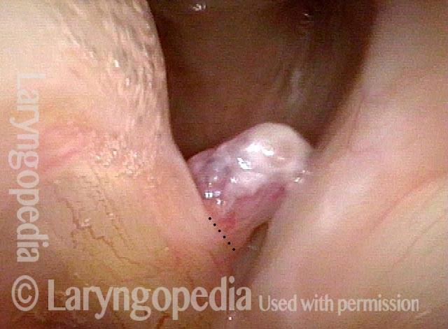
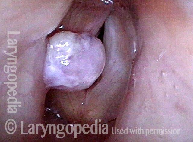
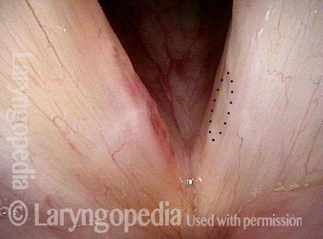
This large polyp resulted from an episode of extremely aggressive voice use six months earlier. In this photo, one cannot tell if the point of attachment covers the same area of the circumference of the lesion, or if it is smaller.

Inspiration (2 of 7)

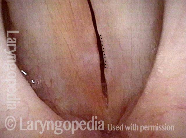
Here, the examiner has elicited rapid inspiration. The rush of air inward pulls the polyp inward and downward, revealing its stalk or peduncle. The attachment is indicated by the dotted line.

Closed position (3 of 7)

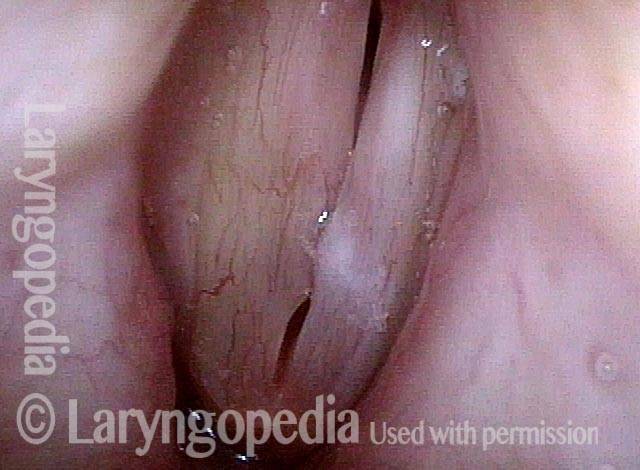
The vocal cords are closed while continuing to draw air in. The polyp is now hidden below the point of closure of the cords.

Phonatory view (4 of 7)

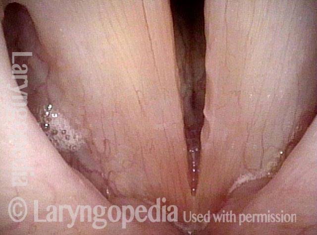
When voice is produced, the polyp flips upwards between the colds and now lies on the upper surface of the vocal colds. None of this movement could happen if the polyp were not pedunculated.

After removal (5 of 7)

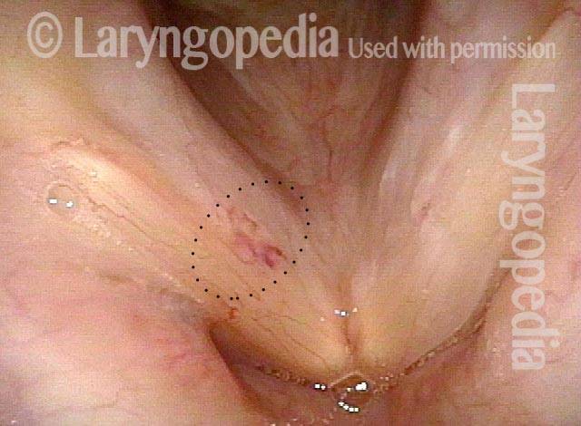
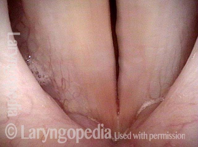
A week after removal of the polyp. Compare with photo 1 to see that the “wound” (area of vascularity) is far smaller than the diameter of the original polyp (dotted line), showing in a second way the idea that the attachment had “pinched in” to a stalk.

Closed phase (6 of 7)

Closed phase of vibration.

Open phase (7 of 7)

Open phase of vibration.

The Operated Side Often Looks Better Than the Unoperated…

Visual Portfolio, Posts & Image Gallery for WordPress

Polyp (1 of 8)

Many months after formation in a low-voiced male, this hemorrhagic polyp has become mobile. Here, inspiration draws the polyp downward towards the undersurface of the left cord (right of photo). When the original bleeding event occurred this polyp would have for some months been more fixed at the free margin, pressing into the left vocal cord (right of photo) at the point of the arrow on the left side (right of photo).

Polyp rides upwards (2 of 8)

As the vocal cords close to produce voice, the polyp rides upwards. The dotted line circles a slight depression, and shows where the polyp originally pressed inward against the left cord (right of photo).

Post-surgery (3 of 8)

A week after vocal cord surgery, the operated side (left of photo) shows residual bruising at the base of the ~ 5mm “wound.” The dotted line on the left cord (right of photo) shows the slight concavity sustained by months of pressing against the polyp, especially during the time before it became pedunculated.

Prephonatory view (4 of 8)

At a prephonatory instant, mild post-surgical swelling of the right cord (left of photo) is seen as subtle convexity. But the slight concavity caused by the pressure of the polyp on the left ( unoperated) vocal cord (right of photo) is seen even more clearly (dotted line).

Six weeks post-surgery (5 of 8)

Six weeks after surgery, voice is normal, including high falsetto. The operated right vocal cord (left of photo) looks better than the unoperated one…

Phonation (6 of 8)

During phonation under standard light, the mild concavity of the left (unoperated) vocal cord (right of photo) is seen more clearly.

Closed phase (7 of 8)

At A4 (440 Hz) under strobe light, closed phase of vibration.

Open phase (8 of 8)

Open phase of vibration at the same pitch. Both sides vibrate equally well, but the operated cord has a straighter margin than the unoperated one.
vocal overuse YT Thumbnail
Play Video

Hemorrhagic Polyp: Before and After

Watch the story of a young man with a hemorrhagic vocal cord polyp. Listen to his voice and see his larynx both before and after surgical removal.
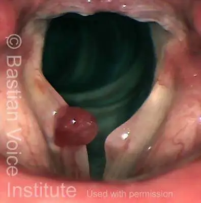
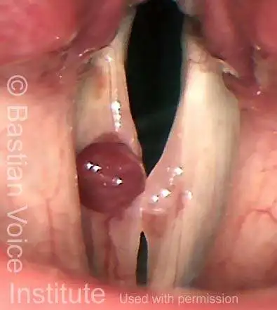
Table of Contents

Subscribe
Notify of

0 Comments
Newest
Oldest Most Voted
Inline Feedbacks
View all comments
0
Click to see all comments.x