Laser surgery is surgery that uses a beam of laser light, rather than other instruments, to cut, dissect, remove, and so forth. The beam of light has advantages over other cutting instruments, such as scalpel or scissors.
First, at the same time that it cuts, it tends to seal off tiny blood vessels and reduce bleeding. Second, it may be especially useful in endoscopic surgery, where there is not a lot of room for instruments. Third, it is very precise. Both the microspot carbon dioxide laser and the RevoLix laser used at our practice have minimum spot sizes of about 1/5 of a millimeter.
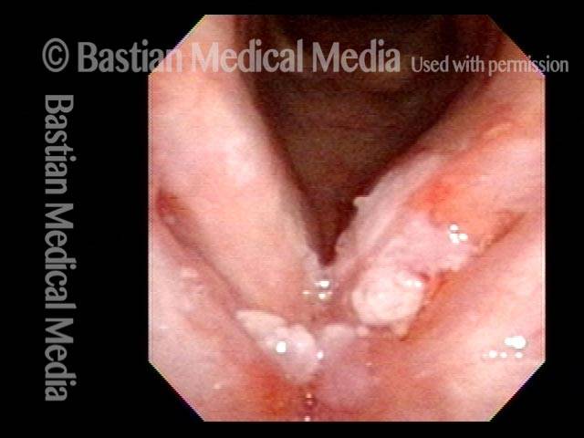
Laser Surgery for Bilateral Vocal Cord Cancer
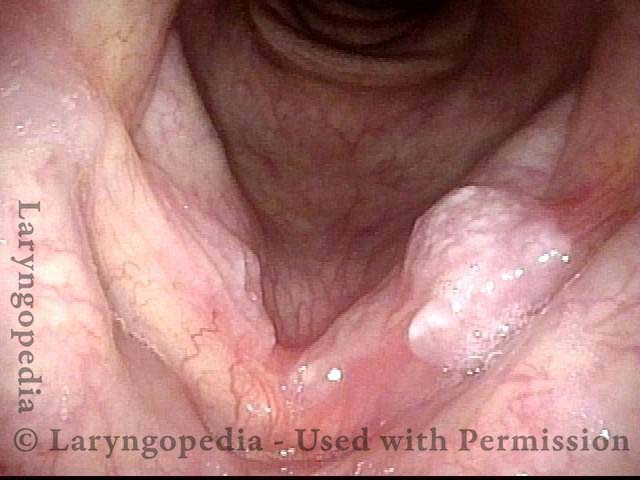
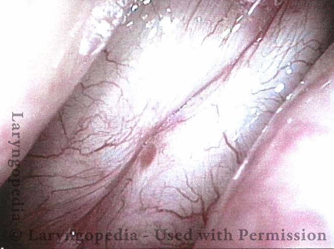
Squamous cell carcinoma (1 of 6)
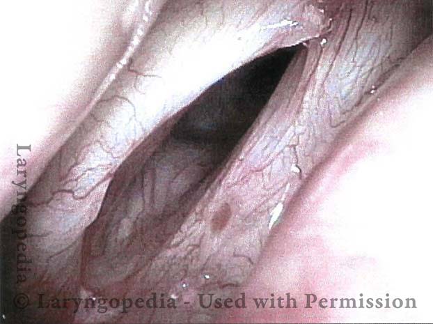
Tumor on the vocal cords (2 of 6)
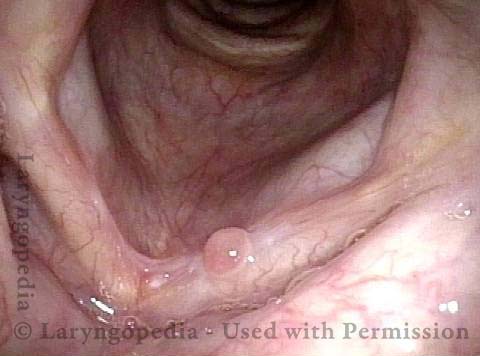
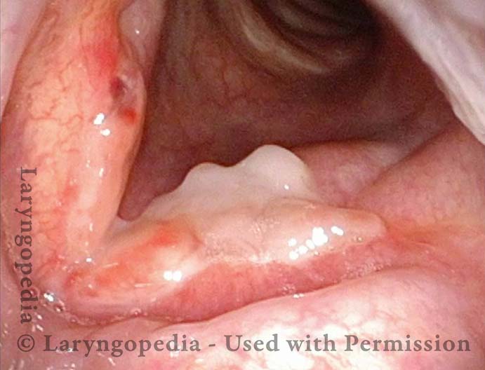
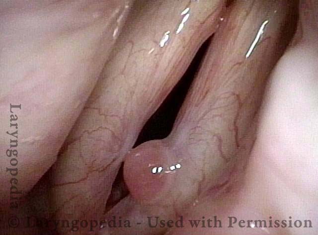
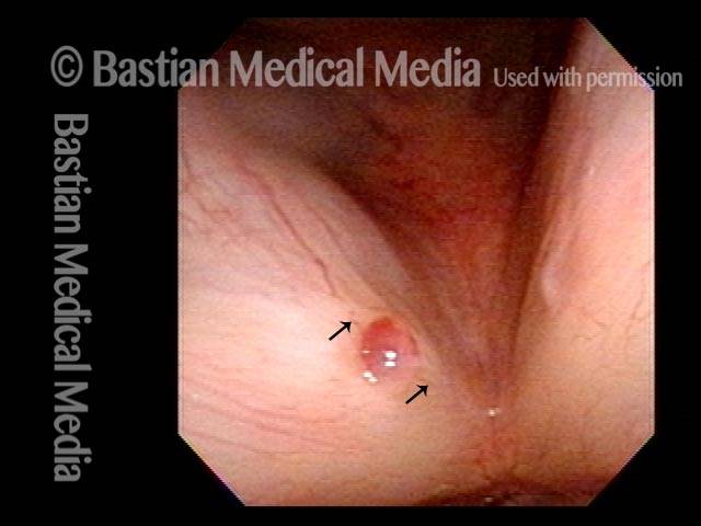
Granuloma delays voice recovery (3 of 6)
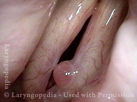
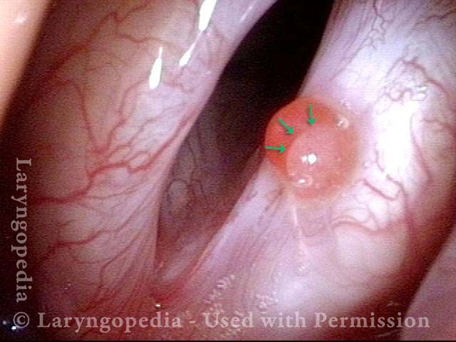
Closer view of granuloma (4 of 6)
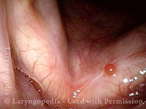
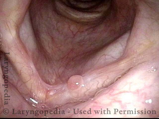
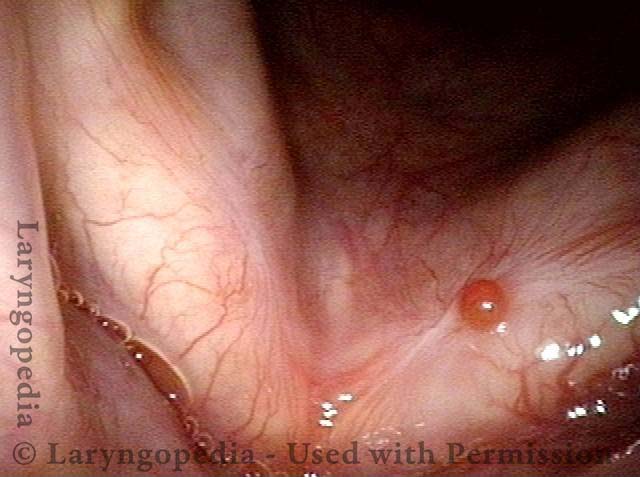
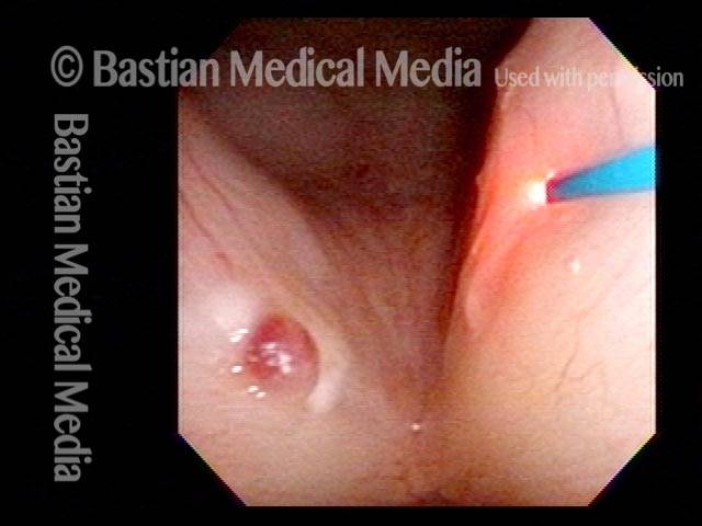
Granuloma is smaller (5 of 6)
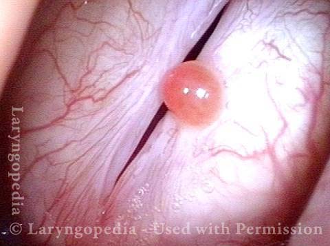
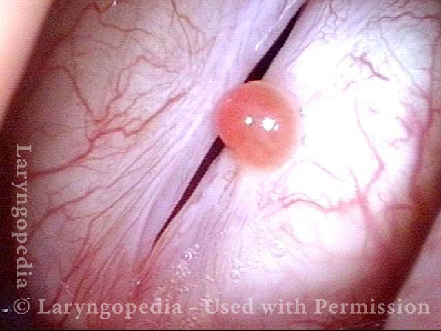
Granuloma doesn’t impede voice (6 of 6)
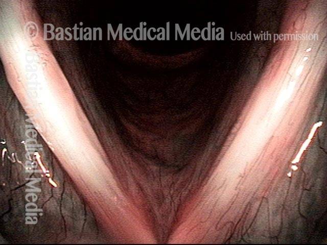
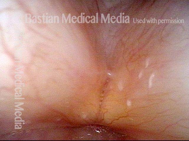
Laser Removal of Vocal Cord Cancer with Bilateral Disease
For treatment of early vocal cord cancer, both laser excision and radiotherapy are in competition as good treatment modalities. See also Early Vocal Cord Cancer: Remove with a Laser, or Radiate? Often, radiation is used when disease is bilateral, in the interest of preserving voice. This is an example of the ability to do fairly extensive laser surgery bilaterally, yet preserving good voice. This man had a friend who had severe difficulty with radiation, and he was therefore opposed to that option.
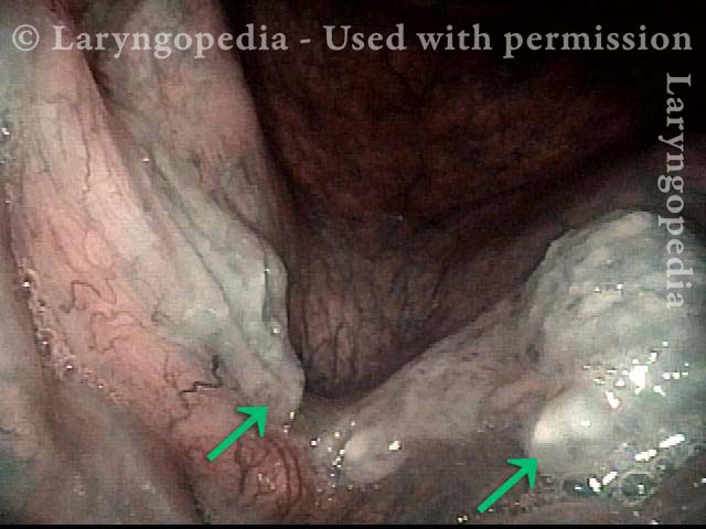
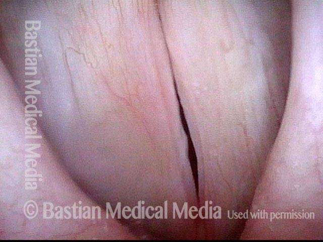
Vocal cord cancer (1 of 10)
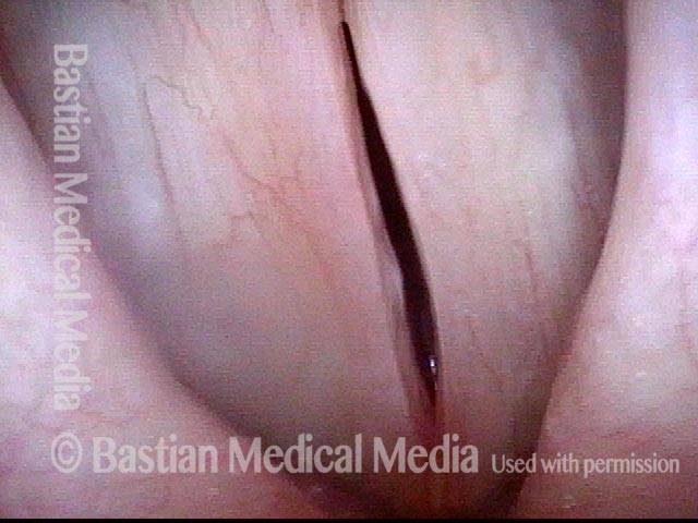
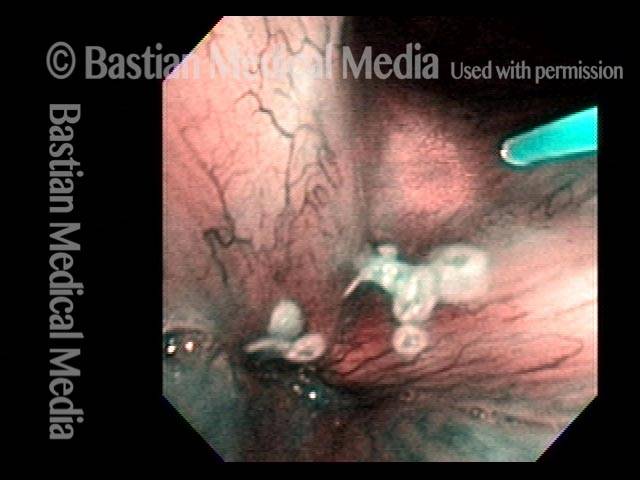
Stippling (2 of 10)
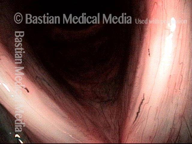
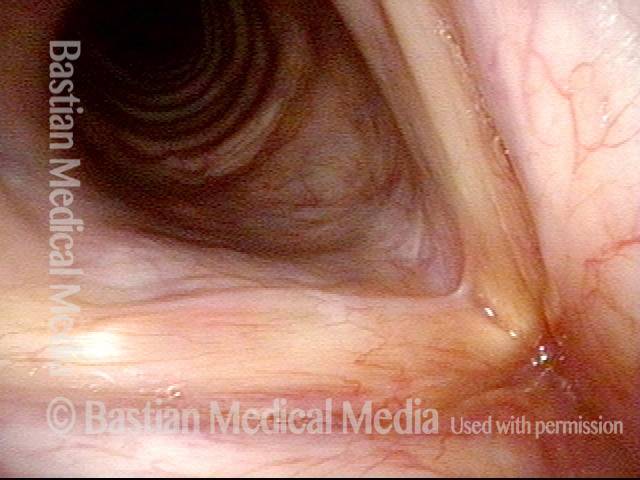
1 week after excision (3 of 10)
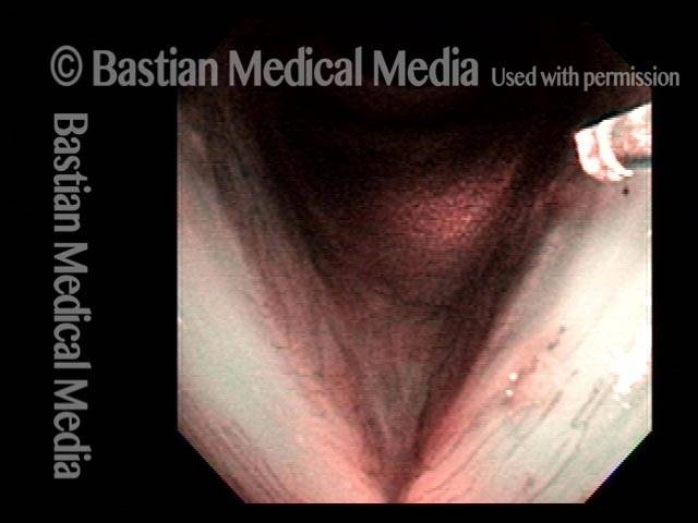
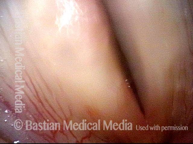
Reparative Granuloma emerges (4 of 10)
Granuloma interferes with voicing (5 of 10)
Granuloma fades away (6 of 10)
Closer view (7 of 10)
Granuloma cleft (8 of 10)
Blood tattoo (9 of 10)
Voice is improved (10 of 10)
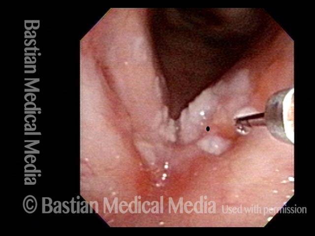
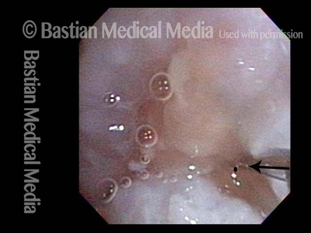
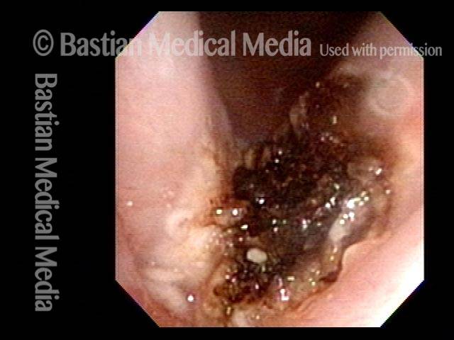
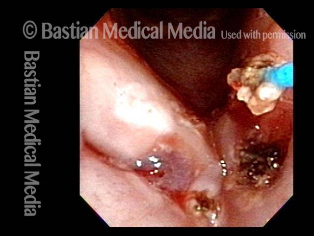
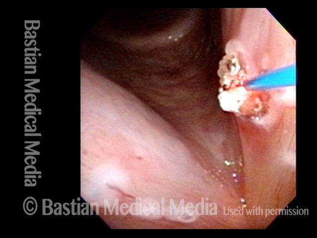
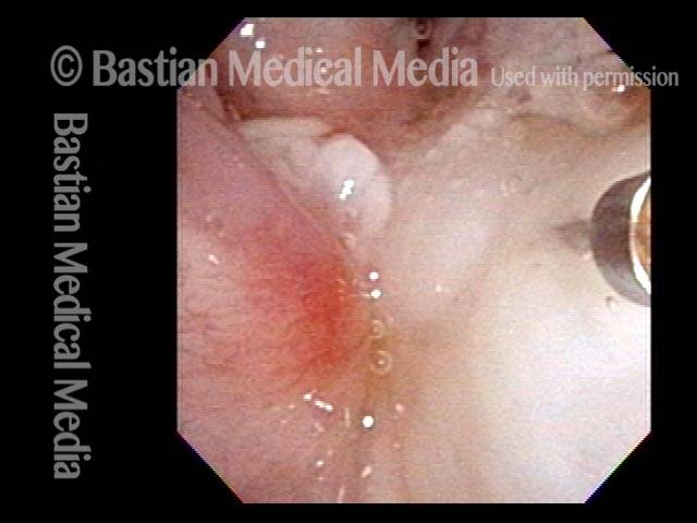
Hemorrhagic Polyp, Treated By Thulium Laser
Hemorrhagic polyp, treated by thulium laser (1 of 8)
Hemorrhagic polyp, treated by thulium laser (2 of 8)
Hemorrhagic polyp, treated by thulium laser (3 of 8)
Hemorrhagic polyp, treated by thulium laser (4 of 8)
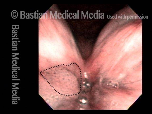
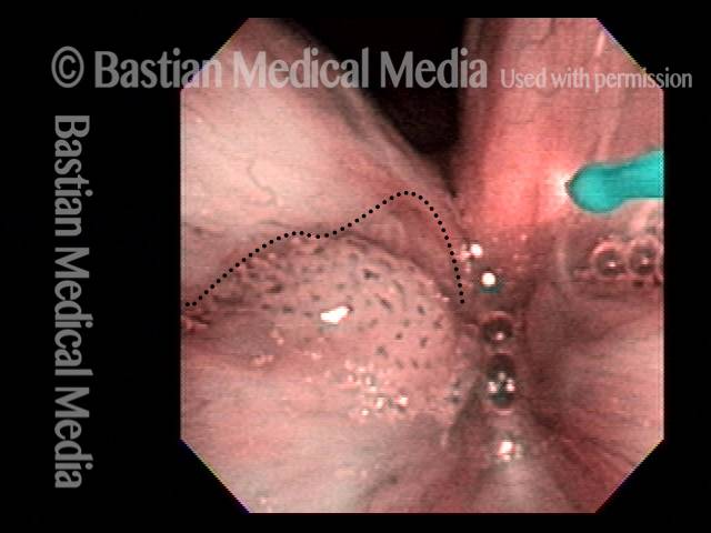
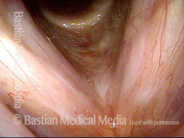
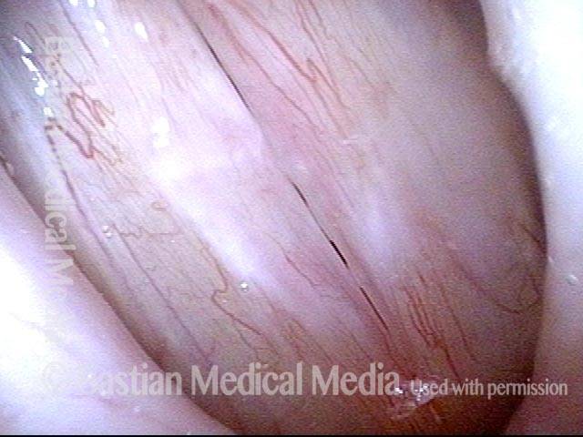
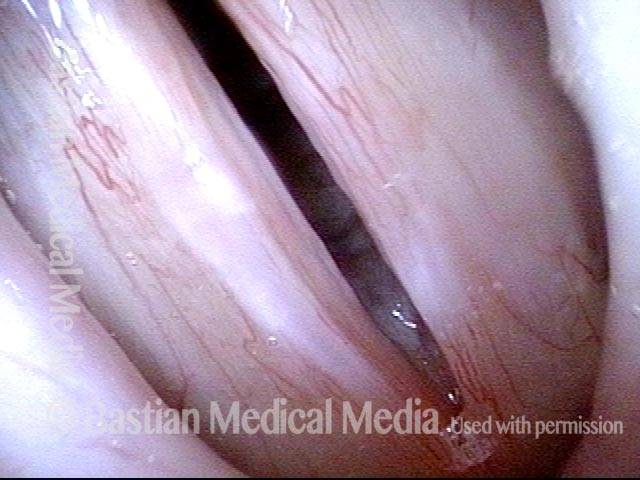
12 weeks after thulium laser treatment (5 of 8)
12 weeks after thulium laser treatment (6 of 8)
12 weeks after thulium laser treatment (7 of 8)
12 weeks after thulium laser treatment (8 of 8)
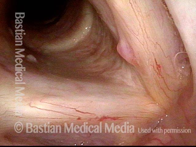
Capillary Ectasia, Before and After Laser Coagulation
Capillary ectasia (1 of 3)
Capillary ectasia, right after laser coagulation (2 of 3)
Capillary ectasia, 6 weeks after laser coagulation (3 of 3)
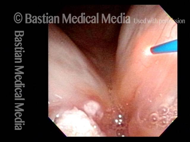
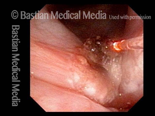
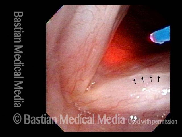
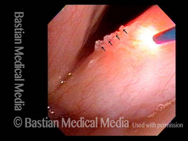
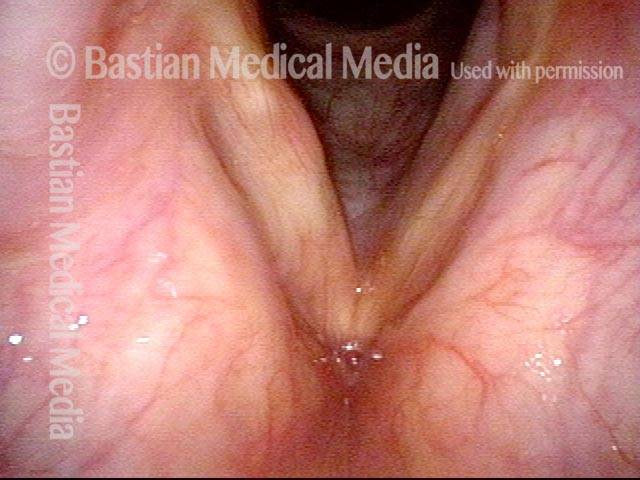
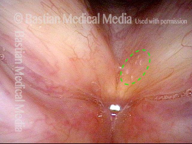
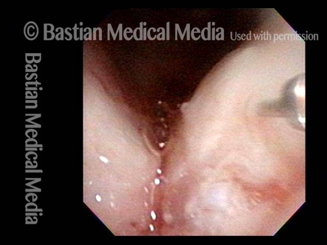
Lidocaine Injection for Aggressive “Office” Laser Treatments
Laser ablations performed in office (1 of 6)
Infiltrating anesthetic (2 of 6)
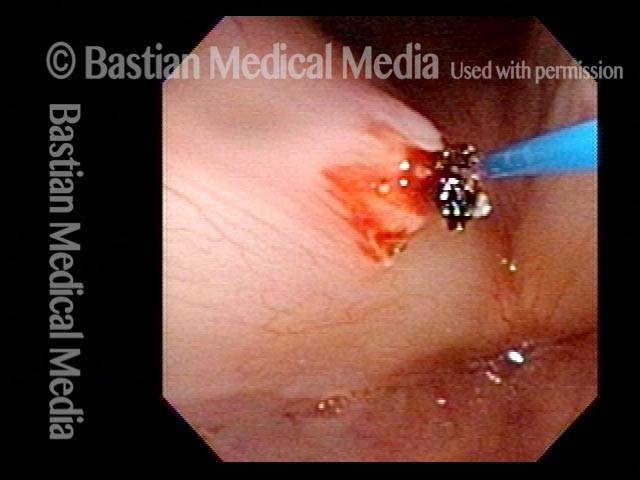
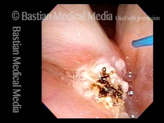
Thulium laser procedure (3 of 6)
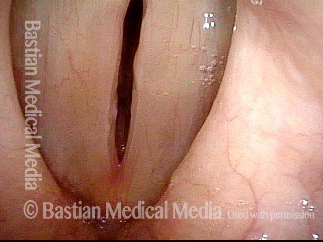
Post-surgery (4 of 6)
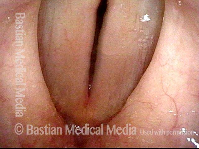
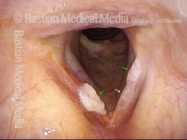
Six weeks post-surgery (5 of 6)

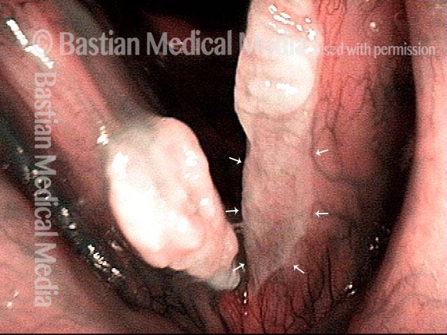
Second laser sugery (6 of 6)
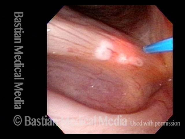
Perfect Candidate for Thulium Laser
Lesion (1 of 4)
Lesion under narrow-band light (2 of 4)
Coagulated with thulium laser (3 of 4)
Finishing up (4 of 4)
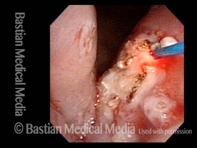
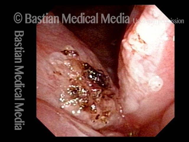
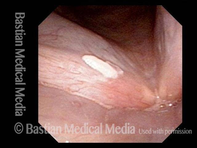
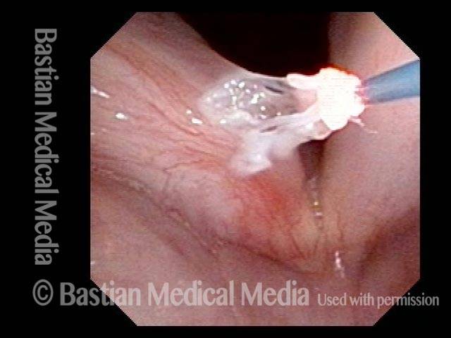
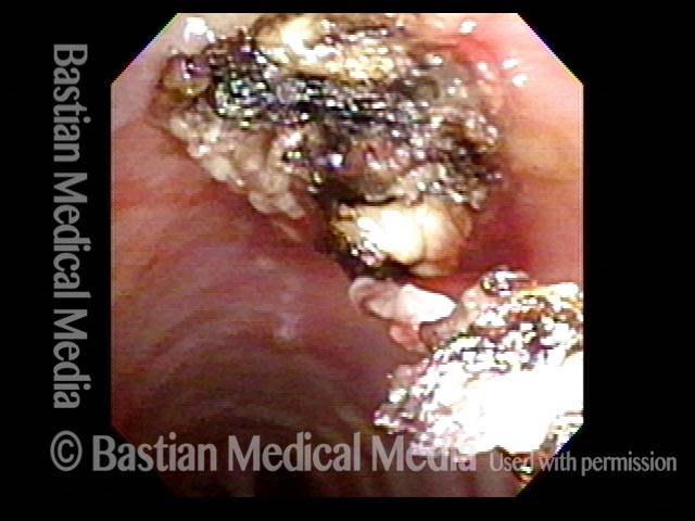
Leukoplakia Battled Over Time
Leukoplakia (1 of 8)
Spilled Milk (2 of 8)
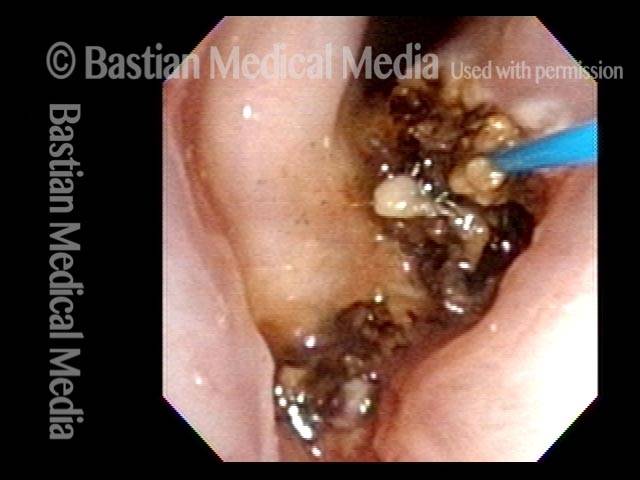
Thulium laser (3 of 8)
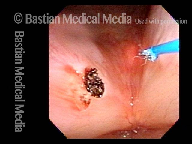
Coagulated tissue (4 of 8)
Leukoplakia (5 of 8)
Detachment (6 of 8)
Superficial vascular pattern (7 of 8)
Coagulated tissue (8 of 8)
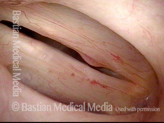
Leukoplakia, Before, During, and After Laser Coagulation
Leukoplakia, not yet seen (1 of 6)
Leukoplakia (2 of 6)
Leukoplakia (3 of 6)
Leukoplakia, coagulated by laser (4 of 6)
Leukoplakia, 3 months after laser treatment (5 of 6)
Leukoplakia, 3 months after laser treatment (6 of 6)
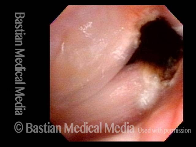
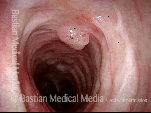
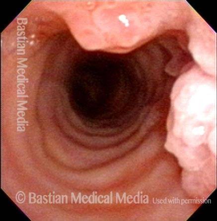
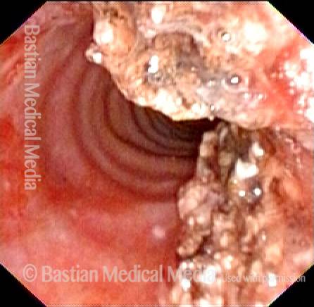
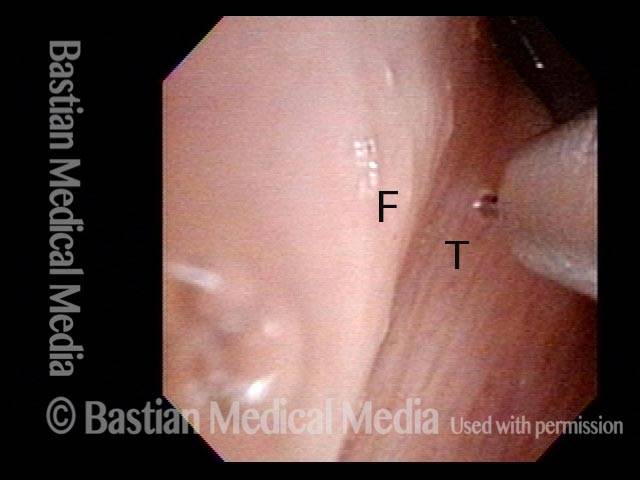
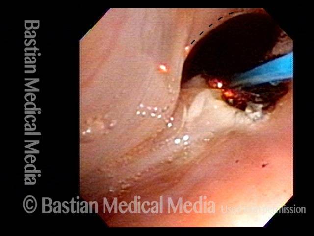
Mid-Tracheal Papilloma, Treated By Thulium Laser
Mid-tracheal papilloma, being treated by thulium laser (1 of 5)
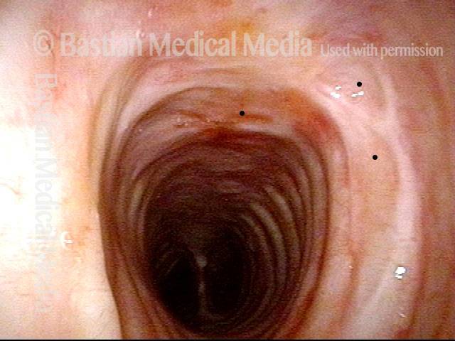
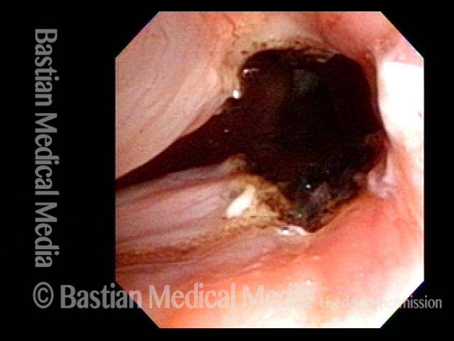
Months after treatment: no papilloma (5 of 5)
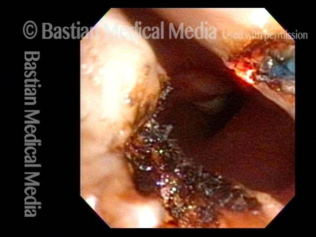
Mid-tracheal papilloma, being treated by thulium laser (2 of 5)
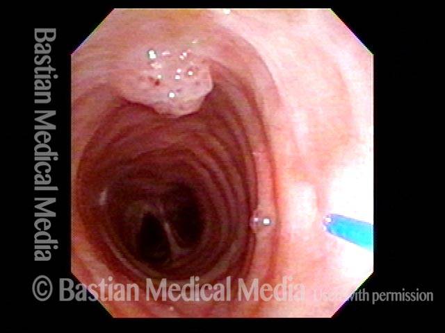
Mid-tracheal papilloma, being treated by thulium laser (3 of 5)
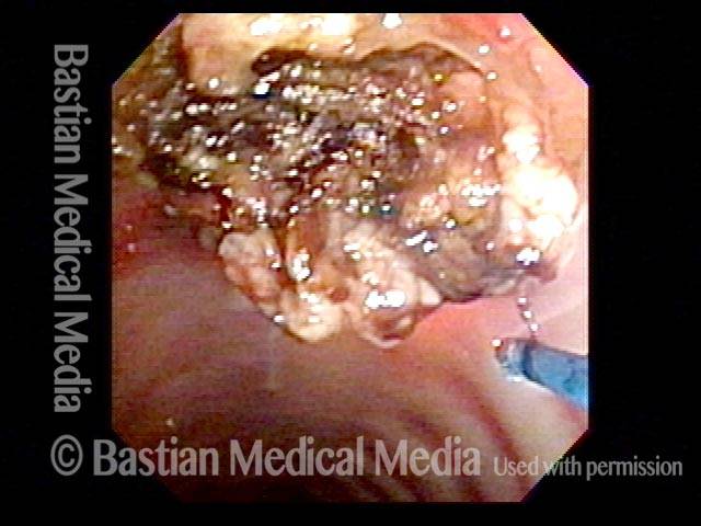
Mid-tracheal papilloma, being treated by thulium laser (4 of 5)
Capillary Ectasia and Hemorrhagic Polyp, Treated by Thulium Laser
Capillary ectasia and hemorrhagic polyp (1 of 7)
Capillary ectasia and hemorrhagic polyp (2 of 7)
Capillary ectasia and hemorrhagic polyp, thulium laser treatment (3 of 7)
Capillary ectasia and hemorrhagic polyp, thulium laser treatment (4 of 7)
Capillary ectasia and hemorrhagic polyp, after treatment (5 of 7)
Vocal cord margin (6 of 7)
Capillary ectasia and hemorrhagic polyp, after treatment (7 of 7)
Thulium Laser Surgery, With Local Anesthetic Injection, to Treat Leukoplakia
Leukoplakia, about to be treated with laser (1 of 4)
Injection of local anesthetic (2 of 4)
Injection of local anesthetic (3 of 4)
Right after thulium laser treatment (4 of 4)
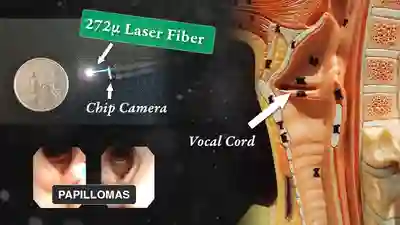
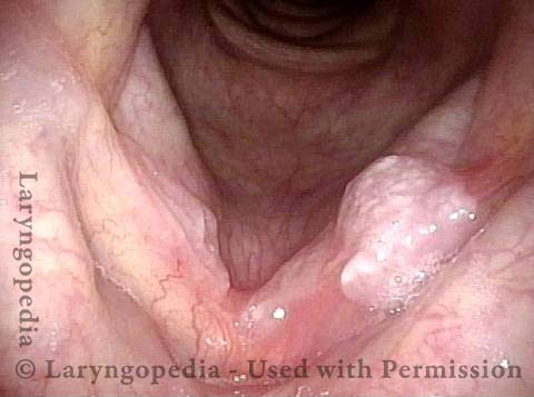
Tracheal Papillomas and the Thulium Laser
HPV 11 (1 of 2)
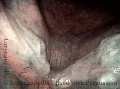
Post laser coagulation (2 of 2)
Office-Based Surgery When General Anesthesia Is too Risky
Involuntary inspiratory voice (1 of 6)
Laser posterior commissuroplasty (2 of 6)
During the commissuroplasty (3 of 6)
Deepening divot (4 of 6)
Inspiratory indrawing decreased (5 of 6)
Phonation (6 of 6)