Miotomia Cricofaringea

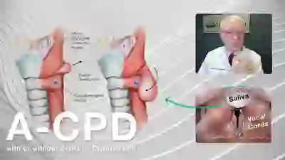
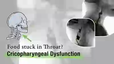
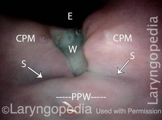
La miotomia cricofaringea (CPM) è una procedura in cui il muscolo cricofaringeo, che forma un “anello” attorno all’esofago superiore, viene diviso o tagliato per rilassarlo. Questo viene fatto nei casi in cui questo muscolo cricofaringeo (cioè lo sfintere esofageo superiore) non riesce a rilassarsi quando si deglutisce (disfunzione cricofaringea anterograda [A-CPD]), con conseguente ostruzione funzionale. Il CPM lascia il muscolo sempre aperto e consente alla persona di riprendere la deglutizione in modo relativamente normale.

In una percentuale di soggetti con mancato rilassamento del muscolo cricofaringeo, le continue alte pressioni della deglutizione possono eventualmente causare una “ernia” nel passaggio della deglutizione chiamata diverticolo di Zenker.

Procedura CPM

La miotomia cricofaringea viene eseguita in anestesia generale, in due modi: la metodologia preferita e più recente viene eseguita per via endoscopica. Utilizzando questo metodo, un “tubo” cavo e illuminato, chiamato esofagoscopio, viene inserito nella gola e nella parte superiore dell’esofago per esaminare l’area e pianificare il passaggio successivo. Quindi, uno speciale cannocchiale laser viene posizionato nell’esofago superiore. Un microscopio consente una visione ingrandita e ben illuminata dell’anello muscolare incriminato.

If a clear view cannot be achieved during esophagoscopy due to difficult patient anatomy (e.g., jaws won’t open well, small lower jaw, short neck, large upper teeth), then we return to a more traditional approach through an incision on the lower left neck. In this case, the muscle is divided from outside in. Depending on a number of issues, an associated Zenker’s diverticulum may or may not be removed at the same time.

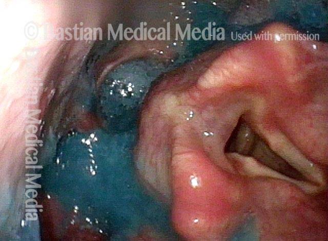
Il laser viene utilizzato per dividere il muscolo e spezzarne la “presa”. Se la sacca di Zenker è evidente, è “marsupializzata”, il che significa che la sua apertura è allargata per essere sicuri che la sacca non trattenga il cibo, ma svuoti invece il suo contenuto direttamente nell’esofago.

A seconda del paziente e dei problemi chirurgici, un drenaggio di aspirazione può essere posizionato all’interno dell’esofago superiore e fatto uscire attraverso il naso. Un secondo tubicino per l’alimentazione può essere inserito attraverso il naso e giù nello stomaco. Una volta posizionate, entrambe le tube vengono rimosse la mattina successiva all’intervento.

Se durante l’esofagoscopia non è possibile ottenere una visione chiara a causa delle difficoltà anatomiche del paziente (ad esempio, le mascelle non si aprono bene, la mascella inferiore è piccola, il collo corto, i denti superiori grandi), si ritorna a un approccio più tradizionale attraverso un’incisione sulla parte inferiore collo sinistro. In questo caso, il muscolo viene diviso dall’esterno verso l’interno. A seconda di una serie di problemi, un diverticolo di Zenker associato può o meno essere rimosso contemporaneamente.

Visual Portfolio, Posts & Image Gallery for WordPress

Cricopharyngeal myotomy (1 of 7)

Upon initial approach to the upper esophagus. The small dark opening in the center is the entrance to the Zenker’s diverticulum or pouch. The point of entry to the esophagus is at the dotted line. The cricopharyngeus muscle lies between the entrances to the Zenker’s pouch and the esophagus.

Cricopharyngeal myotomy (2 of 7)

When the scope is inserted and lifted additionally, the Zenker’s pouch is opened further, and retained food material is seen within. The cricopharyngeus muscle’s contour is seen more clearly (faint dotted line), and the actual opening of the esophagus can be seen at the arrow.

Cricopharyngeal myotomy (3 of 7)

Similar view to photo 2, except that the suction cannula is now inserted into the esophageal opening, further accentuating the contour of the cricopharyngeus muscle.

Cricopharyngeal myotomy (4 of 7)

The food material has been removed from the Zenker’s pouch, and the suction cannula is placed within the esophagus.

Cricopharyngeal myotomy (5 of 7)

Division of the cricopharyngeus muscle is underway, using the CO2 laser. The red aiming beam is visible at the lower end of the incision.

Cricopharyngeal myotomy (6 of 7)

The muscle is now approximately half-divided, along with mucosa lining the anterior wall of the sac (arrow).

Cricopharyngeal myotomy (7 of 7)

The muscle is entirely divided, and the sac marsupialized. The suction cannula (blurry here, but marked by a faint dotted line) now lies within the trough created by the laser. The esophagus is now gaping open; compare with photos 1 through 4 of this series.

Tasso di successo del CPM

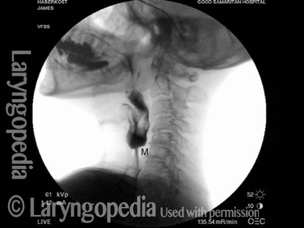
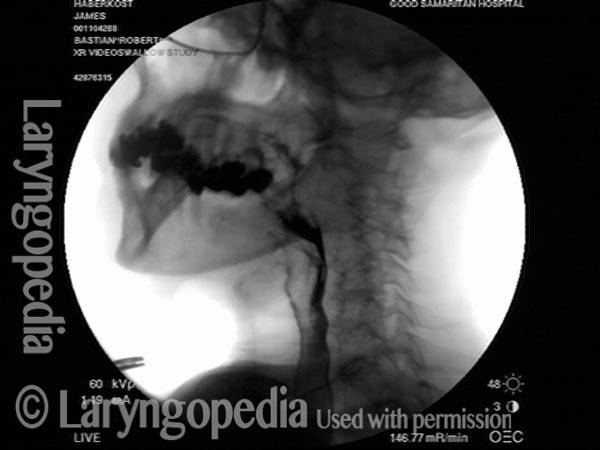
Il tasso di successo può variare a seconda del grado in cui la persona è un candidato ideale per questo intervento chirurgico. Quando la disfagia da cibo solido è il sintomo principale e quando gli studi a raggi X mostrano il mancato rilassamento del muscolo, la procedura può ripristinare drasticamente la capacità di deglutire dell’individuo.

La soddisfazione dei pazienti per i risultati è solitamente molto elevata. “Posso mangiare di nuovo tutto ciò che voglio”, si sente spesso dopo l’intervento chirurgico. Di tanto in tanto un individuo ha più di un deficit di deglutizione, di cui il CPMD è solo uno. In questo caso, la deglutizione potrebbe essere migliore, ma non ancora perfetta dopo il CPM.

Rischi del CPM

A meno che tu non abbia problemi di salute significativi, i rischi sommati insieme sono molto piccoli. I rischi possono includere:

  1. Una reazione al farmaco, un problema cardiaco, ecc., durante l’anestesia generale.
  2. Trauma dentale: ad esempio, dente scheggiato, graffiato, rotto o spostato.
  3. Se l’intervento viene eseguito all’interno dell’esofago esiste un piccolo rischio di un’infezione speciale, potenzialmente grave, chiamata mediastinite. (Non abbiamo riscontrato questo problema con un totale BVI di circa 120 interventi CPM).
  4. Se l’intervento viene eseguito attraverso un’incisione sul collo, esiste un piccolo rischio aggiuntivo di paralisi temporanea – o raramente permanente – di una corda vocale, che viene gestita in modo relativamente semplice.
  5. Il “rischio” o il risultato finale è che il medico non può garantire il grado preciso di miglioramento, ovvero se il risultato sarà “buono”, “molto buono” o “spettacolare”. Ciò dipende in larga misura dalle componenti precise del problema originale; poiché il medico tende a conoscerli prima dell’intervento chirurgico, di solito può fare una stima ragionevolmente affidabile del miglioramento atteso.

Cosa aspettarsi dopo il CPM

Dopo l’intervento, i pazienti tornano a casa più tardi il giorno dell’intervento o la mattina dopo, con rare eccezioni. Naturalmente, la guida deve essere affidata ad un familiare o ad un amico. La maggior parte delle persone ha effetti collaterali minimi dell’anestesia. La nausea, ad esempio, è diventata relativamente rara.

Gli antidolorifici sono costituiti da Tylenol o Tylenol con codeina, salvo allergie o sensibilità. L’acqua può essere ingerita immediatamente dopo l’intervento chirurgico.

I primi cinque giorni la dieta dovrebbe consistere di liquidi e cibi molto morbidi. L’acqua è sempre l’ultima cosa ingerita dopo ogni “pasto”, per “risciacquare” l’area dell’intervento. Alcuni ritengono che cibi/liquidi salati o acidi brucino l’area dell’intervento.

Occasionalmente i denti sembrano un po’ doloranti e allentati, soprattutto gli incisivi centrali superiori.

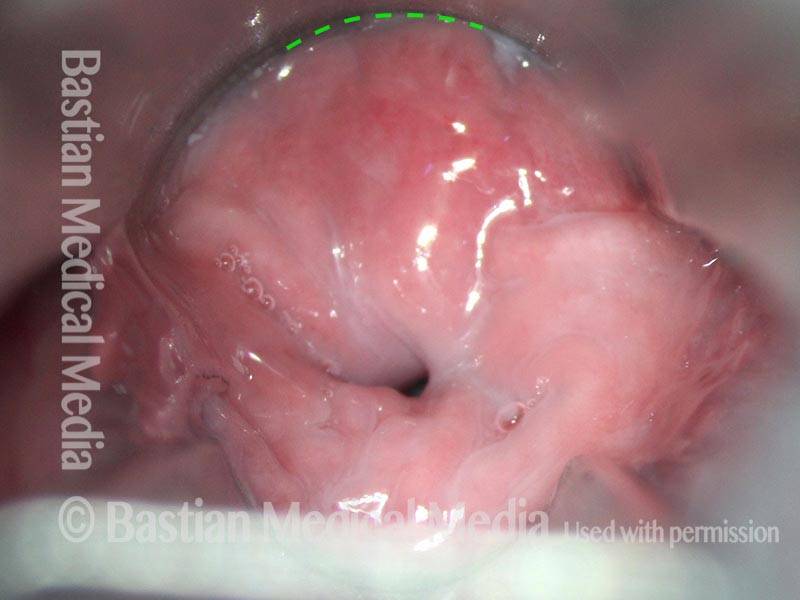
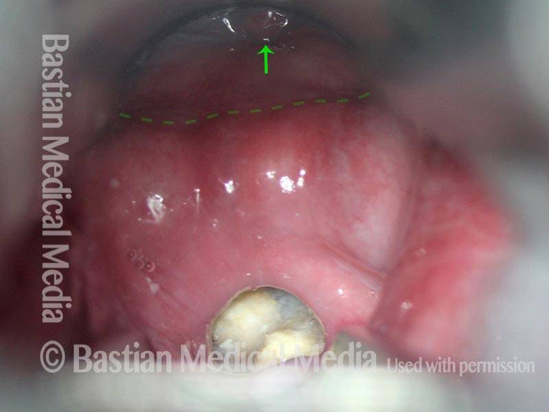
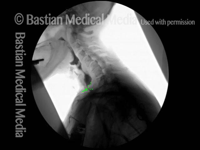
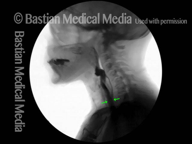
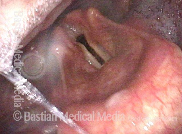
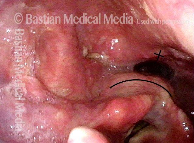
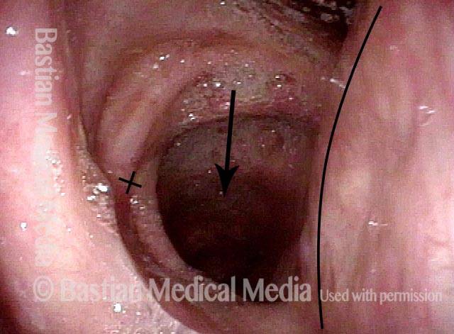
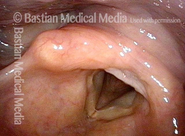
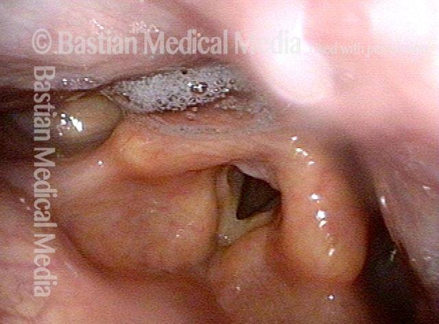
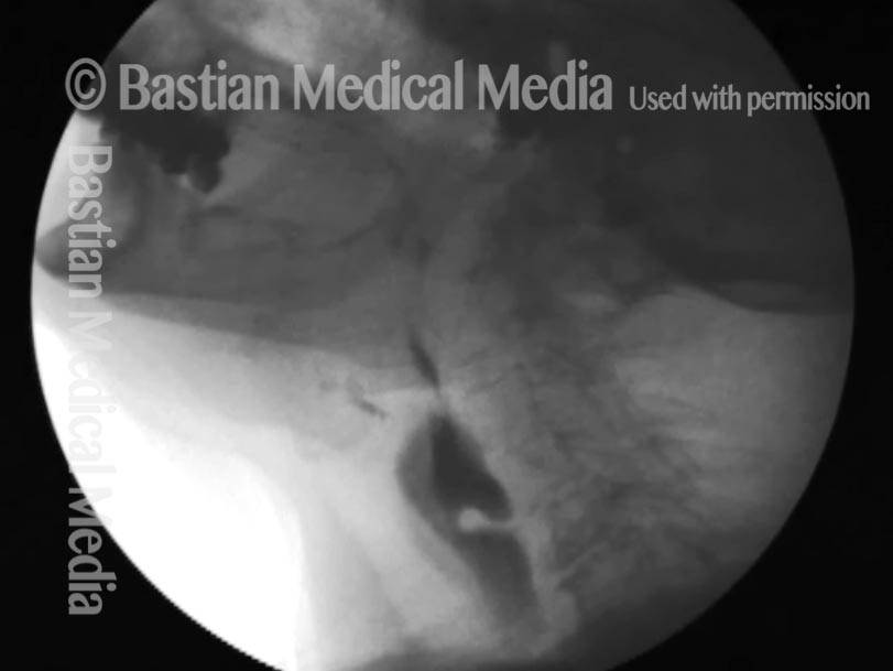
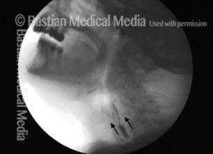
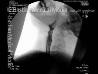
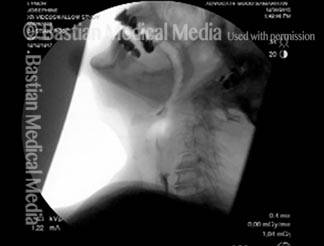
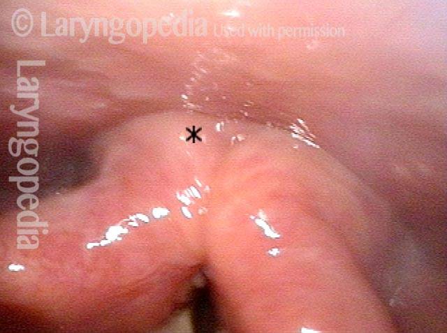
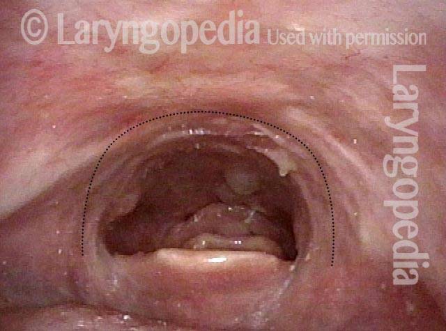
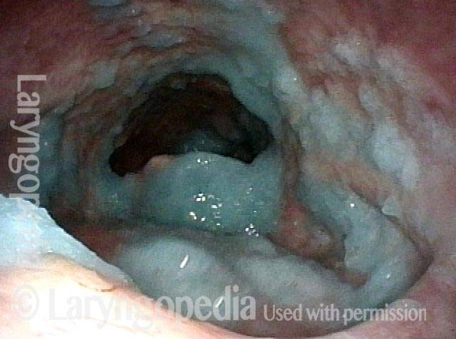
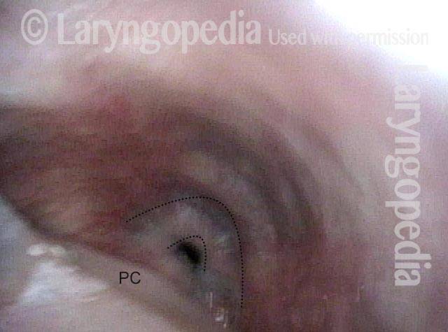
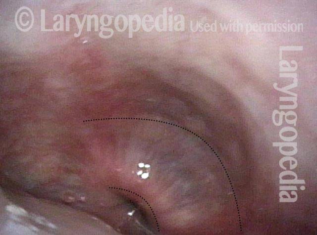
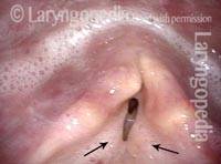
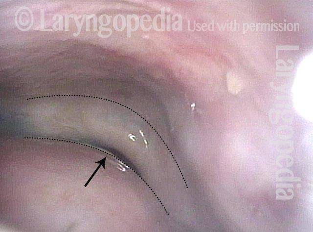
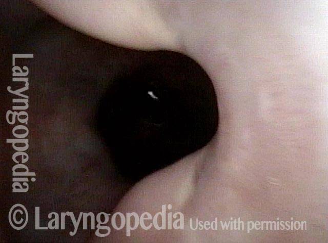
Disfunzione cricofaringea, prima e dopo la miotomia

Visual Portfolio, Posts & Image Gallery for WordPress

Cricopharyngeal dysfunction: before myotomy (1 of 2)

Lateral x-ray of the neck while swallowing barium (seen as a dark column). The non-relaxing cricopharyngeus muscle (light-grey bulge outlined by a dotted line) is causing narrowing of the upper esophageal passageway, as highlighted by the narrowed stream of dark barium at that point (arrow). Liquids and very soft foods can squeak through this narrow opening, but solid foods tend to get stuck.

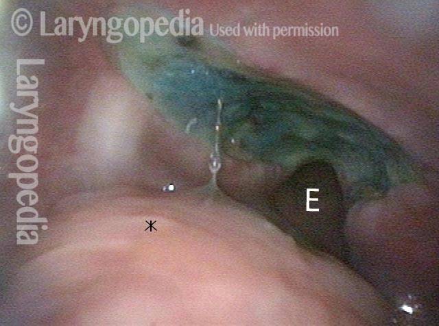
Cricopharyngeal dysfunction: after myotomy, resolved (2 of 2)
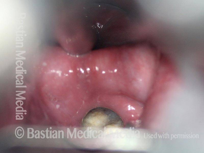
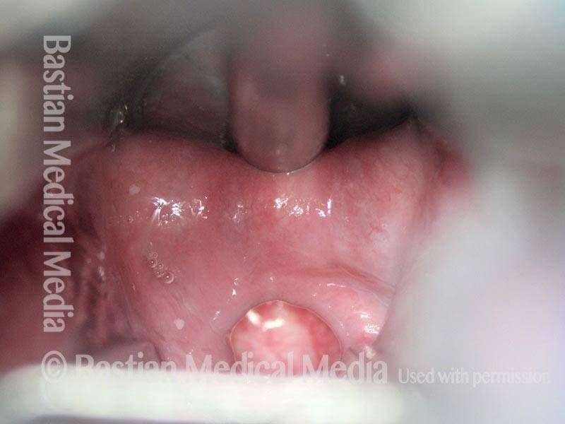
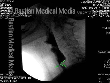
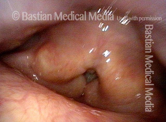
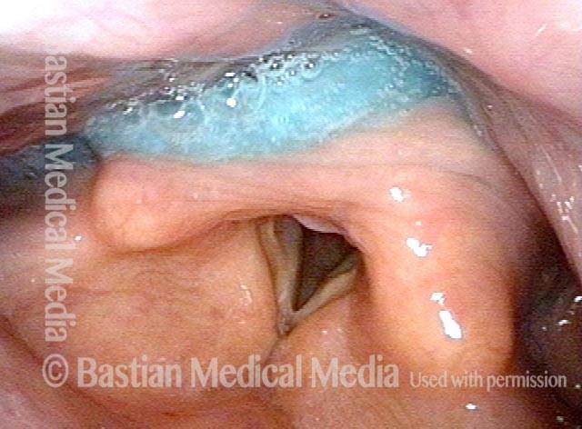
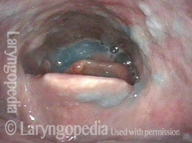
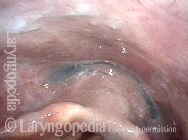
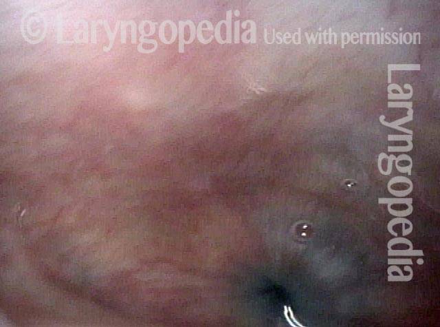
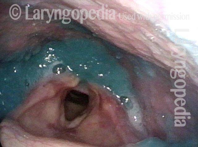
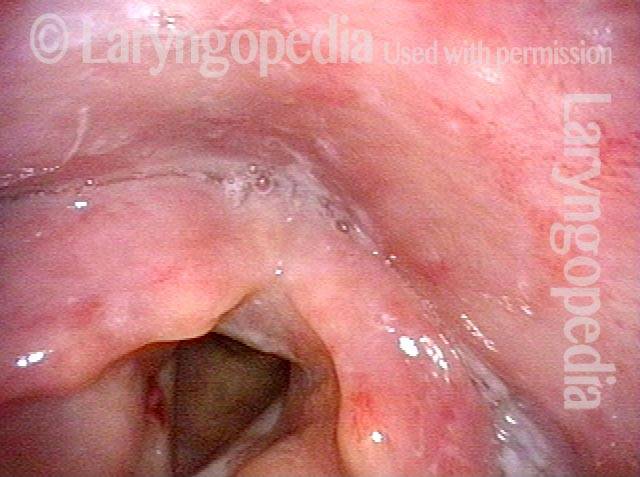
After myotomy. The surgically divided muscle can no longer narrow the upper esophageal passageway, as seen by the widened stream of dark barium at the level of the muscle (arrows).

Esempio 2

Visual Portfolio, Posts & Image Gallery for WordPress

Cricopharyngeal dysfunction: before myotomy (1 of 2)

Lateral x-ray of the neck while swallowing barium (the dark material seen here in the throat). The non-relaxing cricopharyngeus muscle (light-grey bulge outlined by a dotted line) is causing narrowing of the upper esophageal passageway, as highlighted by the narrowed stream of dark barium at that point (arrow). Liquids and very soft foods can squeak through this narrow opening, but solid foods tend to get stuck.

Cricopharyngeal dysfunction: after myotomy, resolved (1 of 2)
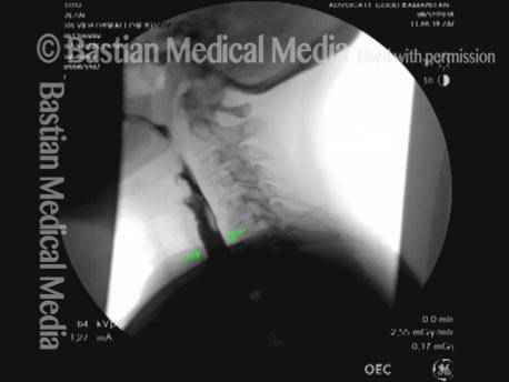
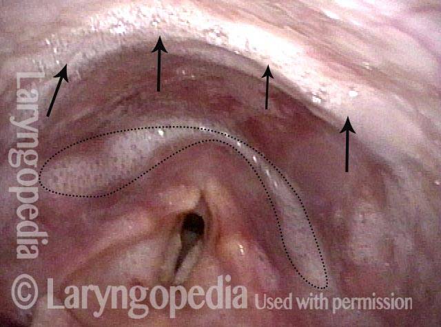
After myotomy. The surgically divided muscle can no longer narrow the upper esophageal passageway, as seen by the widened stream of dark barium at the level of the muscle (arrows).

Esempio 3

Visual Portfolio, Posts & Image Gallery for WordPress

Cricopharyngeal dysfunction: before myotomy (1 of 2)

Elderly patient with nearly a year’s duration of frequent lodgment of solid food at the level of the cricoid cartilage (at the mid-neck level). Note here the cricopharyngeus muscle “bar” which narrows the barium stream (indicated by green dotted line). This narrowing is due to incomplete relaxation of the muscle (aka upper esophageal sphincter) causing a smaller entrance to the esophagus. Liquids and very soft foods can still get through, but solid foods tend to get stuck or to require repeated swallows.

Cricopharyngeal dysfunction: after myotomy (2 of 2)

A month after endoscopic (through the mouth) cricopharyngeus myotomy (division of the muscle with a laser). The patient’s initial swallowing symptoms are completely resolved and the barium stream no longer shows narrowing and the cricopharyngeus bar is no longer seen (see green arrows).

Miotomia cricofaringea per l’alimentazione ricreativa e la gestione della saliva

Visual Portfolio, Posts & Image Gallery for WordPress

Panormaic view, post CPM (1 of 4)

Panoramic view of larynx and hypopharynx in elderly man with both propulsive/ pitcher and receptive/ catcher swallowing problem. Here, after cricopharyngeus myotomy, the remaining, unaddressed propulsive problem is seen as salivary pooling/ clinging. Laryngeal vestibule is unsoiled, however.

Trumpet maneuver (2 of 4)

Trumpet maneuver opens the hypopharynx including at the level of the divided cricopharyngeus muscle. The curved line and ‘X’ are to orient this photo in comparison with the next (photo 3).

Closer view (3 of 4)

Closer view of myotomized and therefore non-functional cricopharyngeus muscle, again during trumpet maneuver. Gravity alone could take secretions and small amounts of food down into the esophagus (arrow).

Well-managed saliva (4 of 4)

After 6 boluses of blue-stained applesauce, intentionally given in rapid-pressured fashion to test limits, laryngeal vestibule remains very clean, and pooling does not tend to be deep enough to easily spill over into the laryngeal vestibule. Still g-tube dependent, this man enjoys some food, and manages saliva better than before myotomy.

98 anni, prima e dopo la miotomia

Visual Portfolio, Posts & Image Gallery for WordPress

Immediately after swallowing (1 of 10)
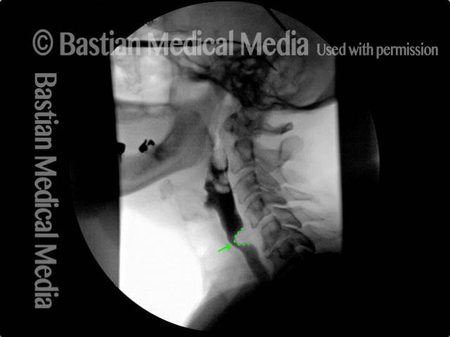
This 98 year-old woman experiences dysphagia that is most noticeable for solids. She is also aware of constant “phlegm” in her throat. In this photo, she has just completed a swallow of her own saliva. See what happens a moment later in the next photo.

Zenker’s (2 of 10)

A second later, saliva emerges from below, as her known Zenker’s diverticulum discharges some of its contents upwards into the hypopharynx rather than downwards into the esophagus.

Dysphagia (3 of 10)

During VESS, part 2, she has just completed a swallow of blue-stained applesauce without leaving any immediate post-swallow residue.

Residue from Zenker’s (4 of 10)

A second later, the applesauce and saliva retained in her Zenker’s diverticulum is pushed upwards from below.

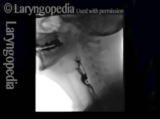
X-ray showing Zenker’s (5 of 10)

An x-ray image showing the Zenker’s diverticulum immediately following her swallow.

Moments later (6 of 10)

A moment later, some swallowed barium has discharged upwards into the hypopharynx.

After myotomy (7 of 10)

A week after endoscopic cricopharyngeus myotomy. The patient says her swallowing has become normal. This view verifies her observation. Here, she has just completed a swallow and after waiting considerable time, no saliva reappears. Compare with photo 2.

No residue (8 of 10)

After not only blue-stained applesauce, but also a cheese cracker, there is no return of material and only a fleck of cracker in the left pyriform sinus (arrow). Compare with photo 4.

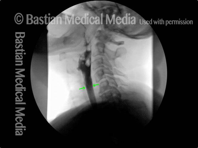
Zenker’s gone (9 of 10)

After myotomy, note that the Zenker’s sac only puddles at its apex, because the rest of the sac has been marsupialized into the esophagus. Patients with this finding have no swallowing symptoms. Compare with photo 5.

No barium in hypopharynx (10 of 10)

While watching throughout the study, no barium ever emerges upwards into the hypopharynx, in constrast to pre-operatively. Compare with photo 6.

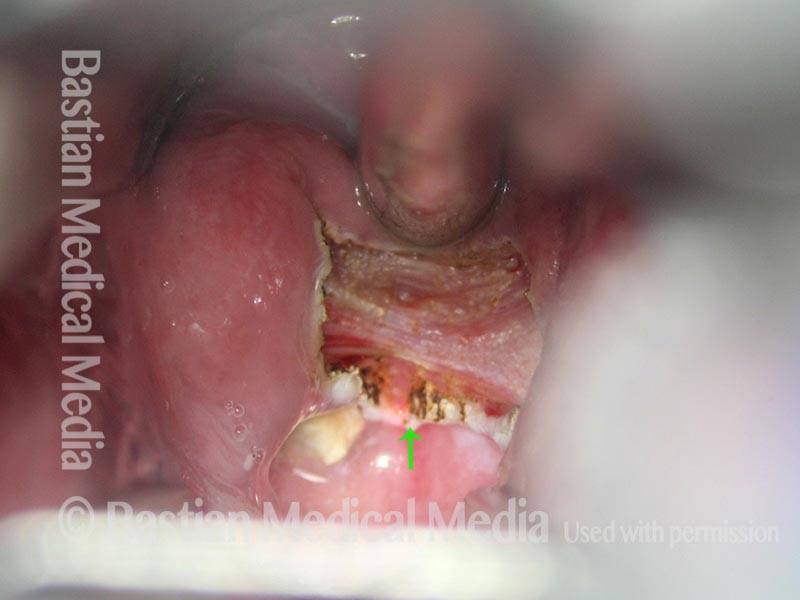
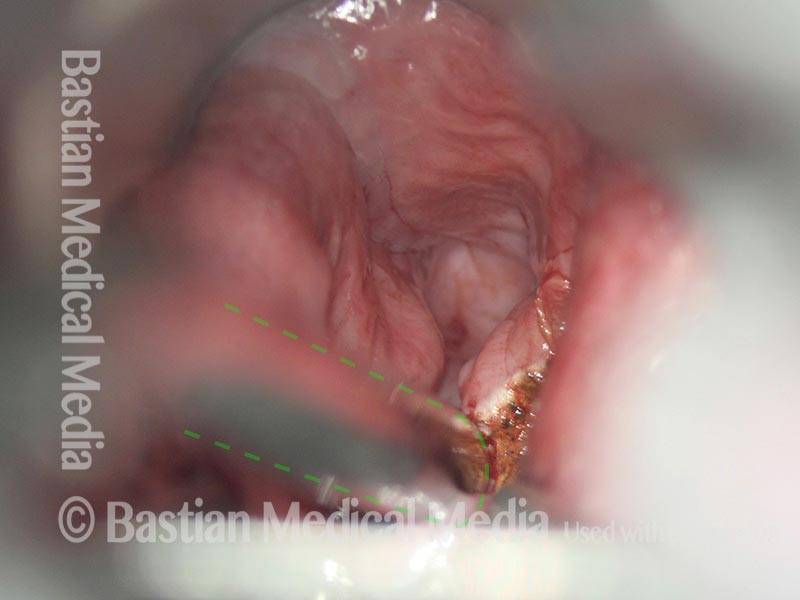
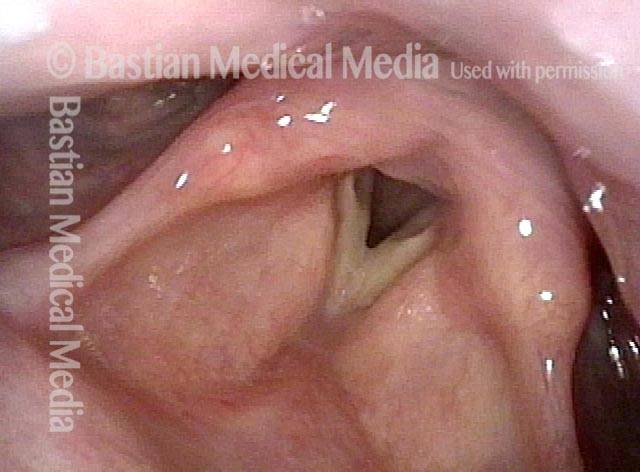
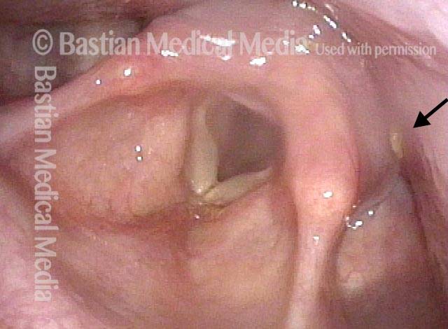
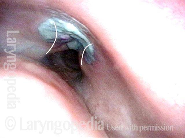
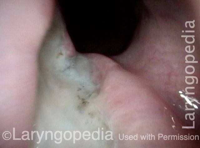
Ottima vista della ferita chirurgica della miotomia cricofaringea fresca

Visual Portfolio, Posts & Image Gallery for WordPress

CPM dysfunction (1 of 4)

After administration of blue-stained applesauce during VESS, small and organized residue in the post-arytenoid area suggests possible cricopharyngeus muscle dysfunction. At this time, the patient noted only occasional pill lodgement.

VFSS six years later (2 of 4)

Six years later, the patient returned saying that swallowing had gradually become extremely difficult. Swallowing pills and eating food were nearly impossible. A VFSS shows narrowing of the barium stream at the arrow, due to a cricopharyngeus bar or “thumb” at *.

Five days post-op (3 of 4)

Five days after endoscopic laser cricopharyngeus myotomy, the patient says that while surgical pain is still significant, she can already swallow pills and solid food easily, a dramatic change from 5 days earlier. The area of surgery is not seen in this resting view. The * is for orientation with the following photo.

Cervical esopagus (4 of 4)

The patient is puffing her cheeks and this is enough to open the cervical esophagus (E). This allows visualization of the raw surface where the muscle was divided with the laser. It is stained by recently administered blue applesauce. The * is for orientation with the prior photo.

Bolus Stream prima e dopo la miotomia cricofaringea

Visual Portfolio, Posts & Image Gallery for WordPress

Pre-myotomy (1 of 2)

This older man has swallowing difficulty with all consistencies, but particularly with solids. Note how the broad bolus stream at the level of the hypopharynx becomes a thin pencil line in the cervical esophagus due to non-relaxation of the cricopharyngeus muscle (M).

Post-myotomy (2 of 2)

Some months after cricopharyngeus myotomy, the bolus width is the same throughout its course.

Risultati VESS dopo la radioterapia

Visual Portfolio, Posts & Image Gallery for WordPress

Narrowed pharyngeal wall (1 of 7)

After radiation and chemotherapy for larynx cancer several years earlier. Note the dry secretions. There is narrowing of the pharyngeal wall (dotted line) due to radiation scarring.

Swallowing applesauce (2 of 7)

After the second bolus of blue-stained applesauce. The propulsive ability (“pitcher of swallowing”) is inadequate, leaving a lot of post-swallow residue.

After sipping water (3 of 7)

After three sips of blue-stained water, much of the applesauce has been washed away.

Gravity aiding in swallowing (4 of 7)

Additional water washes nearly all of the residue in the “swallowing crescent” away–mostly by gravity as seen in the next photo.

Lifting larynx (5 of 7)

Each swallow looks like this. The pharynx “bird swallow” mechanism lifts larynx forward so that the swallowing crescent opens down to the cricopharyngeus muscle, indicated by double dotted lines. (PC = post-cricoid.)

A closer look (6 of 7)

At closer range, the cricopharyngeus muscle bulge is seen more clearly, along with the small opening into the esophagus.

Gravity aiding again in swallowing (7 of 7)

Blue-stained water flowing into the esophagus mostly by gravity.

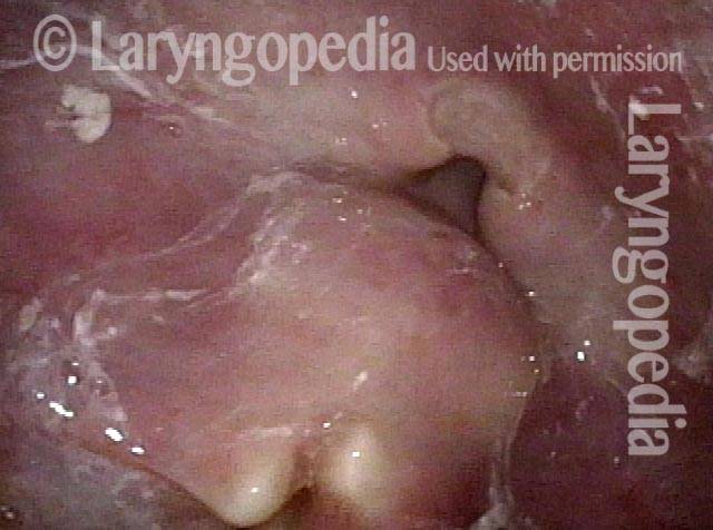
L’evoluzione di una ferita da miotomia cricofaringea

Visual Portfolio, Posts & Image Gallery for WordPress

Difficulty swallowing solid foods (1 of 8)

This ~80 year old man is having considerable trouble swallowing, particularly for solid foods. In this panoramic view at the start of VESS, saliva is noted in the swallowing crescent (outlined) and clinging to the posterior pharyngeal wall (arrows).

Pooled saliva (2 of 8)

At closer range, the pooled saliva in the swallowing crescent is more clearly seen, as is some saliva within the laryngeal vestibule (arrows). Organized pooling of saliva or food / liquid can indicate cricopharyngeus dysfunction (non-relaxation).

Muscle bulge (3 of 8)

The patient has swallowed some water to clear away the saliva, and the pre-myotomy cricopharyngeus muscle bulge (between dotted lines) is seen with only a slit of opening into the esophagus at the arrow.

Residue in swallow crescent (4 of 8)

After many boluses of blue-stained applesauce, the swallowing crescent remains full of residue, but laryngeal vestibule is not soiled. Both propulsive and receptive functions of swallowing are impaired but a significant part is outlet obstruction caused by incomplete cricopharyngeus muscle relaxation.

Three weeks later (5 of 8)

About 3 weeks after cricopharyngeus myotomy, note that the salivary pooling in the swallowing “crescent” is less than pre-operation.

Residual “wound” (6 of 8)

After administering blue-stained applesauce and water, the residual “wound” from the myotomy is stained blue. After myotomy, the cut ends of the muscle retracts laterally as suggested by the curved lines. Compare with the muscle bulge in Photo 3.

Three months post-op (7 of 8)

Nearly 3 months after myotomy, both the patient and his wife say swallowing is much improved. Note the deep “notch” in the muscle bulge as compared with photo 3.

At close range (8 of 8)

At very close range with some clockwise rotation of the view. The muscle can no longer impede passage of food or liquid into the esophagus.

Diverticolo di Zenker una settimana dopo la miotomia del cricofaringeo

Visual Portfolio, Posts & Image Gallery for WordPress

Zenker’s Sac (1 of 3)

Shown in operating position, E = esophageal opening; PPW = posterior pharyngeal wall at the level of hypopharynx; S= residual Zenker’s sac, now marsupialized into the esophagus; CPM = lateral bulges of completely divided cricopharyngeus muscle. The actual wound (W) is stained by the blue applesauce this elderly patient just swallowed.

Zenker’s Sac (2 of 3)

Not only the muscle, but also the elongated mucosal “septum” between sac and esophagus must be divided on both esophageal (anterior) and sac (posterior) surfaces. This view is mostly on the esophageal side. Note the large caliber of the esophageal opening, explaining dramatic resolution of this 90-something year-old woman’s difficulty swallowing.

Zenker’s Sac (3 of 3)

Looking here more directly into the esophagus, one can see that the enlarged esophageal opening as compared with pre-myotomy explains why swallowing pills and solid food is already no longer a problem for this person.

Condividi questo articolo

A-CPD YT Thumbnail
Riproduci video su A-CPD YT Thumbnail

A-CPD può essere trattato con la miotomia cricofaringea

Una piccola percentuale di (soprattutto) persone anziane sviluppa un disturbo della deglutizione progressivo ma curabile chiamato disfunzione cricofaringea anterograda (A-CPD). Inizialmente hanno difficoltà con cibi solidi e pillole.

Con il passare dei mesi e degli anni, la tendenza del cibo a depositarsi in gola aumenta gradualmente. Alla fine, devono limitare la loro dieta a cibi più morbidi e “facili”, sempre più simili agli “alimenti per bambini”. Particolare attenzione è posta su un’efficace procedura laser endoscopica (attraverso la bocca): la miotomia cricofaringea.

Food stuck in throat due to cricopharyngeal dysfunction seen by VFSS YT Thumbnail
Riproduci video su Food stuck in throat due to cricopharyngeal dysfunction seen by VFSS YT Thumbnail

Disfunzione cricofaringea: prima e dopo il CPM

This video shows x-rays of barium passing through the throat, first with a narrowed area caused by a non-relaxing upper esophageal sphincter (cricopharyngeus muscle), and then after laser division (myotomy) of this muscle.

Preoperatively, food and pills were getting stuck at the level of the mid-neck, and the person was eating mostly soft foods. After the myotomy, the patient could again swallow meat, pizza, pills, etc. without difficulty.

Swallowing trouble 101 YT Thumbnail
Riproduci video

Difficoltà di deglutizione 101

Questo video offre una panoramica di come funziona la deglutizione, di come a volte può andare storto (presbifagia o A-CPD) e dei possibili modi per trattare questi problemi (terapia della deglutizione o miotomia cricofaringea).

Cosa succede se i miei denti si allentano dopo il CPM?

Evita di muoverli o di mordere cibi duri finché non si irrigidiscono da soli. Molto spesso ciò richiede circa una settimana.

Circa sei settimane dopo l’intervento è previsto uno studio video-fluoroscopico della deglutizione (VFSS) da confrontare con quello effettuato prima dell’intervento.

Porti con te una copia di quello studio alla visita postoperatoria con il tuo chirurgo più tardi lo stesso giorno in cui è stata eseguita la VFSS.

Tutti i seguenti sono molto improbabili, ma lo sono:

  1. Incapacità di deglutire liquidi.
  2. Dolore al torace o alla schiena che aumenta di gravità dal momento dell’intervento chirurgico.
  3. Febbre o brividi.
  4. Qualsiasi altra cosa che ti preoccupa!

I candidati al CPM dovrebbero accettare entrambi i metodi. In questo modo, il chirurgo può tentare il metodo preferito dall’interno dell’esofago. Se questo metodo non è possibile, passeranno al CPM attraverso un’incisione sul collo.

La maggior parte delle persone, quando escono dalla sala operatoria, vogliono sapere se il problema è stato risolto, “in un modo o nell’altro”.

Le 2 spiegazioni principali che ho riscontrato in una vasta serie:

  1. Il precedente chirurgo non ha eseguito una miotomia completa e/o;
  2. Sono passati molti anni, l’individuo è veramente anziano e ha problemi non con lo sfintere ma con il vigore del lato propulsivo della deglutizione.

Sommario

Subscribe
Notify of

0 Comments
Inline Feedbacks
View all comments
0
Click to see all comments.x