An encyclopedia about voice, swallowing, airway, coughing, & other head + neck disorders.

Presbyphagia

Presbyphagia is a term used to describe swallowing difficulty of the sort that can be associated with the aging process: the process of swallowing as a whole is inefficient and reduced in vigor. Common findings include globally (as opposed to focally) reduced muscle bulk, often seen in vocal cords and not just pharyngeal wall musculature; globally reduced strength of contraction of the pharynx; and tendency for retention or pooling of a part of swallowed food or liquid in the vallecula or pyriform sinuses.

Presbyphagia may be associated with cricopharyngeal dysfunction and, when severe, aspiration.

Is Myotomy Still Effective When the “Pitcher” is Weak?

This 90-year-old person’s swallowing vigor and accuracy are diminished along with her cricopharyngeal function. She has been experiencing swallowing symptoms for nearly 3 years. Initially, she had difficulty swallowing pills, then meat, and now she can only swallow purees and liquids.

We will see that she has not only the “catcher” problem (failure of the sphincter to relax) but also a “pitcher” problem whereby both the strength and accuracy of food propulsion are diminished, leaving a lot of swallowing material “pooled” in the throat and occasionally overflowing into the larynx.

Visual Portfolio, Posts & Image Gallery for WordPress

Presbyphagia – A Weak “Pitcher” (1 of 10)

During VESS, usual symptoms of presbyphagia include: major hypopharyngeal pooling. The blue applesauce clings to base of tongue, pharyngeal walls, pyriform sinuses, and vallecula. There is a significant amount of blue material within the laryngeal vestibule at the arrows, but not below the cords. In other words, vocal cord closure is good enough that she experiences laryngeal penetration but not aspiration. NOTE: Some physicians view this much “presbyphagia” as sufficient to rule out cricopharyngeal myotomy in favor of a feeding tube.

Trumpet Maneuver (2 of 10)

During a trumpet maneuver, the pyriform sinuses inflate to reveal the cricopharyngeus muscle (arrows) but no opening into the esophagus.

After Multiple Boluses (3 of 10)

The applesauce tends to accumulate and further penetrate into the laryngeal vestibule but still without aspiration.

Intraoperatively, Finding the Esophageal Entrance (4 of 10)

The view is inverted in the surgical position. Posterior is now at the lower part of the photo. The cricopharyngeus muscle is so tight and fibrotic, that entry to the esophagus could only be verified by pressing a red rubber catheter anteriorly and having it “seek” the esophageal entrance for orientation purposes.

Laser Division Begins (5 of 10)

Having identified the esophageal opening, the red rubber catheter is now removed. A laser has begun to divide the unyielding muscle.

Laser Division Is Complete (6 of 10)

The muscle is so fibrotic that there has been minimal bleeding. Now the entrance to the esophagus is widely patent, and the almost avascular muscle is no longer an “O” and instead a “U.”

Very Early Postop (7 of 10)

Again in the chair and posterior is at the upper part of the photo. The patient reports large improvement of swallowing, saying she has eaten a whole hamburger with no issue. Here, as the patient performs a trumpet maneuver, you can see the surgical area and esophageal opening. Compare with Photo 2.

Hypopharyngeal Pooling Persists (8 of 10)

The patient still experiences hypopharyngeal pooling from her presbyphagia, but was able to keep up with rapid pressured administration of blue-stained applesauce. Pooling persists, but need for second swallows and laryngeal penetration are both reduced. She has “escaped” the need for a feeding tube…

Cricopharyngeal dysfunction: before myotomy (9 of 10)

Lateral x-ray of the neck while swallowing barium (the dark material seen here in the throat). The non-relaxing cricopharyngeus muscle (light-grey bulge outlined by a dotted line) is causing narrowing of the upper esophageal passageway, as highlighted by the narrowed stream of dark barium at that point (arrow). Liquids and very soft foods can squeak through this narrow opening, but solid foods tend to get stuck.

Cricopharyngeal dysfunction: after myotomy, resolved (10 of 10)

After myotomy. The surgically divided muscle can no longer narrow the upper esophageal passageway, as seen by the widened stream of dark barium at the level of the muscle (arrows).

VESS Demonstrating Presbyphagia, Chin Tuck Maneuver, Hypopharyngeal Pooling, Laryngeal Penetration and Effective Cough
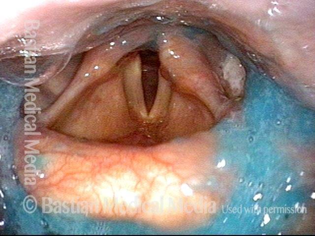
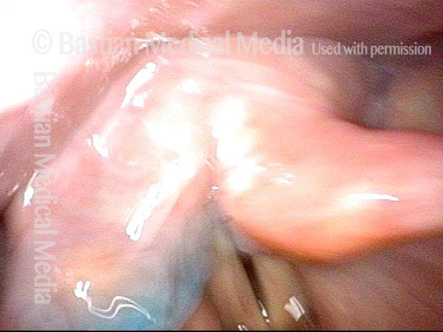
Visual Portfolio, Posts & Image Gallery for WordPress

VESS (1 of 5)

The patient swallowed a bolus of blue-stained applesauce to verify basic (though abnormal) capability. This photo follows 4 additional boluses delivered in a rapid, pressured fashion, intentionally seeking the patient’s “limits.” Note pooled blue applesauce, but without soiling of the laryngeal vestibule.

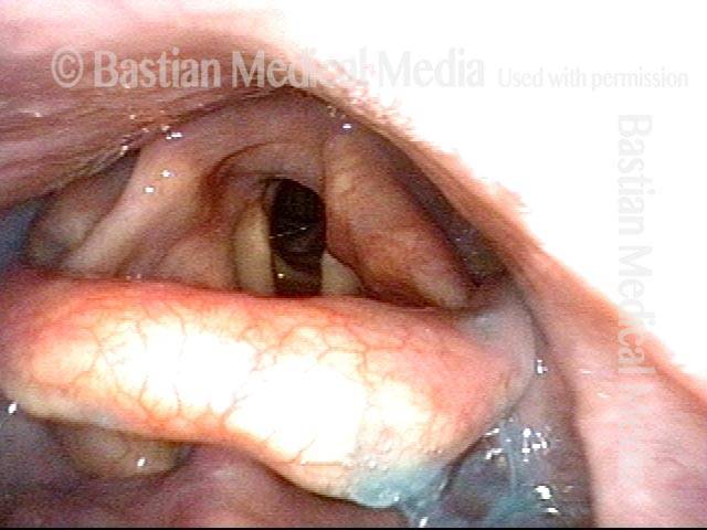
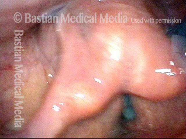
Chin tuck maneuver (2 of 5)

Moments later, the patient was asked to swallow again, but with chin tucked down towards chest. Note how effective this maneuver was in clearing away the residual material seen in the prior photo.
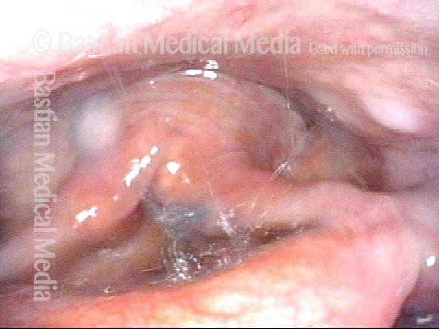
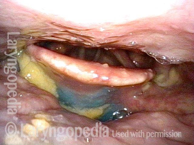
Aspiration (3 of 5)

Due to its lower viscosity, blue-stained water flows more quickly than applesauce, and enters the laryngeal vestibule. Fortunately, the patient is closing the vocal cords simultaneously, so that aspiration does not occur.

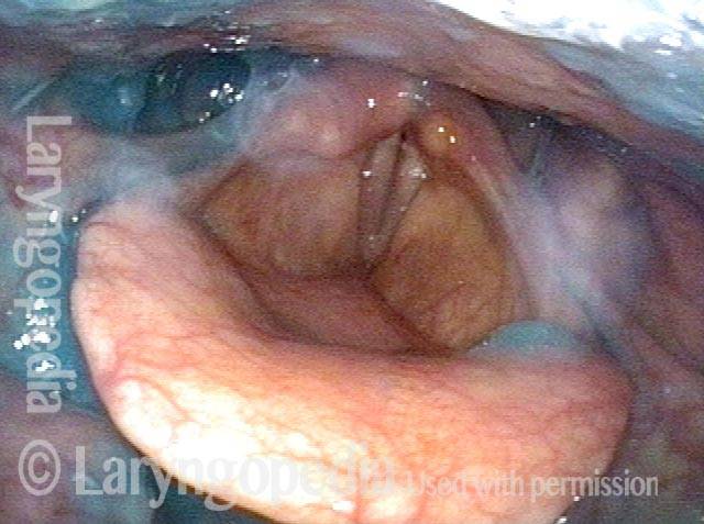
Laryngeal penetration (4 of 5)

Just after the swallow is completed, one can see a trace of blue-stained water just above the not-yet-opened cords. This is technically penetration, and not aspiration.

Air is blasted out of vestibule (5 of 5)

Using “stored” pulmonary air, this trace of penetrated water is “blasted” up and out of the laryngeal vestibule, and is never aspirated.”

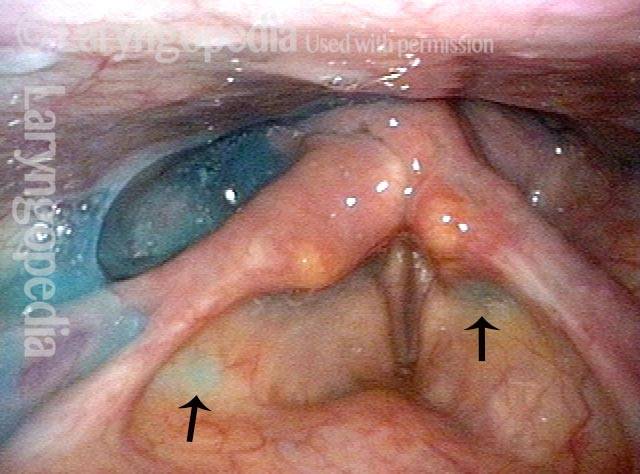
Secretional Pooling Predicts Swallowing Function

Visual Portfolio, Posts & Image Gallery for WordPress

Pooling (1 of 4)

This man coughs frequently especially when drinking. While his palate elevation, pharynx squeeze, and vocal cord functions are intact (as determined during VESS part 1A), note the pooling of saliva especially in the right pyriform sinus (left of photo) and on the pharyngeal wall (arrows) as well as within the laryngeal vestibule (bottom, left arrow). This predicts that his swallowing of blue-stained applesauce, water, and cracker will also be abnormal.

Residue (2 of 4)

As predicted by the information in photo 1, after administration of blue-stained applesauce, we see here an amount and location of residue that mirrors that of the saliva in photo 1. Greater residue in the right pyriform sinus (left of photo) is often seen with right-sided pharynx weakness, but that is not the case here.

Cracker (3 of 4)

After administration of cheese cracker, some of it remains in the vallecula, where it has displaced some of the applesauce.

Blue-stained water (4 of 4)

After administration of blue-stained water, most of cracker and applesauce are washed through. Some minimal blue-staining of the laryngeal vestibule explains why coughing tends to occur when drinking (especially thin liquids).

Share this article

Swallowing trouble 101 YT Thumbnail

Swallowing Trouble 101

This video gives an overview of how swallowing works, how it can sometimes go wrong (presbyphagia or cricopharyngeal dysfunction), and possible ways to treat those problems (swallowing therapy or cricopharyngeal myotomy).

Subscribe
Notify of

0 Comments
Newest
Oldest Most Voted
Inline Feedbacks
View all comments
0
Click to see all comments.x