Stroboscopy means to look at something using strobe rather than continuous illumination. At our practice, we use strobe lighting to allow apparent slow motion videodocumentation of the vibration of the vocal cords. When, for example, the vocal cords produce the pitch of “middle C,” they vibrate at 252 hertz, or cycles, per second; hence, under ordinary illumination, this rapid vibration of the vocal cords is a blur. Under a common setting for the stroboscope, however, the vocal cords appear to be vibrating at just 2 cycles per second, regardless of the actual rate of vibration, which allows the vibratory dynamics to be observed.
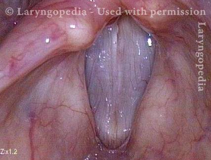
Vocal cords (1 of 7)
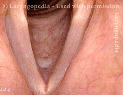
The line of sight is from directly above these open (for breathing) vocal cords, and downward in the line of the trachea. The difference in vocal cord width is not real, but a function of the angle of viewing. This is the position of the vocal cords while breathing.
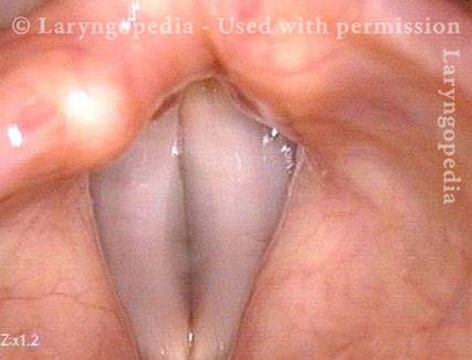
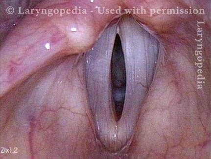
Vocal cords during voice (2 of 7)
Here the vocal cords are producing sound at G2 (98Hz). Under standard light, vibration is so rapid that it is perceived as a blurring of the margins.
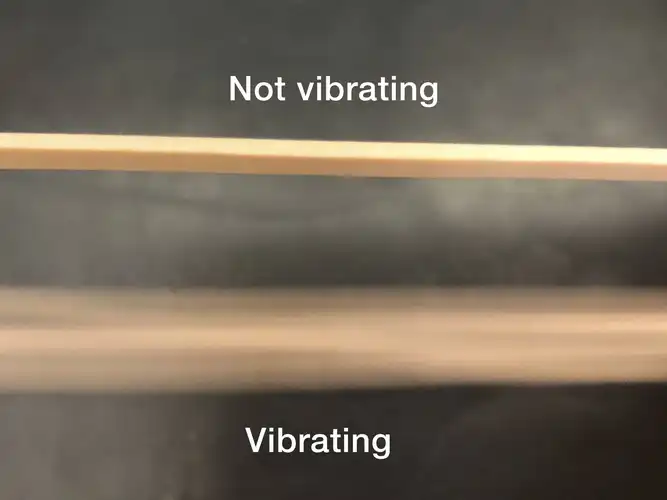
Vibration in rubber bands (3 of 7)
Twang a thick rubber band and you will see the same kind of blurring phenomenon because vibration is so rapid.
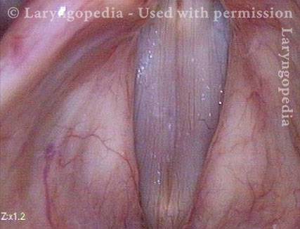
Phonation under strobe light (4 of 7)
During voicing (phonation), under strobe light, to provide apparent slow motion view of vibration. This is the closed phase of vibration at A2 (110 Hz, or 110 vibration cycles per second).
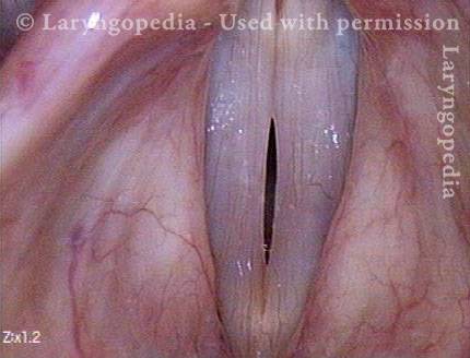
Open phase of vibration (5 of 7)
The open phase of vibration, also at A2. For emphasis: This cycling between open and closed phases of vibration occurs 110 times per second at A2!
Closed phase of vibration (6 of 7)
Now at A4 (440 Hz), this is one of 440 closed phases of vibration per second.
Open phase (7 of 7)
One of 440 open phases of vibration that occurs per second, again as seen under strobe light.
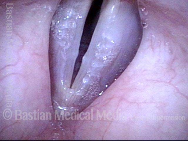
Vocal Nodules, Spicule-shaped
Vocal nodules (1 of 3)
Open phase of vibration just as vocal cords are also parting (to assume breathing position), strobe light. Note the small “spicule” nodules; these are at the other end of the continuum from “fusiform” or “broad-based” nodules.
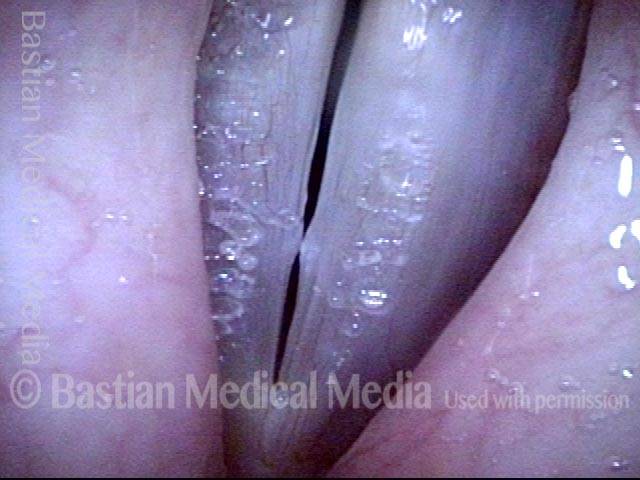
During phonation (2 of 3)
Strobe light, high-pitched voice, showing early contact of the spicule-form nodules.
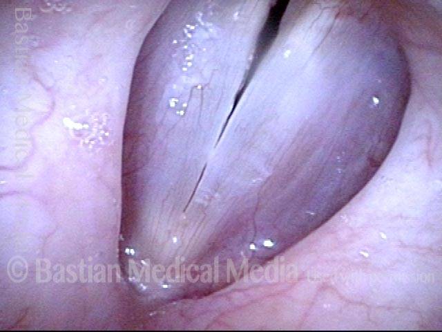
Closed phase (3 of 3)
Closed phase of vibration at lower pitch, strobe light.
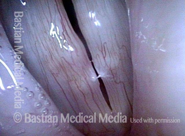
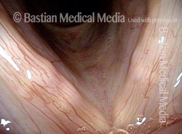
Multiple Rheumatoid Nodules Under 3 Kinds of Light
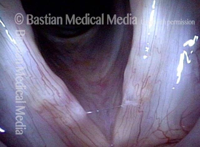
Standard light, submucosal lesions seen (1 of 4)
Young middle-aged woman with chronic severe hoarseness. In abducted (breathing) position under standard light, one can see irregular margins but more importantly, whitish submucosal lesions with the classic appearance of rheumatoid nodules.
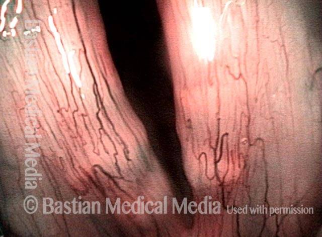
Narrow band light, accentuated capillaries (2 of 4)
Under narrow band light, the overlying capillaries are accentuated, and submucosal masses remain obvious.
Strobe light, nodules (3 of 4)
During open (breathing) position, but under strobe light, another view of these classic, multiple, medial-to-lateral “bamboo” nodules.
Strobe light during phonation (4 of 4)
During phonation also under strobe light, the submucosal masses dramatically inhibit oscillatory flexibility of the mucosa, and prevent accurate margin match at the same time.