Valsalva maneuver is the transient, somewhat forceful exhalation of air against an intentionally blocked airway. In a common variant of this maneuver, a person blocks the exhaled air by sealing the lips and plugging the nose, which forces air up the Eustachian tube and “pops the ears”; this variant is often performed when on a plane that is descending for landing.
In a second variant, a person blocks the exhaled air by closing the vocal cords; this variant is often performed sub-consciously when a person lifts a heavy weight. This second variant of the Valsalva maneuver is also sometimes elicited by a physician during a cardiac or neurological evaluation.
Upper Airway Wheezing, as a Kind of “Skill”
At the level of the vocal cords (1 of 8)
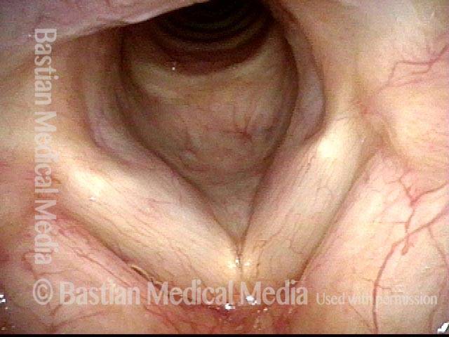
Vocal cords, in breathing position, in a person with wheezing who has been diagnosed elsewhere with asthma. Nothing seen here to explain the wheezing.
At the level of the vocal cords (1 of 8)
Vocal cords, in breathing position, in a person with wheezing who has been diagnosed elsewhere with asthma. Nothing seen here to explain the wheezing.
In the upper trachea: Valsalva maneuver (2 of 8)
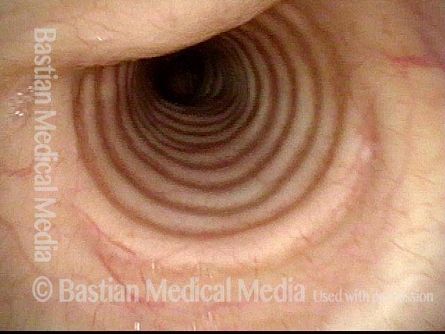
Now descended into the upper trachea. As the patient performs (upon request) a semi-Valsalva manuever, one can see mild inward bulging of the membranous trachea (top- left of photo). This is not sufficient to explain the patient's wheezing.
In the upper trachea: Valsalva maneuver (2 of 8)
Now descended into the upper trachea. As the patient performs (upon request) a semi-Valsalva manuever, one can see mild inward bulging of the membranous trachea (top- left of photo). This is not sufficient to explain the patient's wheezing.
In the mid-trachea: quiet breathing (3 of 8)
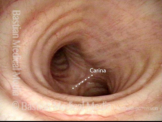
Descended further, into the mid-trachea, with the carina (dotted line) seen in the distance, as the patient breathes quietly.
In the mid-trachea: quiet breathing (3 of 8)
Descended further, into the mid-trachea, with the carina (dotted line) seen in the distance, as the patient breathes quietly.
In the mid-trachea: Valsalva maneuver (3 of 8)
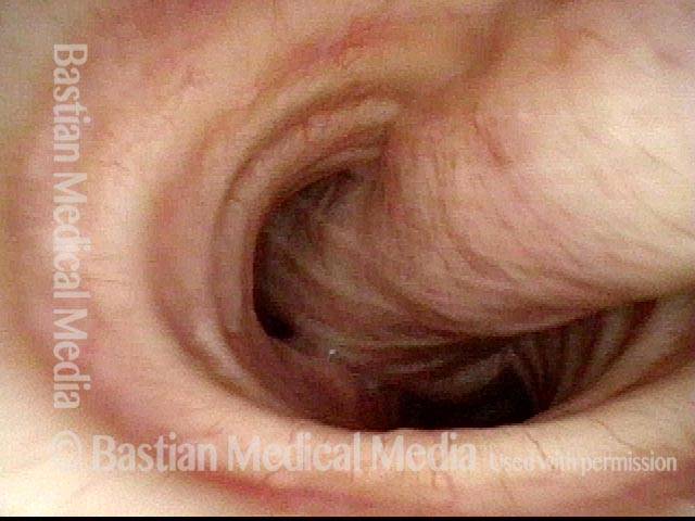
Same view as photo 3, but during another semi-Valsalva maneuver. The membranous tracheal wall bulges inward again, but much more noticeably here than in the upper trachea (photo 2), especially down by the carina (which is now hidden from view).
In the mid-trachea: Valsalva maneuver (3 of 8)
Same view as photo 3, but during another semi-Valsalva maneuver. The membranous tracheal wall bulges inward again, but much more noticeably here than in the upper trachea (photo 2), especially down by the carina (which is now hidden from view).
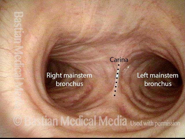
In the lower trachea: quiet breathing (5 of 8)
Down further yet, almost to the level of the carina (dotted line). On each side is the entrance to each of the mainstream bronchi.
In the lower trachea: quiet breathing (5 of 8)
Down further yet, almost to the level of the carina (dotted line). On each side is the entrance to each of the mainstream bronchi.
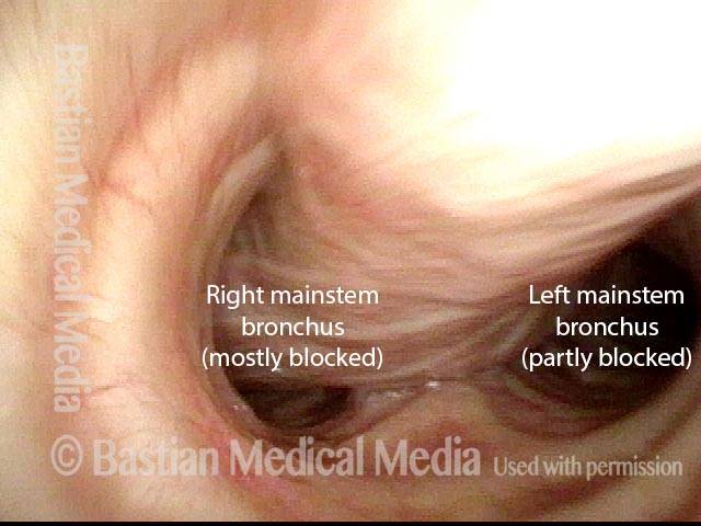
In the lower trachea: Valsalva maneuver (6 of 8)
Same view as photo 5, but during another semi-Valsalva maneuver. The membranous tracheal wall bulges inward again to partly block the airway, especially the right mainstem bronchus. This patient's wheezing sounds loudest over the sternal notch and manubrium; softer wheezing is also heard in the distal lung fields, and as expected from this photo, is louder on the right than the left.
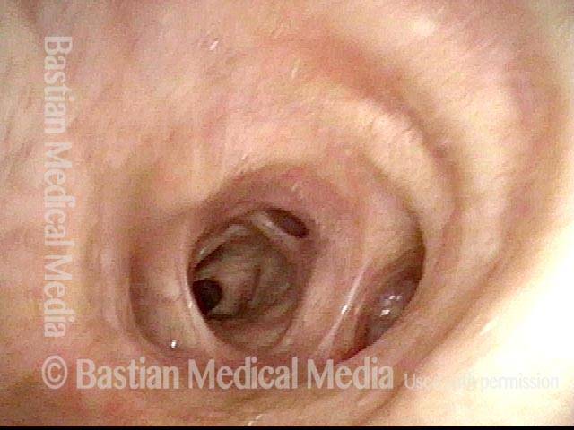
In the right mainstem bronchus: quiet breathing (7 of 8)
Further down yet, now looking directly into the right mainstem bronchus.
In the right mainstem bronchus: quiet breathing (7 of 8)
Further down yet, now looking directly into the right mainstem bronchus.
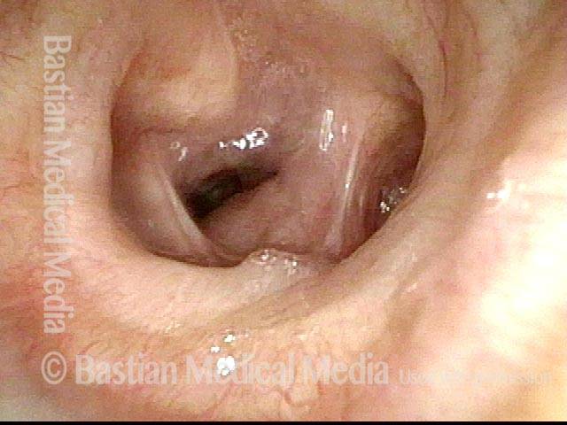
In the right mainstem bronchus: Valsalva maneuver (8 of 8)
Same view as photo 7, but during another semi-Valsalva maneuver. Note compression as well of lobar bronchi, also partly responsible for the patient’s wheezing.
In the right mainstem bronchus: Valsalva maneuver (8 of 8)
Same view as photo 7, but during another semi-Valsalva maneuver. Note compression as well of lobar bronchi, also partly responsible for the patient’s wheezing.
Tagged Anatomy & Physiology, Education
