An encyclopedia about voice, swallowing, airway, coughing, & other head + neck disorders.

Fracture of Larynx

Fracture of larynx is a break—with or without displacement—of the thyroid or cricoid cartilage. Decades ago, a common source of larynx fractures was car accidents, with a person’s neck striking the steering wheel. In this age of protective airbags, the primary source is athletic injuries (e.g., an elbow to the neck while playing basketball). Many larynx fractures are treated conservatively, but occasionally they require repair.

Is There a Difference Between a Fractured Larynx and a Bruised Larynx?

Ten different people with a larynx fracture could or would manifest very differently. It depends on exactly where the larynx (voice box) was fractured; how severe the fracture was and whether it remained displaced or had returned to its original position, and how much the soft tissue within the voice box was bruised, torn, or detached. Some with a larynx fracture experience a lot of bruising and swelling but there is no need to operate and the voice returns virtually to normal. Others require surgery to reattach soft tissue that was torn away from the cartilage.

Each fracture is a particular injury. You might think of the analogy with three people who have each broken an arm. One might only need to wear a sling for a couple of weeks. Another might just need a cast. The third might need open surgery to put in a plate and screws.

The particulars of a larynx fracture should still be evaluable with the combination of a high-resolution CT scan and an exquisitely-detailed video examination such as are represented below.

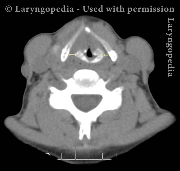
Thyroid Cartilage Fracture and Dislocation of the Right Cricothyroid Joint

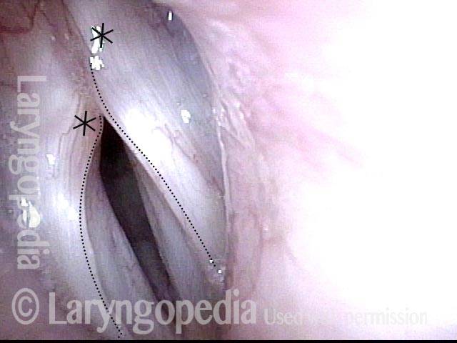
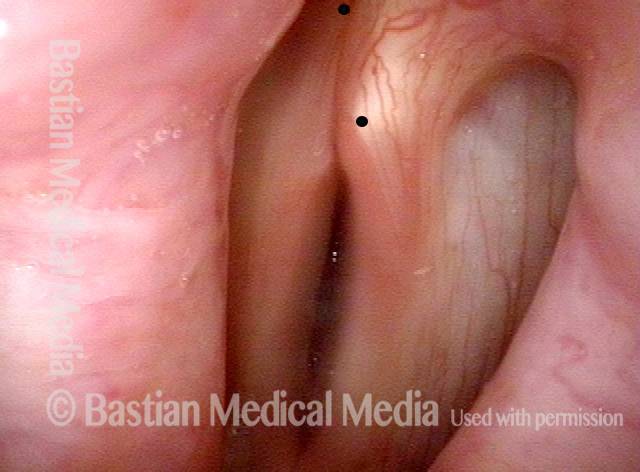
This middle-aged woman fell and hit the front of her neck on a large bottle she was carrying. Voice was altered immediately, though not completely gone. There was no significant hemoptysis but it did hurt to swallow. She also developed subcutaneous air in her neck. The CT scan showed a nondisplaced thyroid cartilage fracture anteriorly. But the previously-overlooked injury was disruption of the right cricothyroid joint. The right thyroid ala was positioned farther posteriorly on the cricoid cartilage, foreshortening the right vocal cord.

Following our rule of thumb that the repaired larynx must be better than the injured larynx, and that the surgical repair trauma could not be worse than the injury trauma, it was decided to leave this problem unrepaired. That’s because the recurrent nerve on the right side would be put at great risk by trying to reattach the cricothyroid joint, which would become ankylotic anyway.

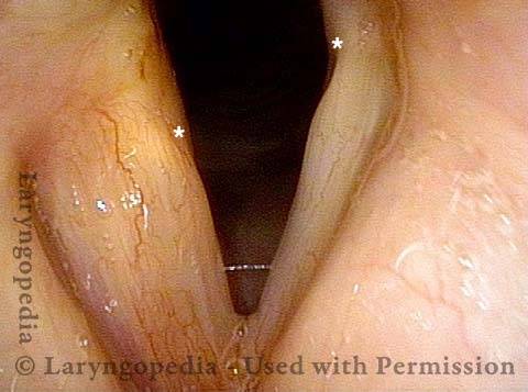
At original examination, bruising, swelling of the right cord is seen, but fairly good mobility of that cord for phonation and breathing. Part of the reason for the convexity is not only swelling but also foreshortening of the right cord as compared with the left.

At a follow up examination several weeks later, speaking voice was extremely functional though lacking in expected upper range due to loss of mobility at the cricothyroid joint. The patient found this result acceptable compared with proceeding to repair with such uncertain results.

Visual Portfolio, Posts & Image Gallery for WordPress

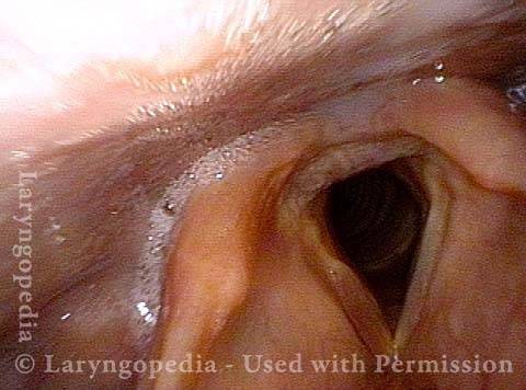
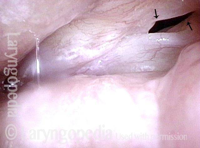
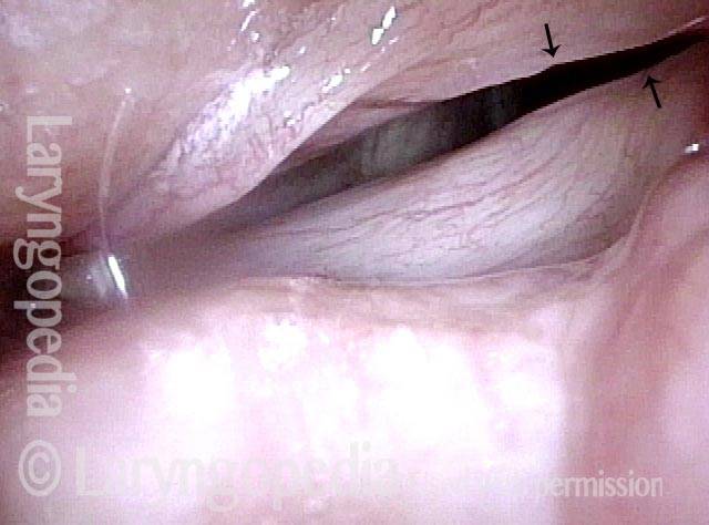
Larynx fracture (1 of 10)

Panoramic view shows bruising and marked swelling of the right arytenoid eminence, as well as the right false vocal cord.

Larynx fracture (2 of 10)

View of vocal cords shows marked edema and bruising right vocal cord.

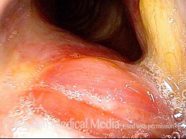
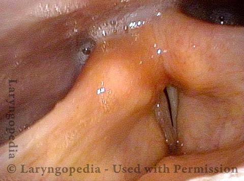
Larynx fracture (3 of 10)

Similar view, during phonation.

Larynx fracture (4 of 10)

View from within subglottis, and down into the trachea shows bruising.

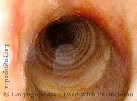
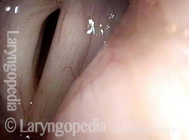
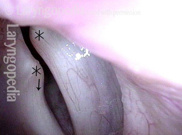
X-ray of Fracture (5 of 10)

This x-ray is from some time before the vocal cord images above. At the time of the original vocal cord images, there was no remaining air in the neck. The key finding is not the cartilage disruption anteriorly at * That is because the anterior commissure (where the vocal cords meet anteriorly) remains attached to the overlying cartilage. The most important injury is disruption of the right cricothyroid joint at the long bracketed line (left of photo). Compare this with the intact cricothyroid relationship at the short bracketed line (right of image). To repair the joint would put the recurrent nerve at great risk. Consequently the rule that “if the risk of the surgery exceeds the potential benefit, do not operate” is invoked and there was no surgical repair.

Bruising largely resolved (6 of 10)

Three weeks later, the patient’s pain on swallowing had subsided and she had what was described by the physician that day as “a nearly normal woman’s voice at low pitch.” Upper voice was markedly deficient and thankfully, this patient was not a singer. You can see that the bruising of the right arytenoid has largely resolved. The right cord abduction (left of photo) is present but not as complete as the left cord’s abduction. Compare with photo 1.

Anterior subluxation (7 of 10)

At closer range, abduction of the uninjured left cord (right of photo) is greater than on the right (left of photo). The dots show estimated location of the tip of the vocal processes. The right side (left of photo) is positioned more anteriorly due to anterior subluxation of the right cricothyroid joint. Compare with photo 2.

Good approximation (8 of 10)

Phonation is not viewed at as close range as in photo 3, but vocal cord approximation appears to be fairly good.

Resolved cricoid bruising (9 of 10)

Brusing within the cricoid ring has partially resolved as compared with photo 4.

Resolved Bruising (10 of 10)

Several weeks after the examination displayed above in images 1-4, the bruising is mostly resolved. All of the air in the neck has absorbed. The thyroid cartilage has reconstituted mostly, but the separation of the right cricothyroid articulation remains. See the long bracket, compared with short bracket at the normal left cricothyroid joint, right of image.

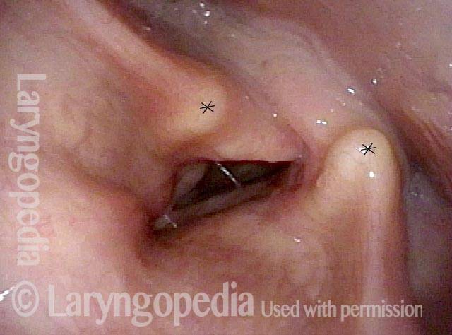
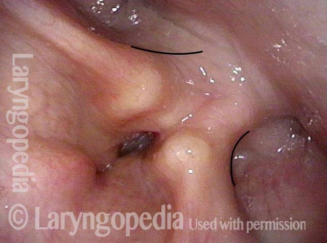
Antero-Superior Fracture of Cricoid Cartilage Containing the Cricoarytenoid Joint

Visual Portfolio, Posts & Image Gallery for WordPress

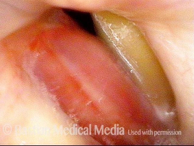
Breathing position (1 of 4)
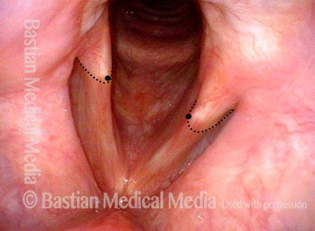
Initial BVI examination, many months after an auto accident with laryngeal fracture. Voice is functional but remains abnormal. In breathing position of the vocal cords, note that tip of the vocal processes of the arytenoid cartilages (denoted by larger dots) are not opposite, but the left one (right of photo) is markedly farther anterior.

Mis-match (2 of 4)

As the vocal cords approach adducted voice-making position, one can again see dramatic mis-match of the tips of the vocal processes.

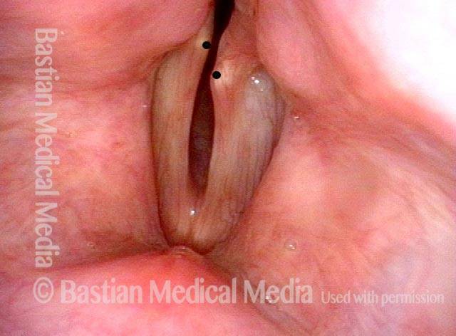
Prephonatory view (3 of 4)
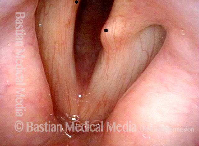
Closer view as the cords approach voice-making position. The dots again indicate the tip of the vocal process, with the right one (left of photo) just out of view.

Phonation (4 of 4)

During phonation, the left vocal process (right of photo) also rides up over top of the right vocal cord (left of photo), and the tip of the right vocal process (left of photo) is again just out of view.

Probable Larynx Fracture Seen Via Endoscopic Cues

This man sustained a sports injury a few months earlier including a blow to the anterior neck. Voice was instantly drastically altered. A fracture is suspected via endoscopic “cues.”

Visual Portfolio, Posts & Image Gallery for WordPress

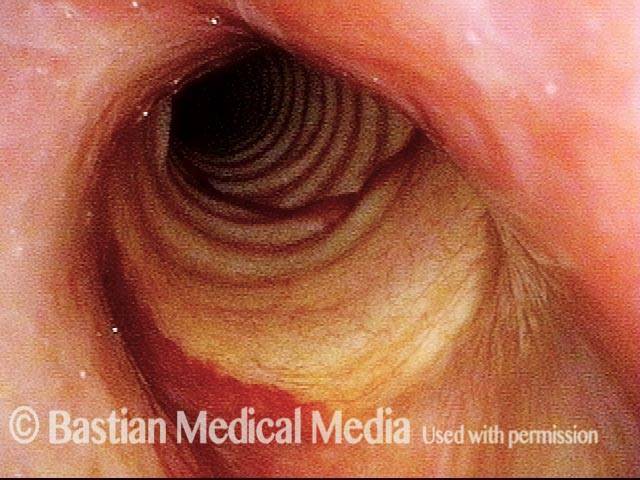
Fracture suspected (1 of 5)

Here a distant view during breathing shows anterior displacement of the arytenoid apex on the right (left of the photo). Compare the two * for reference.

Phonation (2 of 5)

Still viewing from a distance but now while making voice. The arytenoid apices take a more symmetrical position, but the line of the medial wall of pyriform and post-arytenoid area is flattened on the right (left of photo).

Convex margin (3 of 5)

A shortened vocal cord tends to develop a convex margin, and can either mean the arytenoid is displaced anteriorly, or it could be that the thyroarytenoid muscle has pulled away from its attachment to the arytenoid cartilage. Note here as well that the posterior ventricle seems have been displaced anteriorly on the right (left of photo, at —). The tip of the arrow suggests where the tip of the vocal process ends.

Closed phase (4 of 5)

Under strobe light, closed phase of vibration with some phase shifting. To the left of each arrow seems to be where flexible mucosa begins. The foreshortened convex, and more flaccid vibrating segment of the right (upper) vocal cord overlaps the left.

Open phase (5 of 5)

Open phase of vibration, with arrows again indicated the junction between vibrating and adherent (cartilaginous glottis) mucosa.

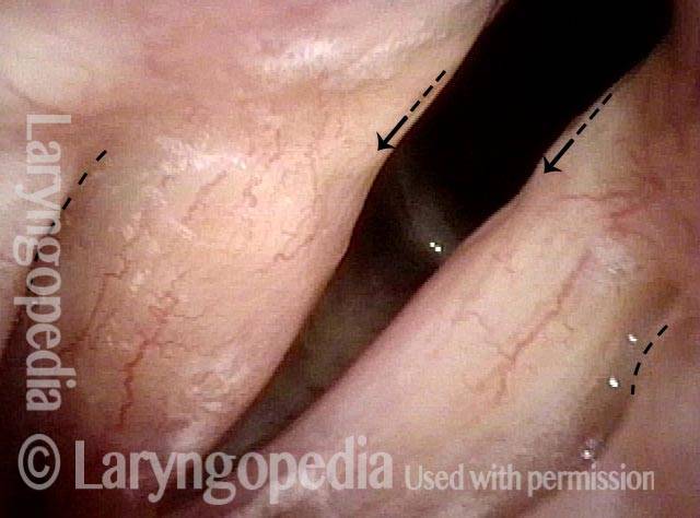
Larynx Fracture—Endoscopic Nuances Are Fascinating!

Twenty years earlier, during a hockey game, this man’s anterior neck was hit by a puck. His voice has never been the same, and he was told there was a “voicebox injury.”

Visual Portfolio, Posts & Image Gallery for WordPress

Hockey injury (1 of 4)

Here, note that both vocal cords abduct fully, suggesting that the crico-arytenoid joint is not likely disrupted, and the PCA muscle is intact. The “smaller-looking “ right vocal cord (left of photo) cannot be due to a disturbed growth plate, as he was in his middle 20’s when the injury occurred. The anteriorly-displaced arytenoid (arrow, left of photo) provides a clue, however, that is explained in photo 4.

Phonation (2 of 4)

During phonation, the right cord (left of photo) appears bowed and atrophied. And is that lateral turning of the vocal process that we see? Is this TA + LCA paresis? Read on…

Phonation under strobe light (3 of 4)

The answer is revealed when the vocal cords approach midline for phonation (under strobe light): Here’s the answer: The tip of the right vocal process (left of photo) is displaced anteriorly as compared with the left. This can be seen by comparing the *’s. The apparent lateral turning of the right vocal process is not in fact of the vocal process, but of the soft tissue of the membranous part of the vocal cord.

Foreshortening of soft tissue (4 of 4)

Open phase of vibration under strobe light shows that the amplitude of vibration is greater on the right (left of photo). To see this more clearly, compare faint dotted lines along the margin of each cord. This “flaccidity finding” is not due to paresis, but instead to foreshortening of the soft tissue (TA muscle and ligament) of the vocal cord. CT of the larynx suggests that the upper lamina of the cricoid is fractured forward on the right, carrying with it an intact and functioning cricoarytenoid joint. Hence, full mobility, yet anterior displacement of the right arytenoid.

Share this article

Table of Contents