Hypopharynx is the inferior-most part of the pharynx, made up of the pyriform sinuses, the lowest part of the posterior pharyngeal wall, and the post-arytenoid/post-cricoid areas.
Reflux Into Hypopharynx, Characteristic of Cricopharyngeal Dysfunction
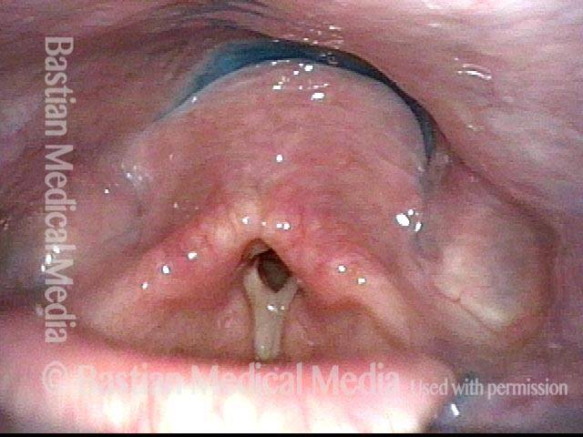
Reflux into hypopharynx (1 of 3)
The patient has swallowing problems typical of cricopharyngeal dysfunction. This swallow study reinforces that impression as well as the likely presence of a Zenker's diverticulum. In this photo, blue-stained water has just been swallowed, and the vocal cords are beginning to open. At this point, the hypopharynx contains no residue.
Water flows into the swallowing crescent (2 of 3)
One second later, the blue-stained water begins to emerge from just above the cricopharyngeus muscle into the "swallowing crescent".
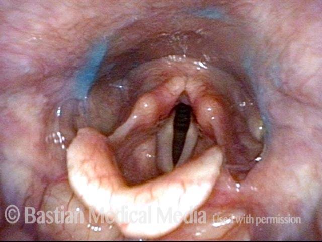
Larynx opens up (3 of 3)
Another two seconds later, the larynx has fully opened post-swallow. The post-swallow hypopharyngeal re-emergence of the blue-stained water is apparent.

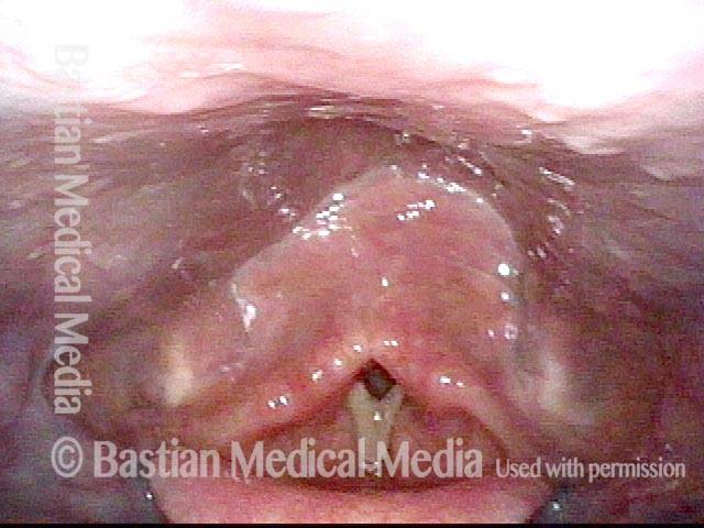
Hypopharynx Pooling After Swallow
Hypopharynx pooling after swallow (1 of 1)
Shows trace of blue-stained applesauce remaining behind after the patient has swallowed.
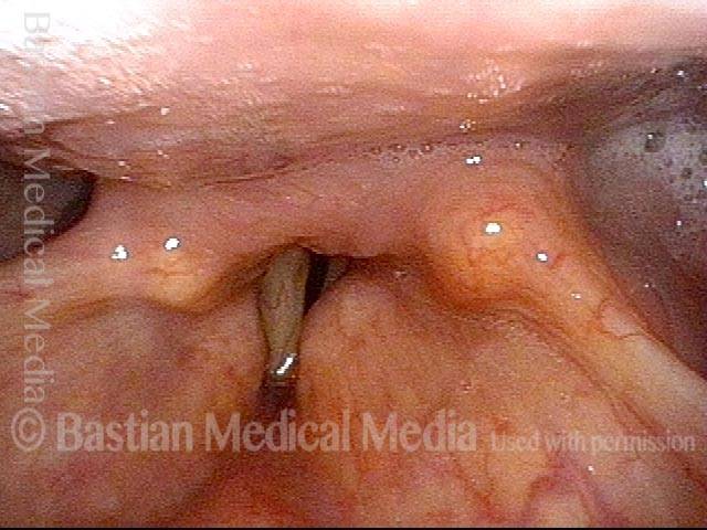
Cervical Osteophyte
Cervical osteophyte (1 of 1)
Panorama of the hypopharynx and larynx. The posterior pharyngeal wall protrudes forward and seems to contact the posterior surface of the arytenoid cartilages.