Pharyngocele is a dilated outpouching from the normal contour of the pharynx. It can be thought of as a throat “hernia.” It is seen in persons who are producing a Valsalva kind of maneuver such as when lifting something heavy except that the resistance to outflow of air (back-pressure) is not the glottis (vocal cords), but instead the lips or something external to the lips.
Think of someone playing a brass instrument at high intensity, a glass blower, or if there were such a thing as a job blowing up stiff balloons all day for many years. The popular image of Dizzy Gillespie playing his horn provides an analogy, except that his cheeks rather than pharynx seemed to be the primary outpouching caused by the backpressure of his music-making.
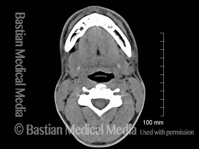
X-ray view at vocal cord level during quiet breathing [without Valsalva] (1 of 9)
A 20-year-old man complains that his neck expands and that he has pain while playing the trumpet. This radiographic image is at the level of the vocal cords, during quiet breathing; at this point, the pharyngeal dilation and pharyngocele are not yet seen. Compare with image 2.
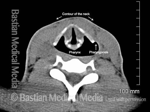
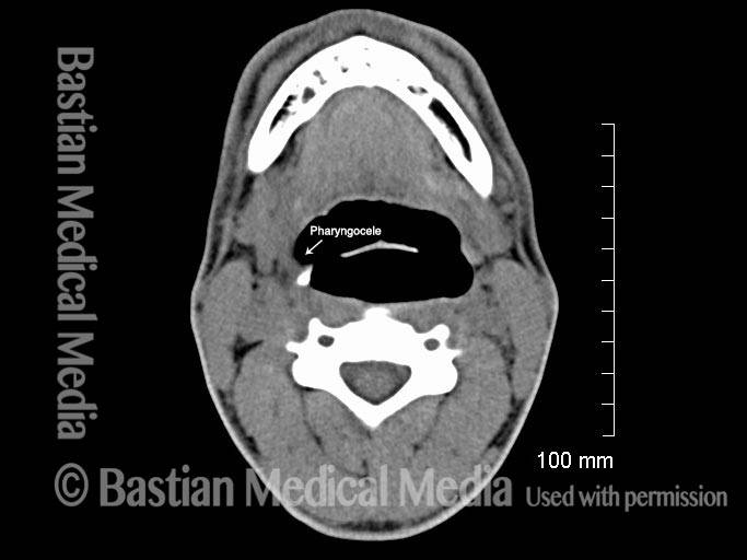
Another X-ray view at vocal cord level [with Valsalva] (2 of 9)
Same view as in image 1, except that the patient is performing a Valsalva maneuver with a lip-stop, to simulate trumpet playing. Whereas in image 1 the pharynx is completely collapsed, here it is inflated with air. A true pharyngocele, seen on the right side of the image, is beginning to develop. Also, compare the neck’s surface contour between this image and image 1.
Endoscopic view of hypopharynx (3 of 9)
The patient is simulating the backpressure of playing the trumpet. This is “inflating” his lower throat (hypopharynx). Black dashed line outlines the upper border, superior cornua, and posterior margin of the right thyroid cartilage. Blue arrows indicate further dilation of pharynx under the hyoid bone (grey dashed line). AEF = aryepiglottic fold. A = arytenoid apex.
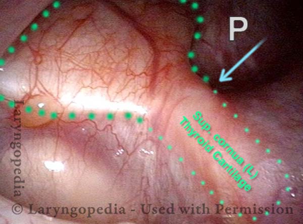
Endoscopic view of left thyroid cartilage outline (4 of 9)
A view of the left thyroid cartilage outline (green dashed line). P = ballooning of the hypopharynx around the posterior border of the left thyroid cartilage.
Endoscopic view of left thyroid cartilage outline (4 of 9)
A view of the left thyroid cartilage outline (green dashed line). P = ballooning of the hypopharynx around the posterior border of the left thyroid cartilage.
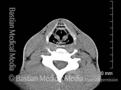
Pharyngocele / pharynx dilation not evident in this X-ray view during quiet breathing (5 of 9)
This view is at the supraglottic level, during quiet breathing, and shows mildly prominent pyriform sinuses, but not dilated as seen in image 4.
Pharyngocele / pharynx dilation not evident in this X-ray view during quiet breathing (5 of 9)
This view is at the supraglottic level, during quiet breathing, and shows mildly prominent pyriform sinuses, but not dilated as seen in image 4.
Another X-ray view at supraglottic level (6 of 9)
The patient again performs a lip-stop Valsalva maneuver, during which the pharynx dilates dramatically. Compare with image 3.
Another X-ray view at supraglottic level (6 of 9)
The patient again performs a lip-stop Valsalva maneuver, during which the pharynx dilates dramatically. Compare with image 3.
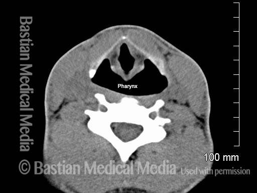
A X-ray view at base of tongue level, quiet breathing (7 of 9)
Higher view yet, at the base of the tongue opposite the tip of the epiglottis, during quiet breathing. Compare with image 6.
A X-ray view at base of tongue level, quiet breathing (7 of 9)
Higher view yet, at the base of the tongue opposite the tip of the epiglottis, during quiet breathing. Compare with image 6.
Another X-ray view at base of tongue level (8 of 9)
The patient again performs a lip-stop Valsalva maneuver, during which the hypopharynx expands dramatically; the beginning of a true pharyngocele can be seen again, this time on the left of image. If this young man were to continue playing trumpet, one would expect the pharynx to dilate more and more over time.
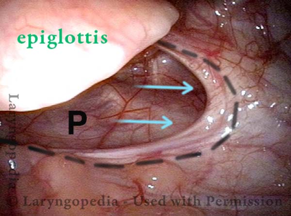
Endoscopic view of left vallecula (9 of 9)
Viewing the left vallecula (between base of tongue and epiglottis. A kind of “herniation” in the direction of the arrows is seen under the left lateral glossoepiglottic fold (dashed line).
Endoscopic view of left vallecula (9 of 9)
Viewing the left vallecula (between base of tongue and epiglottis. A kind of “herniation” in the direction of the arrows is seen under the left lateral glossoepiglottic fold (dashed line).
Tagged Airway disorders, Disorders, Photos

