Glottis

The glottis is often described as the triangular space between the vocal cords, during breathing. During breathing, therefore, a person has an “open glottis.” In common usage, it often includes the cords as well. A “glottal stop” is the sound made when the vocal cords close abruptly to stop the voice.

The true vocal cords that define the lateral boundaries of the glottis are comprised of two main parts: The membranous cord/glottis (anterior 2/3 of its length) and the cartilaginous cord/glottis (posterior 1/3).

Membraneous Glottis vs. Cartilaginous Glottis

The membranous glottis/cords (M) are what vibrate to produce voice. The cartilaginous glottis/cords (C) do not vibrate, because this part of each vocal cord is inhabited by the arytenoid cartilage with only a thin covering of tissue. The inside of the larynx is often looked at with a mild fisheye camera, and depending on the angle of viewing, the proportions of membranous and cartilaginous segments of the cords may seem different from the usual two-thirds vs. one-third.

Disorders of the Glottis

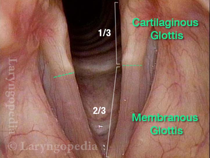
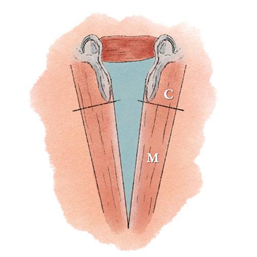
How does disorder of the membranous glottis come to attention? Through a change or loss of voice, most commonly due to overuse vibratory injury such as swelling, nodules, or polyps. Or in the context of infection (viral, bacterial, or fungal) causing laryngitis. Or due to growths like papillomas, or cancer due to tobacco use.

If the cartilaginous glottis cords make no positive contribution to voice, how does it come to the attention of a patient or doctor? Mainly when it suffers injury, most commonly due to a longterm breathing tube in a mechanically ventilated patient. When injured, the perichondrium may respond by generating granulation tissue.  See also contact granulomaAnd when severe, movement of the cricoarytenoid joint may be impaired. Tumors of the cartilaginous glottis/cords are rare. See also post-intubation phonatory insufficiency.


Bilateral Vocal Cord Fixation

Visual Portfolio, Posts & Image Gallery for WordPress

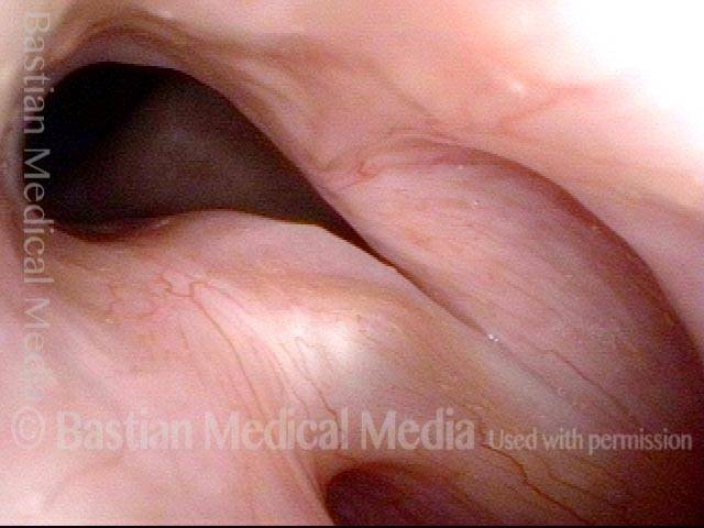
Bilateral vocal cord fixation (1 of 2)

After bilateral posterior cordotomies, “keyhole” shaped glottis. Size of opening at posterior commissure is exaggerated by the wide-angle lens of the distal-chip video-endoscope.

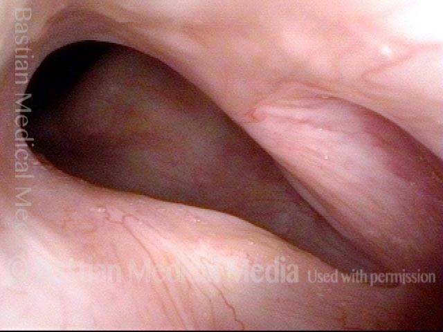
Voice is still good (2 of 2)

Persistent posterior opening, but musculomembranous cords come into good contact and have not been damaged by either the original endotracheal tube or the posterior cordotomies, and voice is very serviceable.

Introduction to Larynx, Pharynx, and Airway Anatomy

In this presentation Dr. Robert Bastian provides an introduction to larynx, pharynx, and airway anatomy. This information is meant to serve as an educational resource.
YouTube