Breathing Tube Injury, not Vocal Cord Paralysis
This middle-aged woman was injured severely in an auto accident as a teenager. Recovery involved a long stay in ICU, and ventilation via a breathing (endotracheal) tube for a few weeks prior to tracheotomy.
Fifteen years earlier, a posterior commissuroplasty was done by me on the left side. Severely short of breath before that procedure, she said the improvement was such that she was able to do most activities of daily living remarkably well for many years. While still much better than prior to the posterior commissuroplasty, she has felt a little more limited in the past few years and wants now another similar airway-widening procedure. Speaking voice can easily pass for normal, though she thinks it is occasionally a little rough.
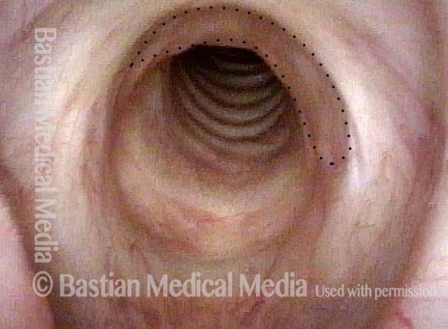
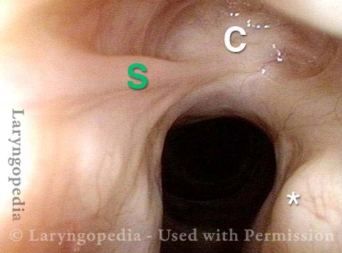
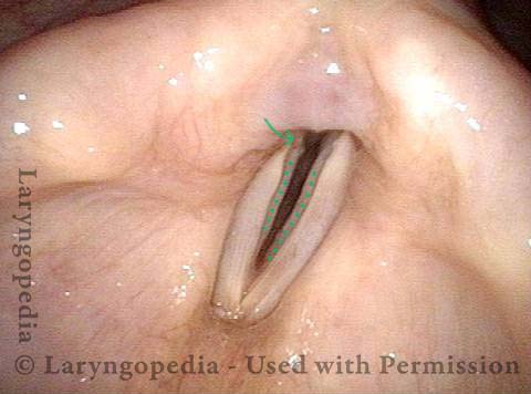
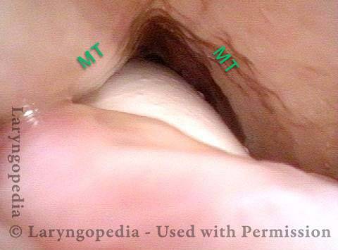
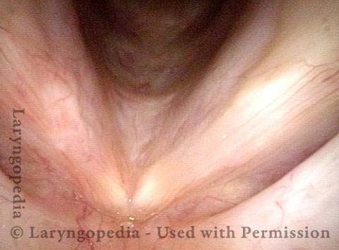
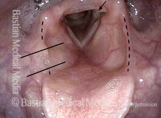
Aperture is very narrow (1 of 6)
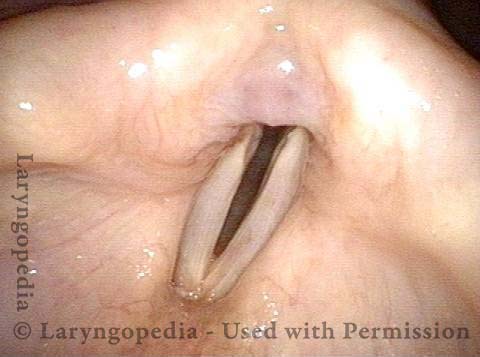
Involuntary inspiratory phonation (2 of 6)
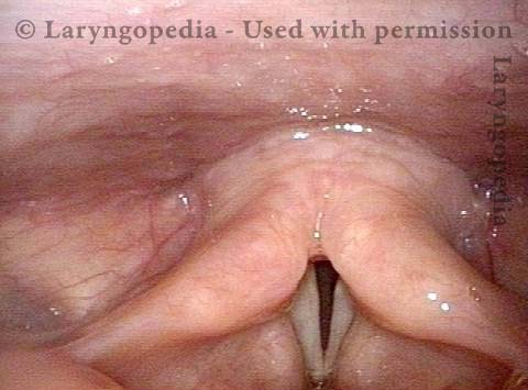
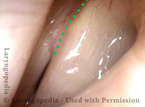
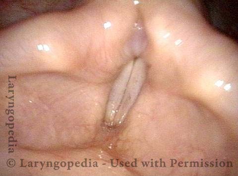
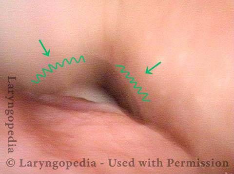
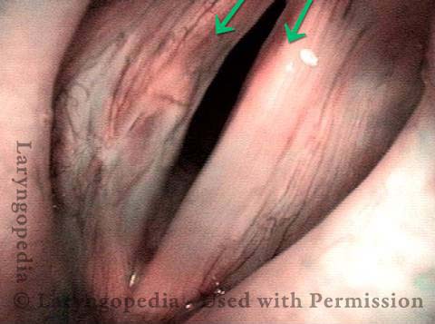
Divot on left vocal cord (3 of 6)
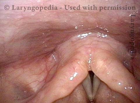
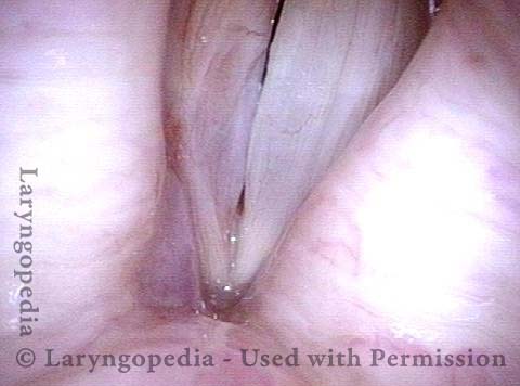
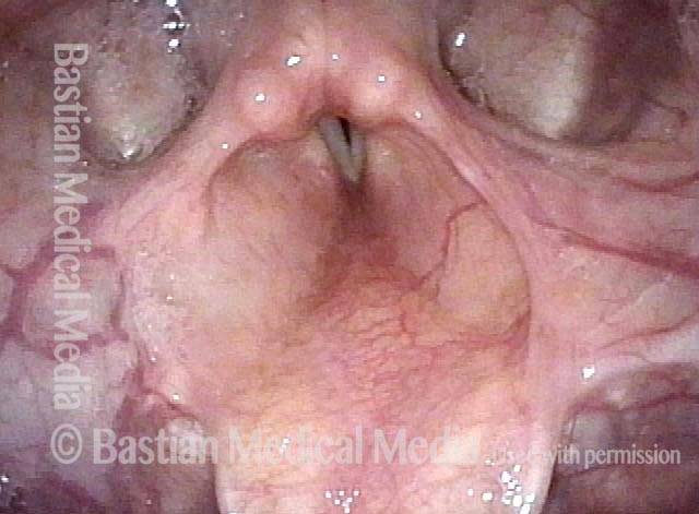
Endotracheal tube injury (4 of 6)
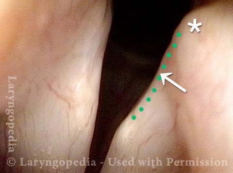
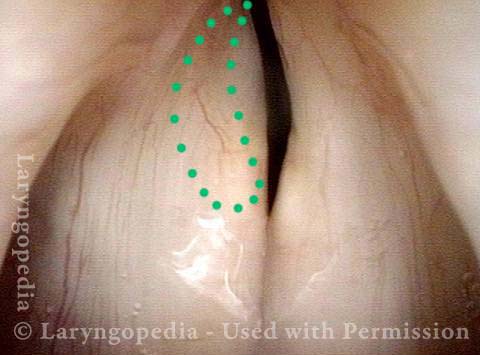
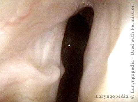
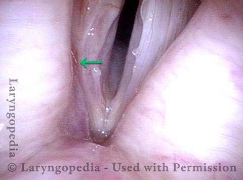
Laser cookie bite (5 of 6)
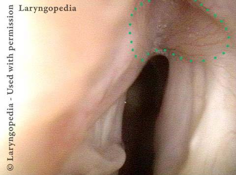
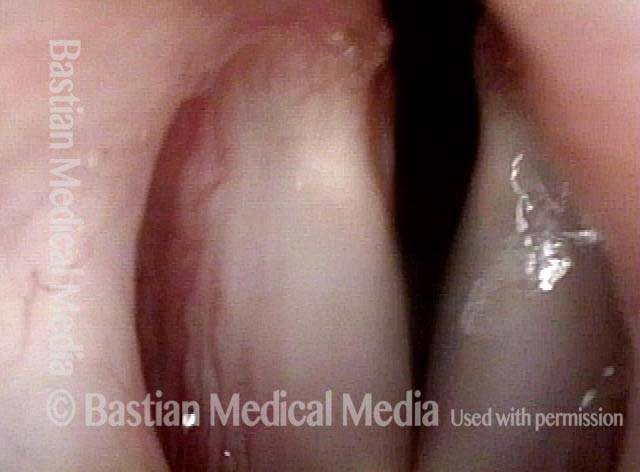
Surface scarring in the tracheotomy (6 of 6)
Breathing Tube Injury may Correlate with Side of Mouth
This person was intubated for 2 weeks due to complications after major surgery. He was sent for evaluation due to hoarseness, with a diagnosis from elsewhere of vocal cord paralysis. During the consultation, he described himself as a very quiet person (a “2” on the 7-point talkativeness scale), and living a quiet life.
After the examination below, his wife was asked “Was the tube taped to the left corner of his mouth? Or do you not remember?” She replied, “Definitely, the left corner.” This history raises the question: Is the diagnosis of paralysis from elsewhere correct, or is the actual diagnosis vocal cord fixation from intubation injury?
Vocal cord fixation (1 of 4)
Paralysis (2 of 4)
Injury from intubation tube (3 of 4)
Mismatch of levels (4 of 4)
Nuances of Endotracheal Tube Injury Distinguished from Paralysis
This woman with high-risk comorbidities of diabetes and obesity, was in ventilated in ICU more than a month for pulmonary complications of Covid-19 infection. She had an orotracheal tube in place for 3.5 weeks, and then a tracheotomy tube was placed. Now at her first visit a year later, she remains tracheotomy-dependent, and is told she has bilateral vocal cord paralysis (disproven in the photo series).
Maximum glottic opening (1 of 8)
Undersurface mucosa indraws (2 of 8)
Phonation (3 of 8)
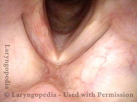
Posterior commissure divot (4 of 8)
Further evidence of scarring (5 of 8)
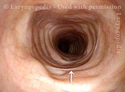
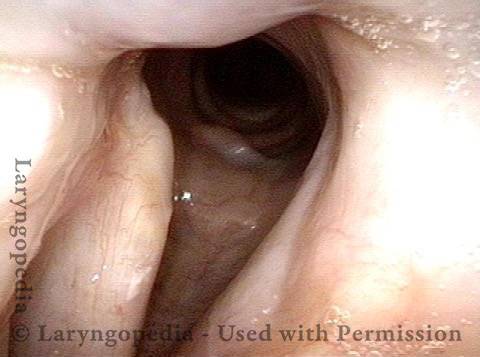
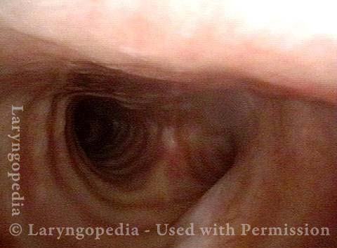
View into trachea (6 of 8)
Vibration of trachea (7 of 8)
Open trachea beyond the tube (8 of 8)
What is the Treatment?
The plan here is posterior commissuroplasty, followed by placement of a smaller trach tube and a trial of plugging. If plugging is tolerated during the day, she will need a sleep study with it plugged at night, given the tracheomalacia and her obesity.
Breathing Tube Injury—A Rare Complication of Intubation for General Anesthesia
Inflamed vocal cord (1 of 5)
Closer view (2 of 5)
Scarring from intubation tube (3 of 5)
Mucosal Injury (4 of 5)
Flaccidity of right vocal cord (5 of 5)
Conclusion: While we try to explain abnormality due to one cause, here, the patient has a mucosal injury and paresis of right TA and LCA muscles, which can also follow intubation. This explains why the initial postop voice was so weak and whispery, and also the rapid partial improvement. This voice will likely continue to improve and be very functional as a speaking voice. Fortunately, this person is not a singer, as clarity especially in upper notes, will likely be remain impaired even after full recovery.
Supraglottic, Glottic, and Subglottic Endotracheal Tube Injury
Breathing tube injury (1 of 4)
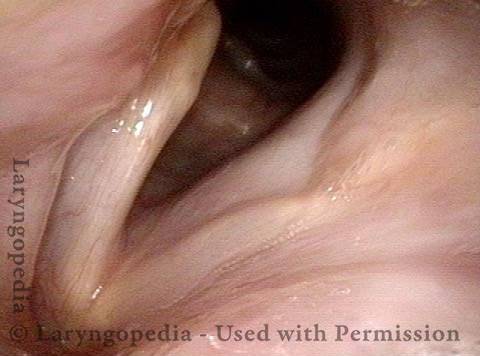
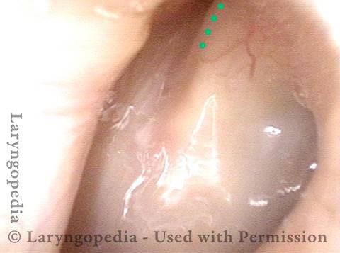
Aryepiglottic cord defect (2 of 4)
Phonation (3 of 4)
Posterior subglottic thickening (4 of 4)