An encyclopedia about voice, swallowing, airway, coughing, & other head + neck disorders.

Vocal Cord Paralysis, Bilateral

Bilateral vocal cord paralysis is a neurological disorder in which nerve supply to both vocal cords is absent. It most commonly results from injury to the recurrent laryngeal nerves, whether due to thyroid surgery, or blunt or penetrating injury to the neck.

Clinical Findings (Symptoms)

On examination, both vocal cords remain in a midline or paramedian position during both voicing and respiration. The key finding is loss of abduction. Because vocal cords can still vibrate well when closely approximated—even if denervated—voice quality is often surprisingly good.

The primary deficit is airway narrowing. Patients may produce an involuntary inspiratory voice when breathing rapidly or during sleep, and they experience reduced exercise tolerance.

Differential Diagnosis: Bilateral Vocal Cord Fixation

A closely related condition is bilateral vocal cord fixation due to scarring—often the result of prolonged endotracheal intubation. The patient’s history often clarifies the diagnosis:

  • Inspiratory phonation and decreased exercise capacity developing immediately after thyroid or goiter surgery suggest bilateral paralysis.
  • The same symptoms following long-term intubation suggest posterior commissure scarring.

Diagnostic Evaluation

In addition to history, an intense examination using topical anesthesia in a voice laboratory can differentiate between the two. Findings of granulation tissue, a posterior commissure divot or divots from pressure necrosis, or a synechia between the vocal processes all support an intubation-related injury.

Management

Some patients already have a tracheotomy to bypass the glottic narrowing. The principal surgical alternative is posterior commissuroplasty, which, in the author’s experience with various techniques, provides the best balance between airway enlargement and preservation of voice.

Compared with transverse cordotomy, arytenoidectomy, or lateralization of one vocal cord using the Lichtenberger needle, posterior commissuroplasty tends to achieve a more physiologic airway while degrading voice quality to a lesser degree. The other methods can restore airway effectively, but often at the cost of increased breathiness or loss of vocal strength.

Posterior Commissuroplasty for Bilateral Vocal Cord Paralysis

This patient had developed multisystem atrophy (MSA) 5 years prior, but it had begun to affect his breathing and swallowing. His noisy breathing especially at night and marked exercise intolerance is the subject of this essay. Physicians elsewhere suggested tracheotomy and CPAP, which the patient rejected. Posterior commissuroplasty was mentioned as an option.

During a few months of patient delay, breathing became much worse than depicted in the initial images below. Posterior commissuroplasty was offered, with the caveat that breathing improvement could be modest and that there might be increased need for his feeding tube.

In almost every scenario, posterior commissuroplasty is performed without tracheotomy. The patient is supported postoperatively with steroids and CPAP to press edema out of the larynx overnight. Most are discharged home the next day. In this patient, almost uniquely, tracheotomy was performed due to his general debilitation and weakness due to his MSA, with the thought that the posterior commissuroplasty would allow him to plug the trach or subsequently fitted mini-trach (in his case, a Montgomerey Cannula). That is in fact what happened.

Visual Portfolio, Posts & Image Gallery for WordPress

Max Abduction (1 of 8)

On initial view (months before the further deterioration led to surgery) his vocal cords abduct about 50% of normal on expiration.

Paradoxical Adduction and Inspiratory phonation (2 of 8)

With rapid inhalation, due to his neurological disease, vocal cords inappropriately adduct, and he makes involuntary inspiratory phonation.

Beginning of Surgery (3 of 8)

In the operating room with inverted view, the laser has outlined the “divots” that will be removed from the cartilaginous (non-vibrating, non-voicing) 1/3 of the glottis.

Not Just Mucosa, But Cartilage (4 of 8)

Medial arytenoid cartilage is included in the divot if possible, dependent on the degree of ossification of the cartilage.

Completed Posterior Commissuroplasty (5 of 8)

The inferior lips of excision need a little trimming. The divots appear very large, but healing will reduce their size often by 50%.

Three Weeks Postop (6 of 8)

During rapid inspiration, the posterior “keyhole” is seen and the divots have not yet mucosalized. Note that the membranous (voice-making) anterior 2/3 of the cords are unaffected.

After Healing, Inspiration (7 of 8)

Compare with photo 2. This larger airway allows the patient to keep his Montgomery cannula plugged 24/7 and doesn’t experience stridor at night, or shortness of breath while working outside.

Side-By-Side Before and After (8 of 8)

In both photos the patient is inspiring rapidly. Note the larger keyhole in the second photo. Even this apparently small difference remarkably increases his airway and reduces his inspiratory noises; he is able to plug his tracheal cannula both day and night.

Bilateral Vocal Cord Paralysis

Visual Portfolio, Posts & Image Gallery for WordPress

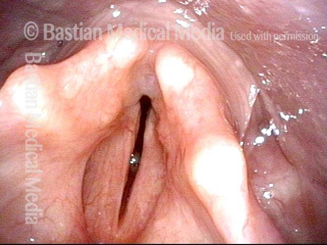
Maximum space between cords (1 of 2)

After thyroidectomy many years earlier, the patient’s exercise tolerance became minimal without becoming short of breath and making loud inspiratory vocal sounds. Eventually, she underwent tracheotomy, which she has continued to wear for more than ten years. This view shows the maximum space between her vocal cords, which paradoxically occurs when she exhales.

View during inhalation (2 of 2)

When asked to inhale with tracheotomy tube momentarily plugged, the passing air causes the vocal cords to indraw slightly and come into vibration, creating “involuntary inspiratory phonation.” Note the faint convexity and grey blur where the mucosa is vibrating.

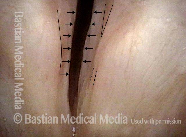
Mucosal Indrawing with Inspiration

Visual Portfolio, Posts & Image Gallery for WordPress

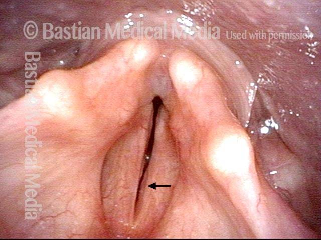
Paralyzed vocal cord (1 of 2)

Paralyzed right vocal cord, with bowing and atrophy. Here, the left vocal cord is maximally but incompletely abducted. Subtle markings are for reference with photo 2.

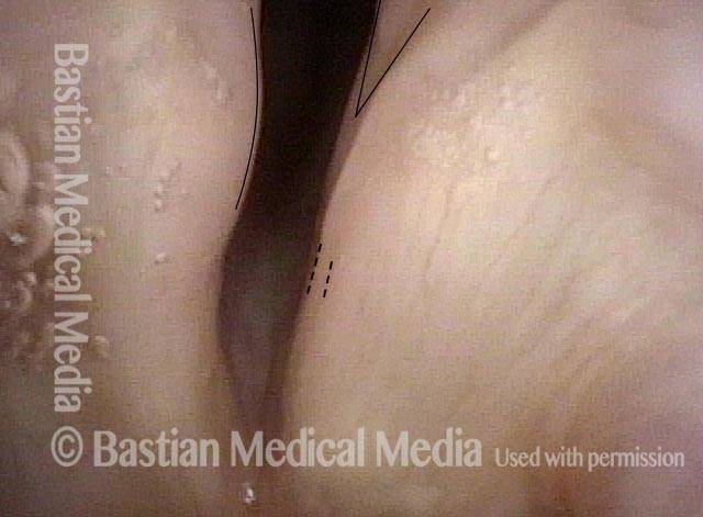
Indrawing with inspiration (2 of 2)

With elicited inspiration, the mucosa of the undersurface of both vocal cords indraws due to Bernouilli effect/ micro-vortices and further narrows the airway. At the same time, the patient involuntarily makes inspiratory voice.

Before and After Posterior Commissuroplasty

This elderly woman had undergone total thyroidectomy approximately 25 years ago. At presentation her inspiratory noises were prominent even at rest. Inspiration between every sentence created involuntary inspiratory phonation. Her husband said he had to use a separate bedroom because she was so loud during sleep.

Very early in Dr. Bastian’s development of posterior commissuroplasty many years earlier, he had performed a unilateral medial partial arytenoidectomy procedure to her great benefit. Now, after many years lost to follow up, she returned, saying that while airway remained much better, she was desiring an additional improvement.

Visual Portfolio, Posts & Image Gallery for WordPress

After Prior Partial Medial Arytenoid Excision—Breathing Out (1 of 10)

This patient has bilateral vocal cord paralysis due to a thyroidectomy many years ago. Although a previous medial arytenoid excision on the right (left of photo) created a permanent divot (arrow) and improved her breathing, she desired better (less noisy) nighttime breathing. The blue lines show the expected degree of opening of the cords in a normal larynx.

Rapid Inspiration and Involuntary Inspiratory Phonation (2 of 10)

Here, the patient is breathing in rapidly per clinician request. Note the vibratory blur of the membranous vocal cords, causing inspiratory phonation, but the persistent posterior gap, which we now want to enlarge further.

Initial Operative View (Endotracheal Tube Briefly Removed) (3 of 10)

Here you can see the posterior cordotomy divot especially of the right cord (arrow). Even with membranous cords fully approximated, this space permits inspiration but also brings the membranous cords into involuntary vibration (voicing).

Partial Medial Arytenoidectomy In Progress (4 of 10)

During brief apnea (endotracheal tube removed), a divot is taken from the left medial arytenoid.

Excision Nearly Complete (5 of 10)

Using a laser, a small “cookie bite” from the posterior left vocal cord (cartilaginous glottis) is removed.

Posterior Commissuroplasty Complete (6 of 10)

Compare with image 3. If desired, a second excision could have been taken from the previously operated (right) side…NOTE that the above surgery was done without a tracheotomy.

Three days post-op (7 of 10)

The patient has a very poor early postoperative voice, as expected. She says breathing is much better and her husband says nighttime breathing is now quiet. More than is usually seen, it appears that the cord has been detached from the arytenoid, but in fact most of the muscle fibers remain attached to the remaining arytenoid.

Years Later: Much Better Breathing and Functional Voice (8 of 10)

After healing, now we have noticeable divots of both vocal cords (arrows). This larger space also tends to “de-activate” the Bernouilli effect (micro-vortices) keeping the membranous cords from “sucking together” with rapid inspiratory flow (seen in next image).

Breathing In Forcefully (9 of 10)

This shows more clearly in comparison with the prior image, the inspiratory indrawing of the left cord margin yet without vibratory blurring or inspiratory phonation.

Phonation after Posterior Commissuroplasty (10 of 10)

It appears that the Bernouilli effect is bringing cords into sufficient vibratory approximation to explain her functional (if modestly weakened) voice.

Resources for Further Reading

Titulaer K, Guntinas-Lichius O. Surgery for bilateral vocal fold paralysis: systematic review and meta-analysis. Front Surg. 2022;9:956338. doi:10.3389/fsurg.2022.956338

Lechien JR, Crevier-Buchman L, Finck C, et al. Management of bilateral vocal fold paralysis: a systematic review. Otolaryngol Head Neck Surg. 2021;164(2):255-263. doi:10.1177/0194599820944892

Hillel AT, Giraldez L, Samad I, et al. Voice outcomes following posterior cordotomy with medial arytenoidectomy in patients with bilateral vocal fold immobility. JAMA Otolaryngol Head Neck Surg. 2015;141(8):728-732. doi:10.1001/jamaoto.2015.1136

Czesak MA, Modrzejewski M, Stręk P, et al. Methods of surgical treatment of bilateral vocal fold paralysis. Endokrynol Pol. 2020;71(1):9-14. doi:10.5603/EP.a2020.0002

eMedicine. Bilateral vocal fold paralysis: practice essentials, history, physical examination. Medscape. Updated March 12, 2024. Accessed November 3, 2025. https://emedicine.medscape.com/article/863885-overview

Hoover WB. Bilateral abductor paralysis: operative treatment by submucous resection of the vocal cord. Archives of otolaryngology–head & neck surgery. 1932;15(3):339-355. doi:10.1001/archotol.1932.03570030357001

Kelly JD. Surgical treatment of bilateral paralysis of the abductor muscles. Archives of otolaryngology–head & neck surgery. 1941;33(2):293-304. doi:10.1001/archotol.1941.00660030296010

Share this article

Support Laryngopedia
Are you a patient or doctor who loves Laryngopedia? If we've helped you, help us help others too!

Table of Contents