An encyclopedia about voice, swallowing, airway, coughing, & other head + neck disorders.

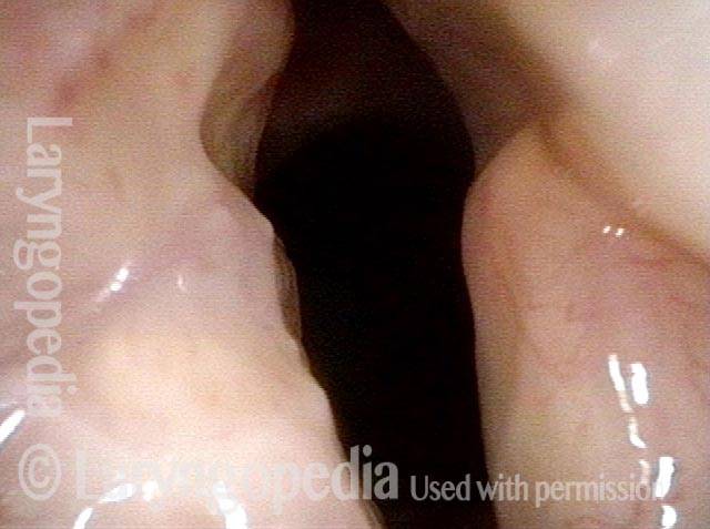
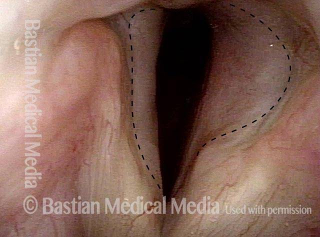
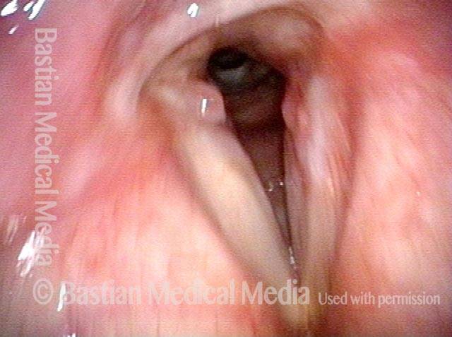
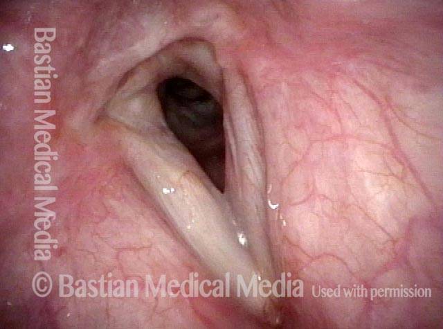
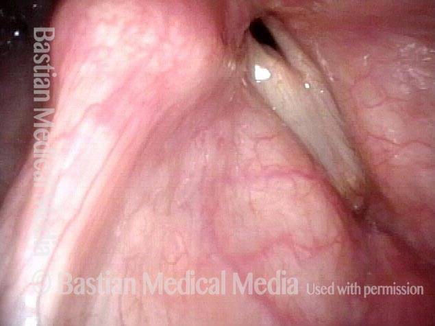
Posterior commissuroplasty is an endoscopic procedure performed for individuals who have difficulty breathing, due to either bilateral vocal cord paralysis or bilateral vocal cord fixation. These individuals’ vocal cords are immobile or fixed in a mostly closed position, which inhibits breathing and often causes noisy inspiration.

In a posterior commissuroplasty, the clinician uses a carbon dioxide laser to take small divots from the posterior ends (membranous glottis) of both vocal cords. These divots create more space between the cords so that, during breathing, air can pass through more easily. This procedure can avoid the need for a tracheotomy. It also preserves the voice’s functionality better than a transverse cordotomy typically does.


Office-Based Surgery When General Anesthesia Is too Risky

Visual Portfolio, Posts & Image Gallery for WordPress

Involuntary inspiratory voice (1 of 6)

This elderly man is tracheotomy-dependent due to inability to open the vocal cords. Here while breathing in, there is a posterior “keyhole” from the divots caused by pressure necrosis of the breathing tube. Still, due to inspiratory airstream, he produces involuntary inspiratory voice. General anesthesia for laser widening of the airway (posterior commissuroplasty) would be very risky due to his diabetes and many other medical problems. Hence, the decision to attempt this with patient awake and sitting in a chair.

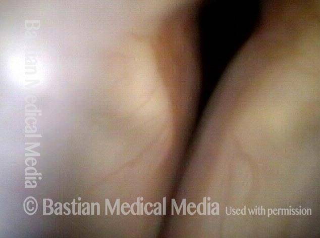
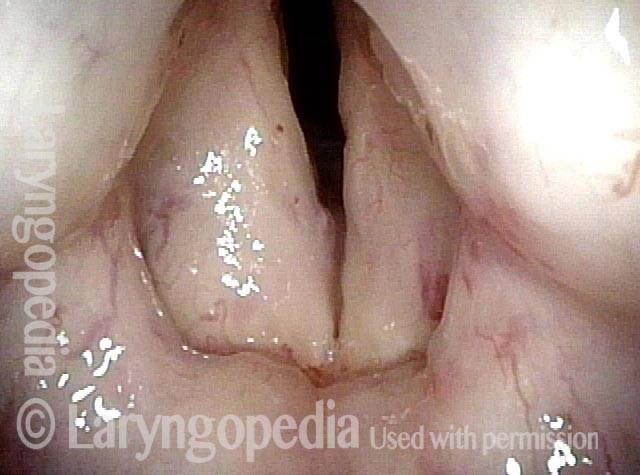
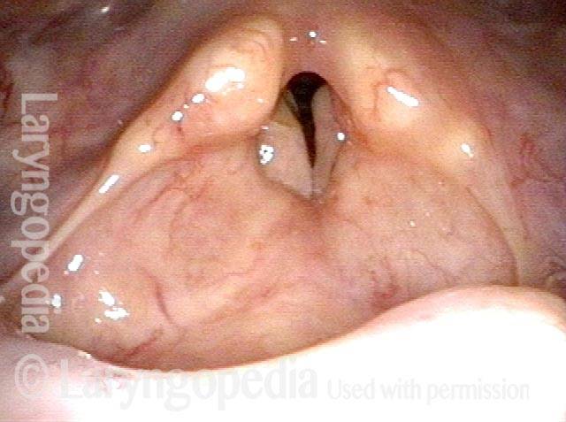
Laser posterior commissuroplasty (2 of 6)

The posterior right vocal cord is injected with lidocaine with epinephrine, in preparation for office laser posterior commissuroplasty. F = false vocal cord. T = true vocal cord, near its posterior end. The left vocal cord is injected similarly prior to the procedure that follows.

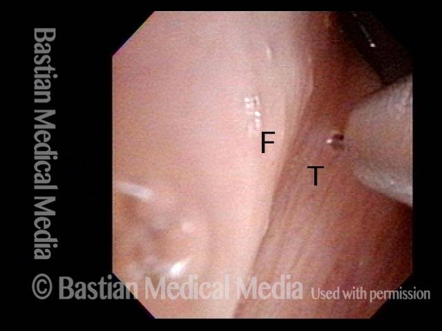
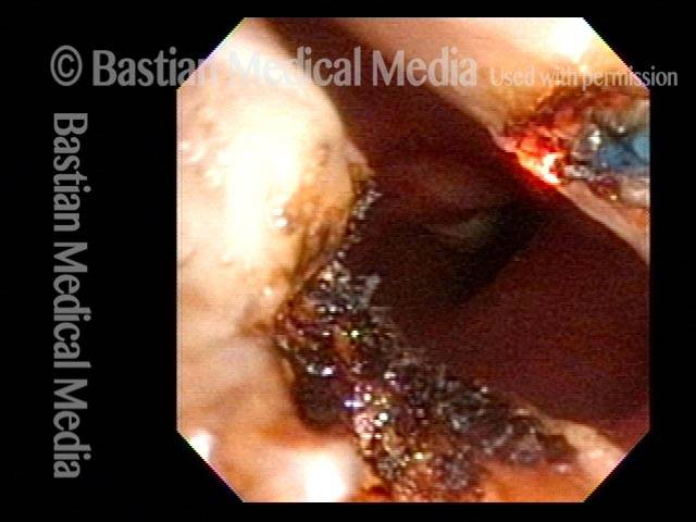
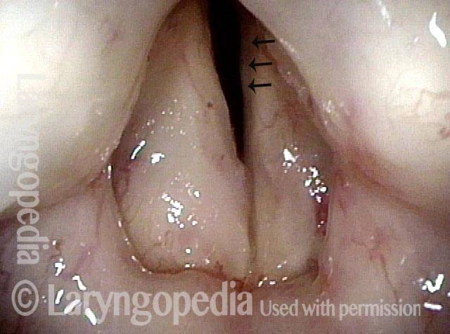
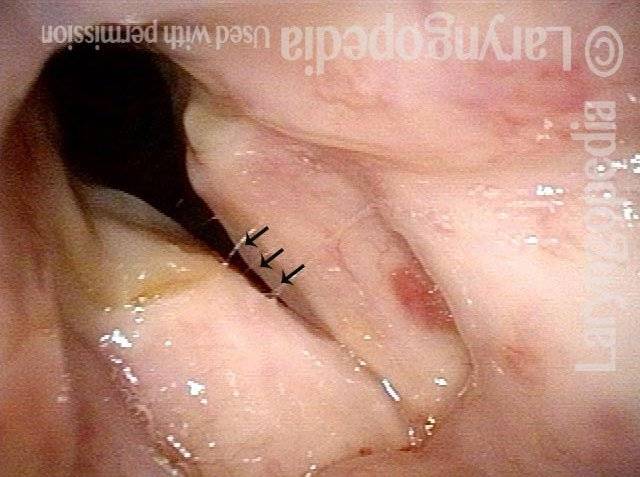
During the commissuroplasty (3 of 6)

The thulium laser fiber is being used to excavate the posterior commissure. Note the existing divot of the opposite (right) vocal cord (dotted lines) which will also be enlarged (next photos).

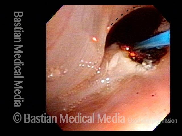
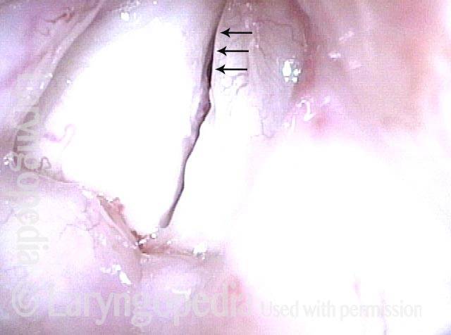
Deepening divot (4 of 6)

With view rotated clockwise approximately 45 degrees, work is commencing to deepen the right vocal cord divot.

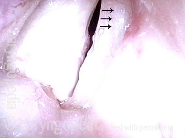
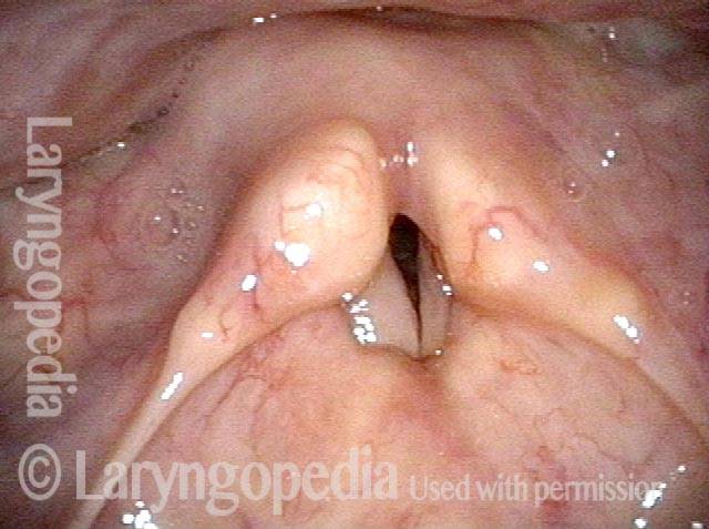
Inspiratory indrawing decreased (5 of 6)

At the conclusion of the procedure. Not only is the ‘keyhole’ seen in photo 1 larger, but inspiratory indrawing of the rest of the vocal cords is greatly diminished.

Phonation (6 of 6)

Now phonating, voice is similar to the beginning of the procedure, because the vibrating part of the vocal cord was not disturbed. Of course, number of words per breath is slightly lower, due to increased use of air through the keyhole—air wasting.

Sometimes you DO Remove Granulation to Avoid Tracheotomy

Visual Portfolio, Posts & Image Gallery for WordPress

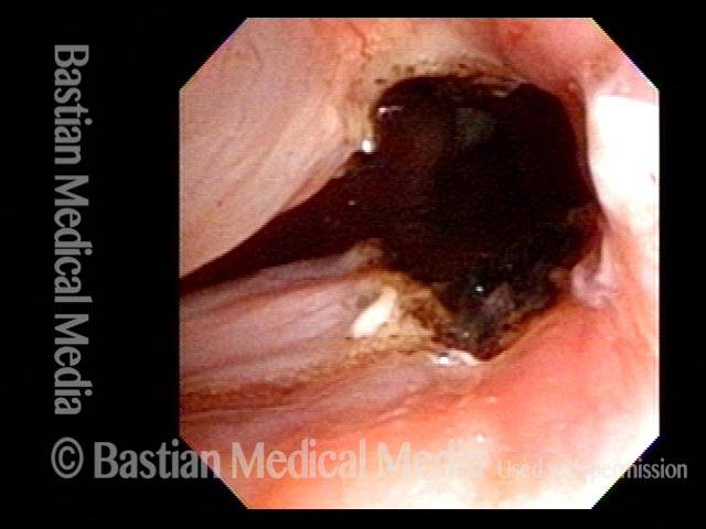
Granulation (1 of 8)

Prior to this first visit, this person suffered extensive burns, was intubated for 10 days, and then underwent tracheotomy, and then was decannulated (tracheotomy removed). She has scarring of the posterior commissure outlined by the dotted line. The granulation extends well down into the subglottis. She is uncomfortable with a marginal airway and noisy breathing. Laser and microdebrider are planned to try to avoid having to reinsert the tracheotomy.

Closer view (2 of 8)

Tip of the iceberg view of granulation and scarred area.

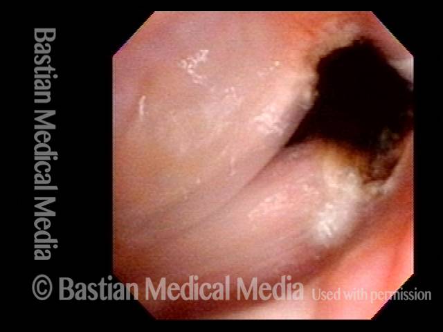
Post microlaryngoscopies (3 of 8)

After a series of microlaryngoscopies purely to improve airway and avoid tracheotomy, the granulation has finally matured. Airway is no longer marginal, but is still very limited for significant activity.

Scarring (4 of 8)

At close range, the area of posterior scarring is again indicated by dotted line; the dark area of the actual airway is narrow and slit-like.

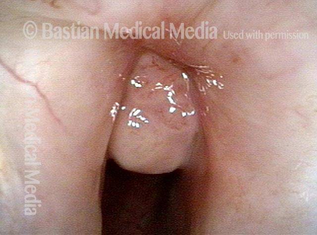
Post posterior commissuroplasty (5 of 8)

A month after posterior commissuroplasty, breathing is improved due to the widened space posteriorly. Compare the dark area for breathing with photo 3.

Breathing improved (6 of 8)

Six months after posterior commissuroplasty, breathing remains much improved. Compare dark airway contour again with photo 3 above.

Closer view (7 of 8)

A closer view of the airway, which is much wider posteriorly than preoperatively (photo 4).

Phonatory view (8 of 8)

When patient makes voice, there is a persistent space posteriorly, where the airway was surgically widened, but again, this has not significantly affected the voice.

Progressive Radiation Fibrosis Effects on the Larynx and a Solution to some of It

Visual Portfolio, Posts & Image Gallery for WordPress

Forty years post-radiation (1 of 8)

This photo is taken forty years after curative radiation for a vocal cord cancer. Four decades of progressive radiation fibrosis (“leatherization”) has taken away arytenoid movement so that this is the maximum opening. The patient is exercise-intolerant and makes loud inspiratory breathing noises while sleeping. Her voice is also very poor.

Involuntary inspiratory voice (2 of 8)

With sudden inspiration, the darker mucosa (at the arrows) indraws and vibrates, making an involuntary inspiratory voice.

Only capable of high pitch (3 of 8)

Other than a stage whisper, she can only make a very high pitch, because the only mucosa capable of vibration is the small segment indicated by the arrows.

Open phase vibration (4 of 8)

Again under strobe light, this is the open phase of vibration, with arrows again indicating the short segment of mucosa that can oscillate.

One week post-commissuroplasty (5 of 8)

A week after posterior commissuroplasty, the patient’s breathing is much improved. Despite the distant view, the “cookie bites” taken from the posterior cords are visible.

Rapid inhalation, closer view (6 of 8)

In a much closer view, the posterior vocal cord divots are seen well. The segment of flexible mucosa is indrawing here as the patient inhales rapidly (at arrows).

Three months post-surgery (7 of 8)

Three months after the laser surgery, the patient continues to say the improvement of breathing is “large.” In this distant view the full reason why is not seen.

Closer view, post-surgery (8 of 8)

In a closer view, as is always the case after complete healing, the divots are smaller than just after surgery.