Phonation is the process of making vocal sound by bringing vocal cords together while a stream of pulmonary air passes between them, causing them to vibrate. Roughly, this means to “make voice.”
Normal Larynx
Vocal cords (1 of 7)
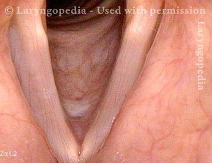
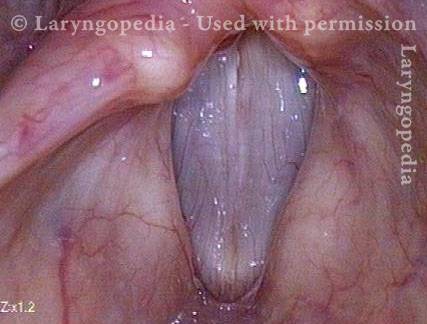
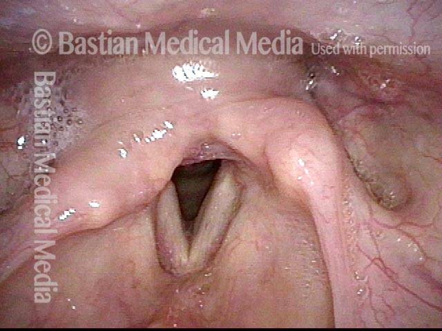
The line of sight is from directly above these open (for breathing) vocal cords, and downward in the line of the trachea. The difference in vocal cord width is not real, but a function of the angle of viewing. This is the position of the vocal cords while breathing.
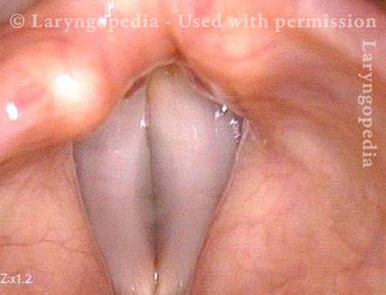
Vocal cords during voice (2 of 7)
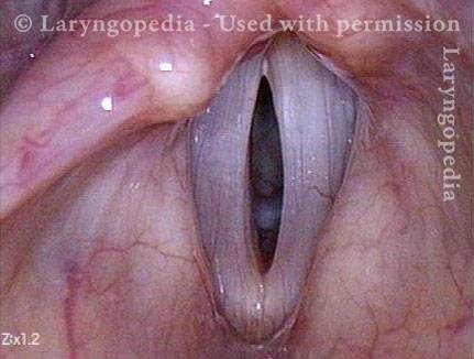
Here the vocal cords are producing sound at G2 (98Hz). Under standard light, vibration is so rapid that it is perceived as a blurring of the margins.
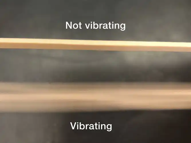
Vibration in rubber bands (3 of 7)
Twang a thick rubber band and you will see the same kind of blurring phenomenon because vibration is so rapid.
Phonation under strobe light (4 of 7)
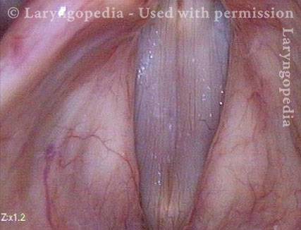
During voicing (phonation), under strobe light, to provide apparent slow motion view of vibration. This is the closed phase of vibration at A2 (110 Hz, or 110 vibration cycles per second).
Open phase of vibration (5 of 7)
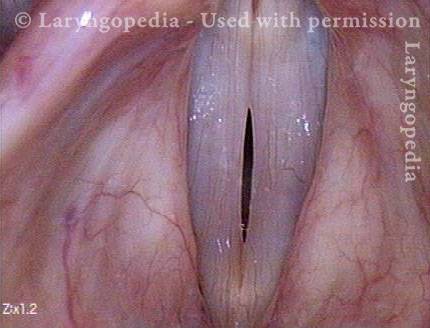
The open phase of vibration, also at A2. For emphasis: This cycling between open and closed phases of vibration occurs 110 times per second at A2!
Closed phase of vibration (6 of 7)
Now at A4 (440 Hz), this is one of 440 closed phases of vibration per second.
Open phase (7 of 7)
One of 440 open phases of vibration that occurs per second, again as seen under strobe light.
Phonatory Insufficiency
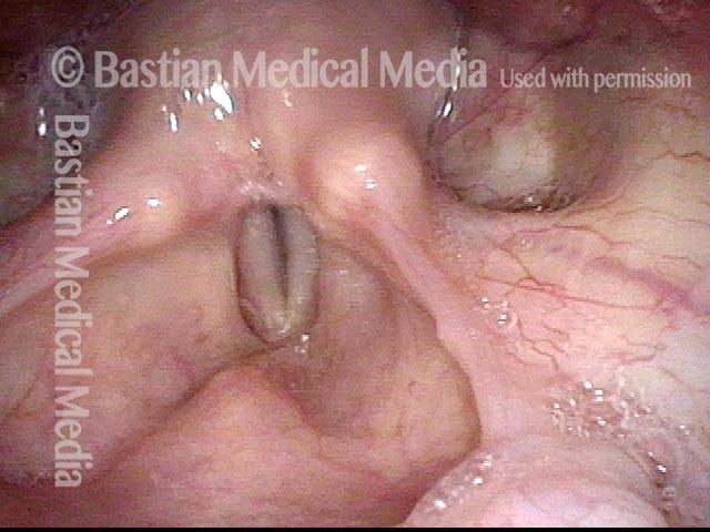
Phonatory insufficiency (1 of 4)
After 15 days of intubation, this voice is sounding both breathy (air-wasting) and pressed. From a distance it appears that the right cord (left of image) is paralyzed. (Compare with image 2)
Phonatory insufficiency (2 of 4)
During phonation, the voice again sounds breathy and pressed.
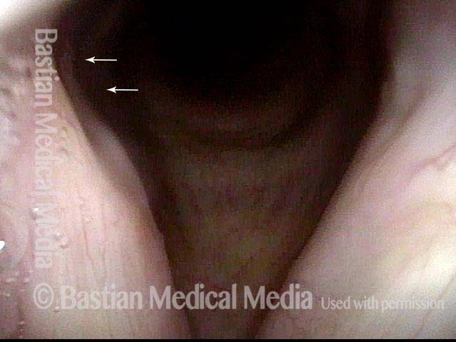
Phonatory insufficiency (3 of 4)
A close up view shows the posterior divot of the right cord (left of image). The absence of atrophy, bowing, or flaccidity, confirms that the problem is right cord fixation due to scarring of the right cricoarytenoid joint, not paralysis.
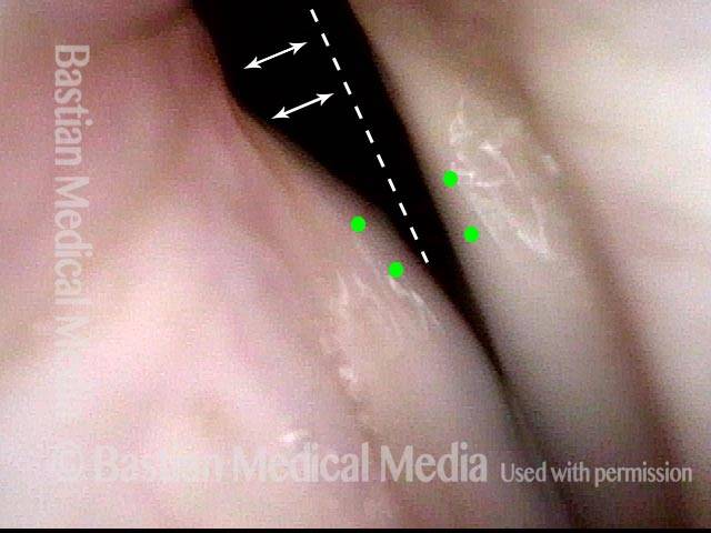
Phonatory insufficiency (4 of 4)
During phonation, the posterior commissure deficit caused by pressure necrosis from the endotracheal tube is seen with the dotted line. The small green circles represent the vocal processes not approximating thus validating the joint injury.