The epiglottis is the flexible, leaf-like cartilaginous structure that sits upright in the pharynx, between the base of the tongue and the larynx. The root or petiole of the epiglottis is inside the upper part of the thyroid cartilage just above the anterior insertion of the vocal cords. During swallowing, the epiglottis bends backward to cover the entrance of the larynx, helping to divert food into the esophagus.
Laryngeal Penetration
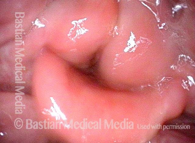
Laryngeal penetration (1 of 1)
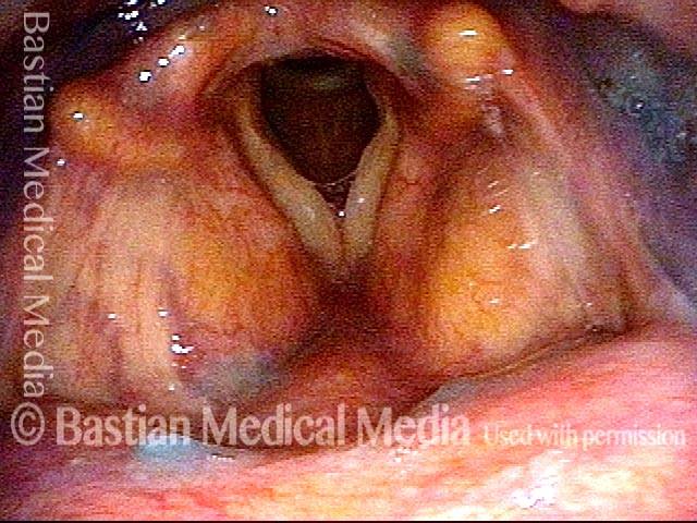
After the patient swallowed several boluses of blue-stained applesauce, there were traces visible on the laryngeal surface of the epiglottis, indicative of penetration into the earliest part of the airway. By itself, soiling of the laryngeal vestibule to this minor degree does not threaten the person’s ability to eat by mouth.
Another Voice without Vocal Cords
Hemilaryngectomy (1 of 4)
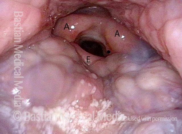
After removal of the anterior larynx (hemilaryngectomy) for cancer that recurred after radiation therapy. Though not well seen here, the vocal cords are surgically absent. The black dot seen is for orientation to the next photo. A = arytenoid; E = epiglottis.
Within the larynx (2 of 4)
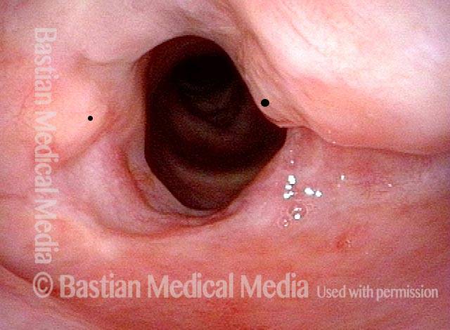
A view within the larynx. Note again that vocal cords are surgically absent, with only the arytenoid cartilages remaining at the level of the cords. The black dot, on the left arytenoid cartilage, orients to the prior photo. The dot is on the right vocal process.
"Wolfman Jack" voice (3 of 4)
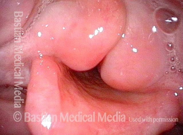
The patient is about to produce his rough, “Wolfman Jack” voice but the arytenoid mounds have not yet started to vibrate.
Arytenoid vibration (4 of 4)
Aggressive voice use brings arytenoid mounds into vibration (notice blurring). With time and practice, this kind of supraglottic voice can serve moderately well, but it is always difficult to be heard in competition with background noise.
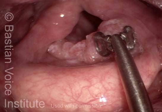
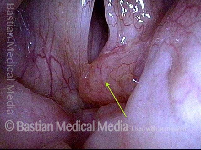
Biopsy
Biopsy, epiglottis (1 of 1)
Biopsy of lesion involving the petiole (low laryngeal surface of epiglottis). The pathology report revealed squamous cell carcinoma, usually caused by smoking.
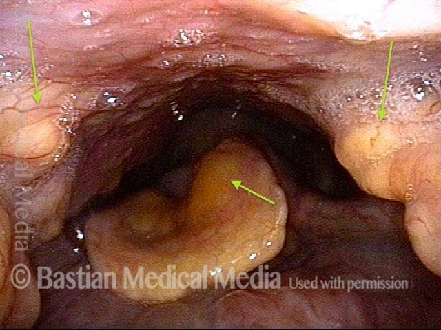
Amyloidosis, before and after Debulking
Amyloidosis, before debulking (1 of 4)
Panoramic view. Submucosal amyloid deposits in pharyngeal walls and epiglottis, with examples at arrows.
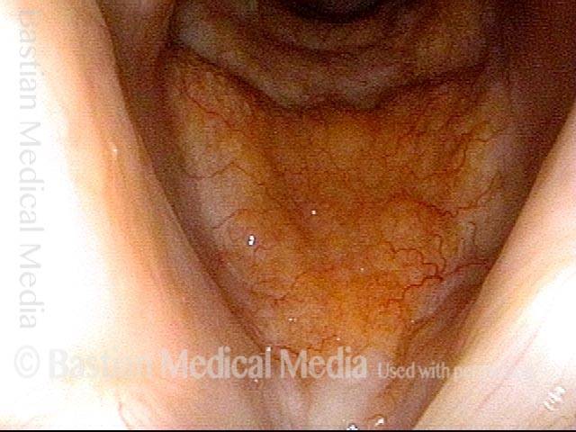
Amyloidosis, before debulking (2 of 4)
View of anterior subglottis shows diffuse infiltration of yellowish, submucosal amyloid.
Amyloidosis, before debulking (3 of 4)
Phonation under strobe light shows deposit at anterior commissure tethering upper surface of left vocal cord, and resultant vibratory asymmetry.
Amyloidosis, after debulking (4 of 4)
Some months later, after laser debulking at short arrow, showing improvement of vibratory closure after tethering reduced. Note increase of nearby supraglottic amyloid deposit does not need to be addressed because it has no functional consequence to the patient, who has other asymptomatic deposits in subglottis, epiglottis, and nasopharynx.
