An encyclopedia about voice, swallowing, airway, coughing, & other head + neck disorders.

Sfintere Esofageo Superiore

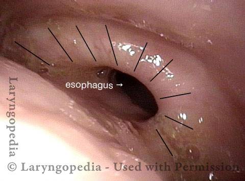
Lo sfintere esofageo superiore è un muscolo sfintere che circonda l’estremità superiore dell’esofago. È quasi sempre in uno stato contratto, anche durante il sonno. La sua azione è come un pugno continuamente chiuso. Questa contrazione chiude l’ingresso dell’esofago.

Ogni volta che una persona deglutisce, il muscolo cricofaringeo si rilassa momentaneamente, aprendo la presa e consentendo al cibo o al liquido di passare ed entrare nell’esofago.

Tipi di disturbi

Il muscolo è soggetto a due tipi di disturbi. La disfunzione cricofaringea è l’incapacità del muscolo di rilassarsi, che causa difficoltà di deglutizione. Lo spasmo cricofaringeo è un’ipercontrazione del muscolo, che provoca la sensazione di un nodo alla gola ma senza interferire con la deglutizione.

Lo sfintere esofageo superiore

Visual Portfolio, Posts & Image Gallery for WordPress

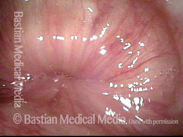
Cricopharyngeus muscle (1 of 4)

Close-up view of the cricopharyngeus muscle. When contracted like this, the muscle squeezes shut the entrance to the esophagus. When a person swallows, the muscle relaxes, and the entrance to the esophagus opens dramatically to let food or liquid enter.

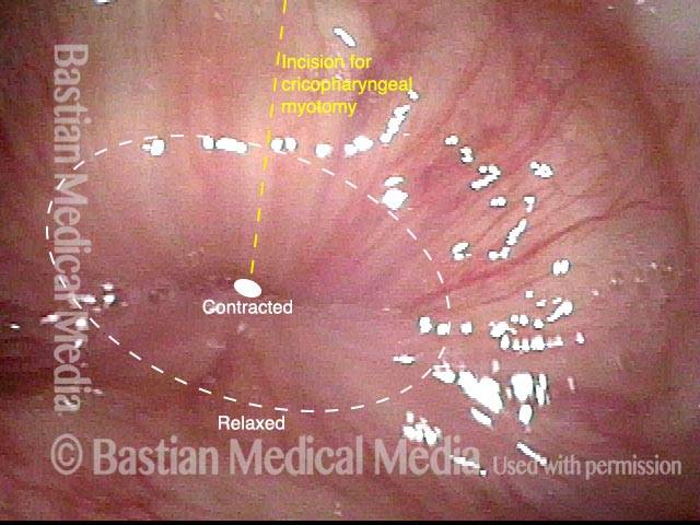
Opening when Contracted (2 of 4)

Same image as photo 1, but a small solid oval marks the center-point of the muscle’s grip, when contracted, and a larger dotted oval shows roughly how large the opening becomes when the muscle relaxes. Also, a yellow dotted line indicates the line of incision used when performing a cricopharyngeal myotomy (not needed in this patient).

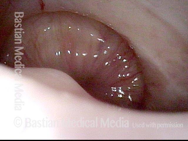
A distant view (3 of 4)

More distant view of the cricopharyngeus muscle, as the camera has retracted upward and is now sitting in the postcricoid part of the hypopharynx, looking downward.

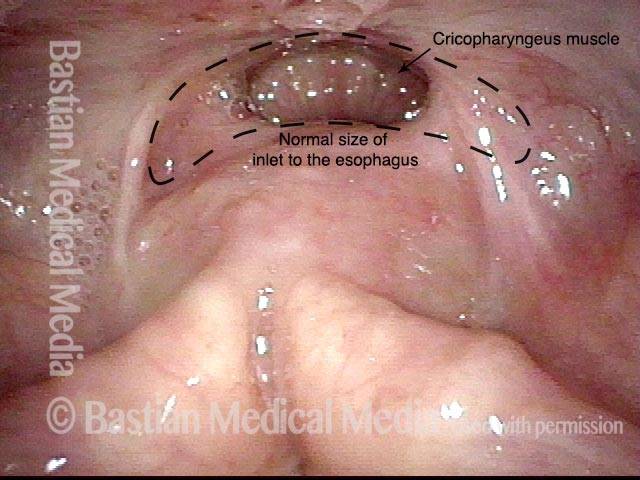
hypopharyngeal inlet (4 of 4)

Much more distant view yet of the cricopharyngeus muscle, now framed within the hypopharyngeal inlet to the esophagus, at the top-center of the photo. Also now in view are the arytenoid mounds, at the bottom of the photo. Incidentally, this patient’s hypopharyngeal inlet to the esophagus is abnormally narrowed, due to post-surgery scarring; black dotted lines indicates the normal size of this inlet.

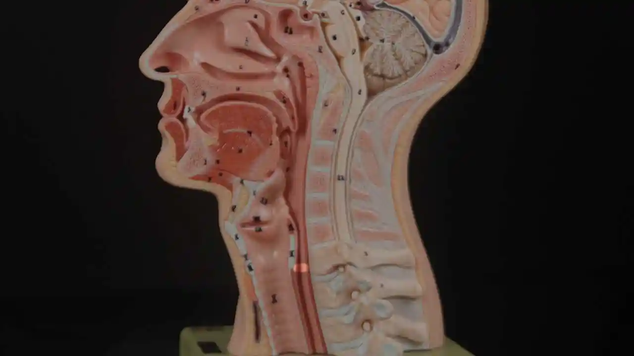
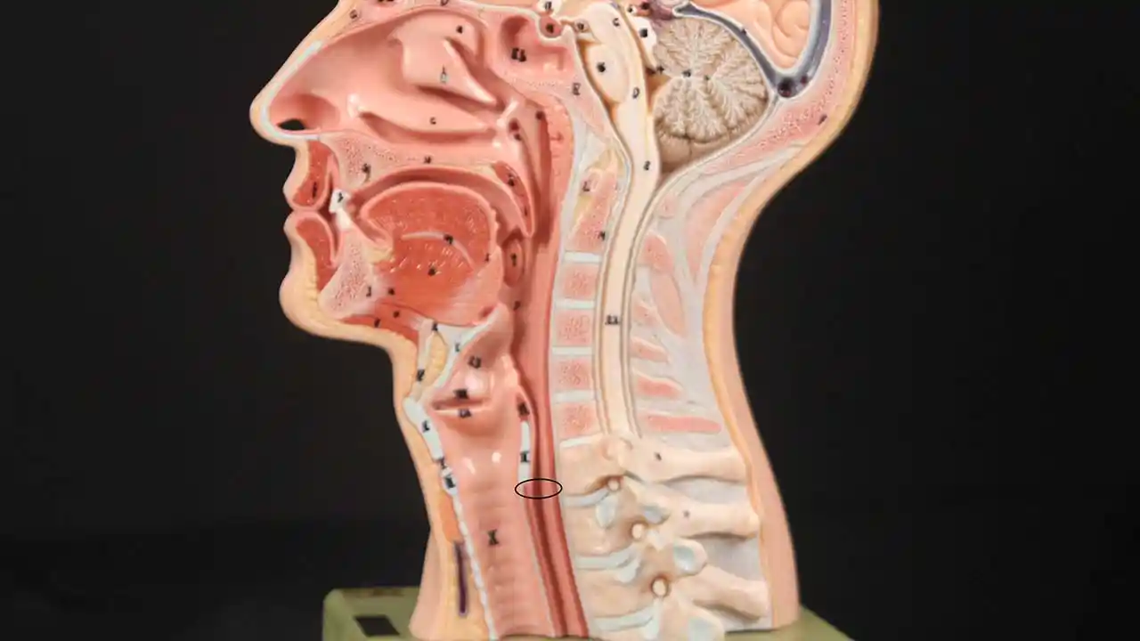
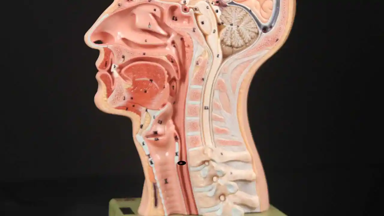
Posizione dello sfintere esofageo superiore

Visual Portfolio, Posts & Image Gallery for WordPress

Cricopharyngeus Muscle (1 of 3)

The highlighted oval represents the location of the cricopharyngeus muscle.

Open Cricopharyngeus Muscle (2 of 3)

The cricopharyngeus muscle in the open position.

Closed (3 of 3)

The cricopharyngeus muscle in the contracted position.

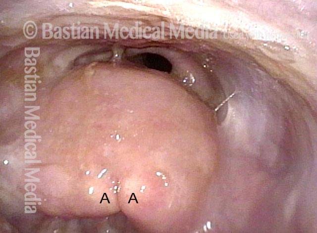
Manovra a tromba Rivela lo sfintere esofageo superiore

Visual Portfolio, Posts & Image Gallery for WordPress

Trumpet maneuver (1 of 4)

This man is performing a trumpet maneuver in order to open the post-arytenoid and post-cricoid area of the hypopharynx. He is able to reveal the opening to the esophagus (dark oval). A = arytenoid mound apices.

The “shelf” (2 of 4)

At closer range, the “shelf” of the cricopharyngeus muscle is seen posteriorly (upper photo). The radial lines on the slightly relaxed / open muscle should be compared with photo 4, where the sphincter is completely closed.

Cervical esophagus (3 of 4)

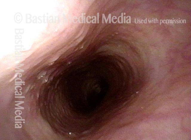
After passing through the sphincter, a view of the open cervical esophagus.

Closed position (4 of 4)

Back again above the sphincter, now in closed (contracted) position.

Sfintere esofageo superiore osservato durante la deglutizione

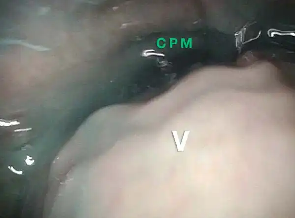
Questa persona ha difficoltà a deglutire a causa di una combinazione di precedenti interventi chirurgici per il cancro della lingua decenni fa e degli effetti delle radiazioni a lungo termine. I cibi solidi sono i più problematici, quindi questa sequenza mostra un tentativo di ingoiare acqua macchiata di colorante alimentare blu.

Visual Portfolio, Posts & Image Gallery for WordPress

Swallowing crescent (1 of 5)

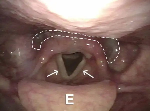
Panoramic view of the hypopharynx and larynx during breathing. E = epiglottis. Arrows point to the open vocal cords with triangular entrance to airway between them. Dotted line indicates the “swallowing crescent” including part of pyriform sinuses and post-arytenoid space.

Swallowing water (2 of 5)

Blue-stained water is collected in the “swallowing crescent” and about to enter the upper esophagus. The larynx is closed anteriorly (lower photo). The V marks the same post-arytenoid location in subsequent photos.

Cricopharyngeus muscle (3 of 5)

The larynx is coming forward, opening the way into the esophagus. The cricopharyngeus muscle is seen in the distance, marked with CPM.

Relaxed CPM (4 of 5)

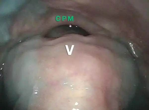
With the blue water now passed into the esophagus, the partially relaxed sphincter is seen more clearly, again marked CPM.

Partially open esophagus due to A-CPD (5 of 5)

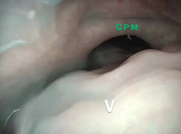
A closer view, showing only partially open esophageal entrance because of antegrade cricopharyngeus muscle dysfunction: it refuses to relax fully and this is part of the explanation for why this person cannot swallow solid foods through a less-than-half-opened sphincter.

Come funziona uno sfintere

Visual Portfolio, Posts & Image Gallery for WordPress

Open (1 of 2)

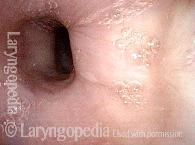
After total laryngectomy, the upper esophageal sphincter is open here for a brief moment, allowing one to see beyond into the esophagus.

Contracted (2 of 2)

A moment later, the sphincter contracts, as though the purse string of a velvet jewelry bag had been tightened.

Disfunzione muscolare cricofaringea anterograda, prima, durante e dopo la miotomia

Visual Portfolio, Posts & Image Gallery for WordPress

Non-relaxing cricopharyngeus muscle (1 of 4)

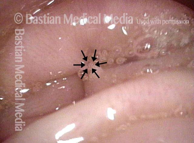
View of hypertrophied, non-relaxing cricopharyngeus muscle, and resultant narrowed entry into the esophagus, at arrows.

Opening the esophageal orifice (2 of 4)

Suction cannula is being used to forcefully stretch open the esophageal orifice, by pressing posteriorly against the cricopharyngeus muscle.

Laser cricopharyngeus myotomy (3 of 4)

Laser cricopharyngeus myotomy in progress. Notice the horizontally-directed fibers of the cricopharyngeus muscle which are being transected. Red dot is aiming beam of the laser.

Cricopharyngeus myotomy nearly complete (4 of 4)
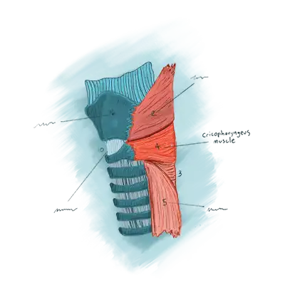
Only a few fibers remain at arrow. Notice widely patent opening to esophagus resulting from myotomy.
Illustration of the cricopharyngeus muscle

Condividi questo articolo
Videoconsulto con
il dottor Bastian
Hai ancora domande? Pianifica una chiamata con il Dr. Bastian.

Sommario

Subscribe
Notify of

0 Comments
Newest
Oldest Most Voted
Inline Feedbacks
View all comments
0
Click to see all comments.x