An encyclopedia about voice, swallowing, airway, coughing, & other head + neck disorders.

Marfan Syndrome is genetic connective tissue disorder caused by a defect in gene FBN1, which codes for abnormal structure of fibrillin-1, a protein crucial for formation of normal connective tissue. Most critical is Marfan syndrome’s effect on heart and blood vessels, which tend to dilate and be at risk of rupture. Connective tissue in bones, ligaments, and other parts of the body is also affected.

Laryngologists may encounter Marfan syndrome because parts or all of the aorta may need to be replaced over time, due to abnormal dilation of the weakened arterial wall, with risk of rupture. When such surgery is done, the left recurrent nerve is at risk of injury, and this would lead to left vocal cord paralysis. With Marfan syndrome, it is rare to live to age 70.
Visual Portfolio, Posts & Image Gallery for WordPress

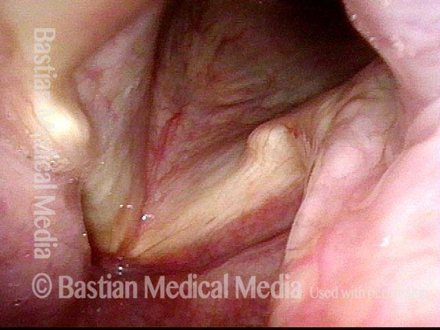
Marfan syndrome (1 of 5)

70-year-old patient whose left vocal cord (right of photo) was implanted 15 years ago with a silastic wedge to rehabilitate paralysis. Voice was reported as "normal" for many years, but in recent months has deteriorated.

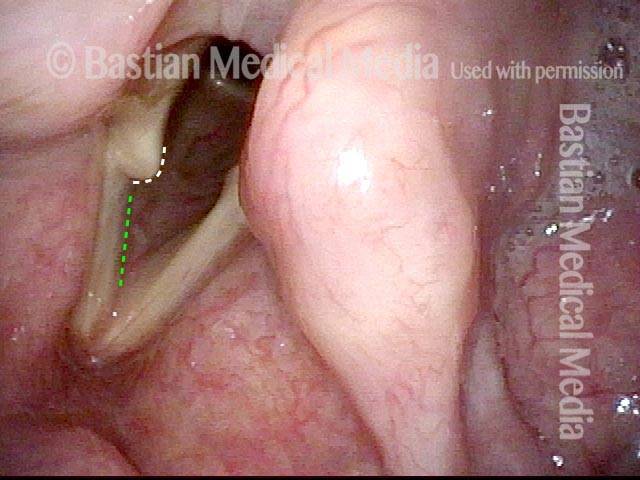
Marfan syndrome (2 of 5)

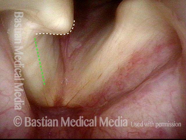
As the right vocal cord (left of photo) moves toward the midline, the arytenoid cartilage (white dotted line) appears to emerge from the right vocal cord without bringing the membranous segment (green dotted line) with it.

Arytenoid cartilage (3 of 5)

At close range, one can see more clearly that the connective tissue attachment between the arytenoid cartilage and soft tissue of the vocal cord itself has been completely lost. As in photo 2 the white dotted line marks the arytenoid cartilage and the green dotted line marks the vocal cord's membranous segment.

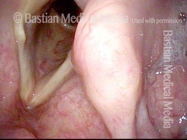
Marfan syndrome (4 of 5)

A different view, showing again the vocal process of the arytenoid cartilage detaching from the vocal ligament/muscle of the membranous portion of the vocal cord.

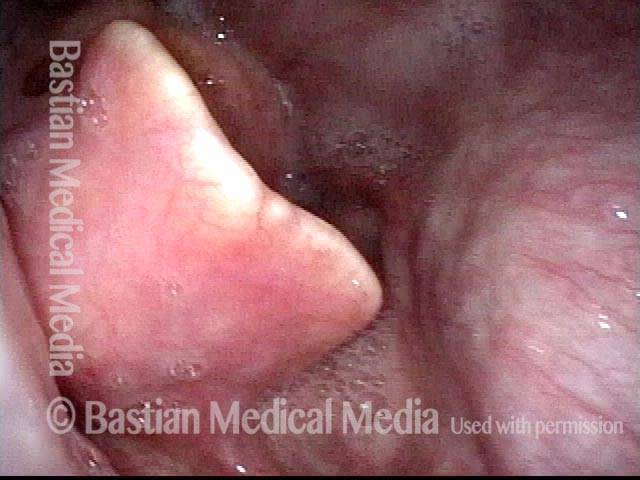
Aneurysmal dilation (5 of 5)

Panoramic view of the hypopharynx, showing aneurysmal dilation of the internal carotid artery (bulge on right side of photo). Epiglottis is at the center and left.

Tags