An encyclopedia about voice, swallowing, airway, coughing, & other head + neck disorders.

R-CPD Esophageal Findings

The details of the following photos may support the R-CPD diagnosis, though they should not be considered diagnostic.

Take note that all photos are non-channel scope images; that is, the scope is not able to insufflate (blow in) air. 

The significance

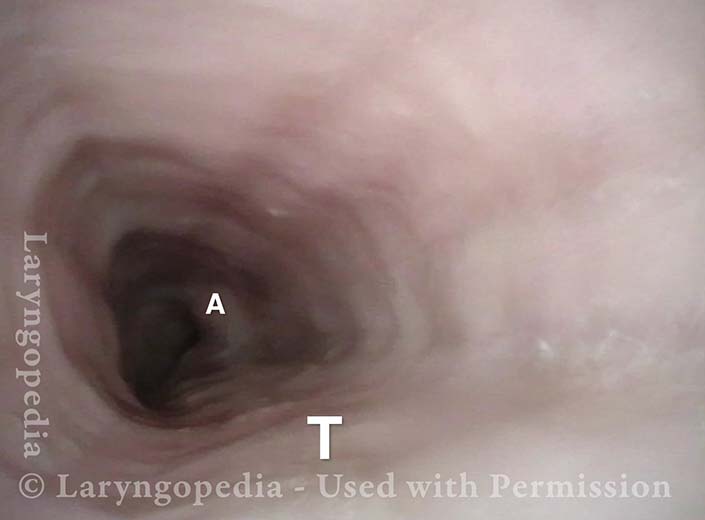
The esophagus is typically collapsed around endoscopes inserted into them and air is pumped in through a tiny channel in order to gently expand the esophagus so that its walls can be seen. Here, the air the patient cannot belch/evacuate is doing that work for us. And the esophagus remains open for extended time, the full duration of the examination.

Four findings are being evaluated and compared with normal esophagoscopy images (also without insufflated air to make the comparison valid):

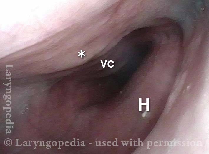
  1. Reflux from the lower esophagus, suggesting damage to the lower esophageal sphincter from constant upward pressure trying unsuccessfully to belch.
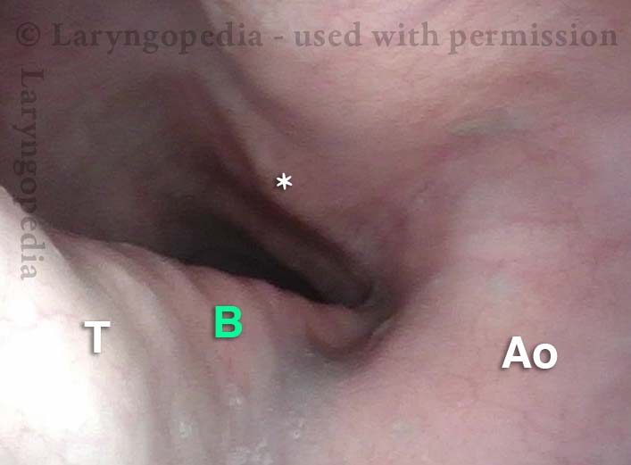
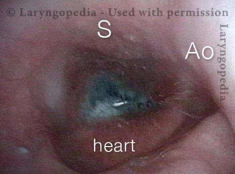
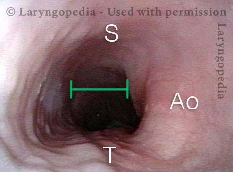
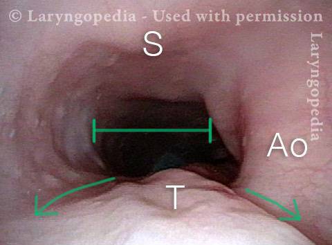
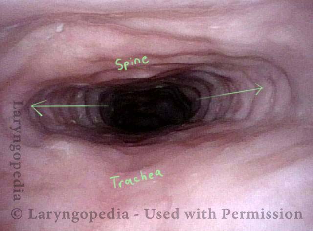
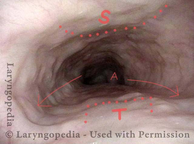
  2. What we call an “aortic shelf,” meaning that rather than an indentation of the medial circumference of the aorta, dilation of the esophagus drapes its mucosa across the upper surface of the esophagus, making a “horizontal shelf.” Keep in mind again that this is without insufflating any air.
  3. Continuous patency with very infrequent, partial “clamping” down of the lumen or, often no closure at all, suggesting that there is sustained opening pressure of unbelchable air and/or that the contractile ability of the esophagus is reduced, in similar fashion to what happens to an overly-distended urinary bladder.
  4. Upper esophageal dilation in a medial-lateral axis so that the upper esophagus becomes stretched in an exaggerated “oval” rather than a more gentle oval or even “circle.”

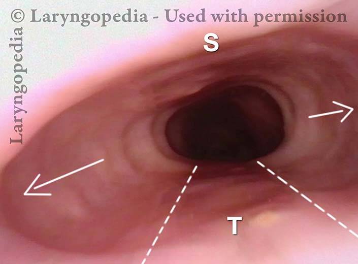
Dramatic lateral stretch of the esophagus

Swallowed air (a fraction of every human swallow) must either be burped, absorbed, or (after some time) passed as flatulence. In a person with retrograde cricopharyngeus dysfunction (R-CPD: defined syndromically as inability to belch, gurgling, bloating, flatulence, etc.) the esophagus will eventually dilate.

This esophageal stretching can hurt, especially during hiccups. And the esophageal wall muscle thins out and its ability to contract weakens. The lower esophageal sphincter (LES) can also fail, leading to reflux of stomach acid from stomach up into the esophagus. Standard manometry typically describes low esophageal and LES pressures and slow transit. These findings are not the diagnosis, but instead are findings that result from the fundamental diagnosis: R-CPD.

Visual Portfolio, Posts & Image Gallery for WordPress

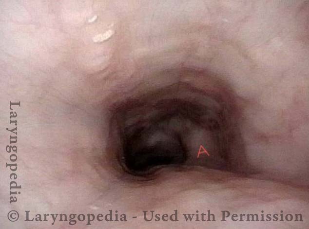
Normal Esophageal View (1 of 3)

Expected view inside the a “general population” esophagus, when using an ENT scope. Such as scope is normally ineffective for esophagoscopy. That is because the diameter of the scope is tiny (3.7mm) and it does not have a channel to insufflate air (blow it out the tip of the scope from a compressor, to expand the esophagus). Consequently, the walls of the esophagus are collapsed and can’t be seen clearly. The arrow points to the collapsed lumen.

Reflux in stretched esophagus (2 of 3)

In this R-CPD patient, an ENT scope works extremely well to visualize the esophagus, because of the retained air the individual cannot burp out. That un-burped air keeps the esophagus in an “always open” state, and so the ENT scope can see the walls well. They are smooth/unwrinkled, denoting full expansion and even “stretch.” In this view of the lower esophagus, H = the posterior surface of the beating heart. W= blue water the patient has swallowed, pooled in the lower esophagus and occasionally refluxing upward.

Stretched Esophagus (3 of 3)

This view is in the mid-esophagus. The tracheal “mound” is indicated by the dotted lines and “T.” Neither trachea nor spine (“S”) is compressible. Thus, the un-burped air under pressure bulges the esophagus laterally. The esophagus of this patient is estimated to have stretched laterally (arrows) to 3 or 4 times the diameter of the trachea!

Dramatic esophageal dilation from R-CPD, even in a young person!

The central problem for persons with R-CPD is (obviously) the inability to burp. Bits of air travel through the upper esophageal sphincter (cricopharyngeus muscle) which closes behind each swallow.

Even as the air builds up, expanding the esophagus and sending increasing signals of the need to burp, the air is unable to “get back out” via burping.  Initial discomfort from stuck burps-in-waiting may elicit reflexive “extra” swallows that only add more air that cannot be burped.

Gradually this dilates the esophagus (and eventually the stomach, and as air traverses the GI tract, the intestines all the way to the rectum, where it must be released as flatulence). Even in young people, esophageal dilation can be dramatic, causing chest discomfort.

The images below are from the office examination of a teenager sitting in the examination chair with a small ENT scope in his esophagus. The column of air he cannot burp up is holding the esophagus widely open. No air has been insufflated through the scope. The stretched esophagus throws structures outside it into high relief as seen below.

Visual Portfolio, Posts & Image Gallery for WordPress

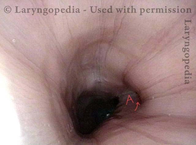
Stretched esophagus (1 of 3)

In this view, the lower third of the esophagus is in the “always open” position seen routinely in R-CPD, due to his unburped column of air. His heartbeat can be seen from its posterior wall (H). Unusually well seen is the ridged vertebral column (VC).

Mid-esophagus (2 of 3)

In the mid-esophagus, at the lower end of the trachea (T), the segmented left mainstem bronchus (B) turns laterally to pass under the Aorta (Ao).

Stretching esophagus (3 of 3)

Again at the lower trachea, but with the view rotated to the patient’s left, note that since the trachea (T) is not compressible, the esophagus tries to stretch around it, bringing even the side of the trachea into view.

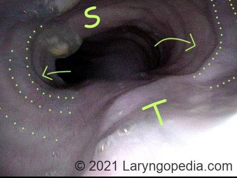
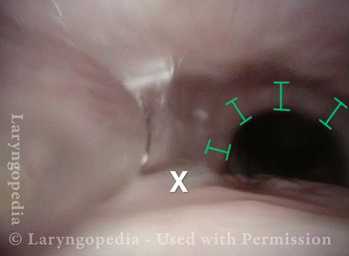
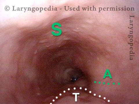
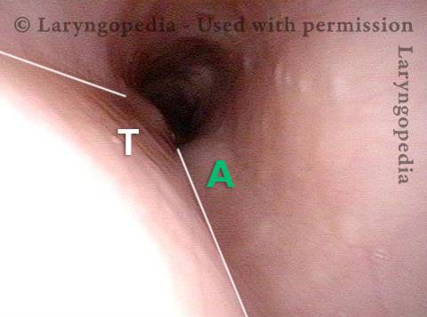
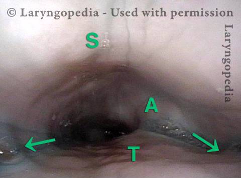
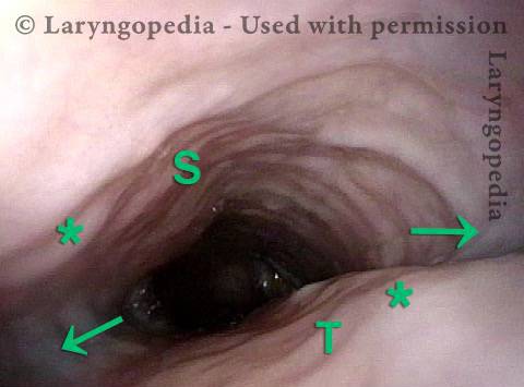
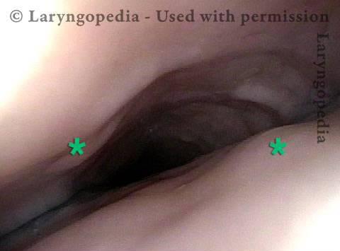
Airway, Foodway, Cricopharyngeus Muscle, and Esophagus in R-CPD

This young woman has never been able to burp normally. She experiences typical, severe R-CPD symptoms of daily bloating, loud gurgling noises, and excessive flatulence.  Botulinum toxin injection of the cricopharyngeus muscle is planned to rid her of the syndrome of R-CPD in the short term, and to teach her how to burp “permanently” (even after the botulinum toxin has worn off).

Visual Portfolio, Posts & Image Gallery for WordPress

Laryngopharynx (1 of 5)

Panorama of the laryngopharynx shows the collection “funnel” of the airway (circumscribed by dotted lines), directing air between the cords and into the trachea below. The “foodway” collection crescent, aka “swallowing crescent” is circumscribed by the dashed lines. The foodway is closed because the airway is open. When swallowing rather than breathing, the esophagus would open at the arrow.

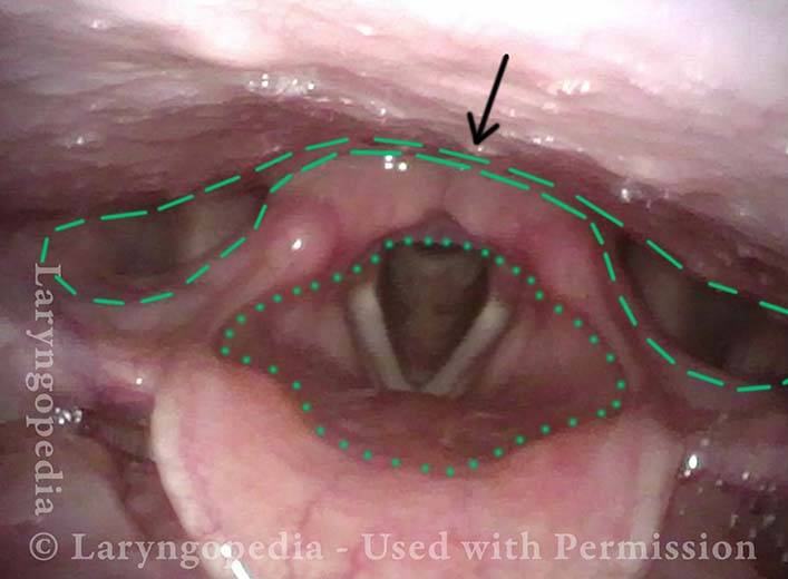
Esophageal opening (2 of 5)

Closer view shows where the esophageal opening will be in subsequent photos. “X” is marked for reference with the next photo.

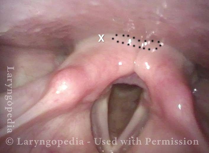
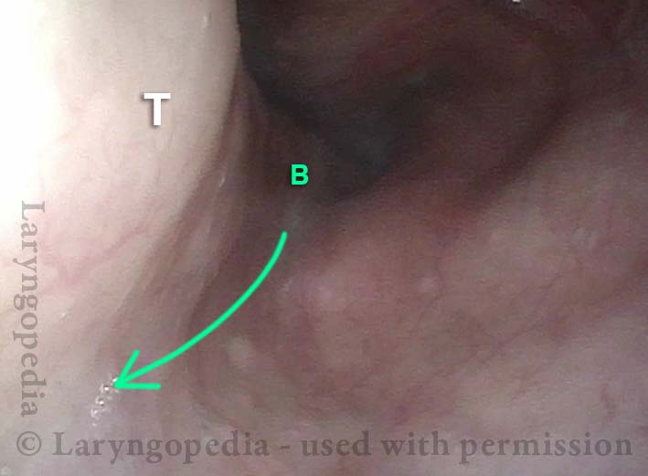
Ridge of UES (3 of 5)

The foodway (essentially esophagus) is opening here for a swallow of air. “X” is in approximately the same location as in the prior image. The cricopharyngeus muscle (UES) is mostly relaxed, but its ridge is still visible at the asterisks (*).

Ridge of UES (4 of 5)

The entrance to the esophagus is now fully open with rounder, less oval opening. The cricopharyngeus muscle is even more relaxed. The esophagus below is in darkness.

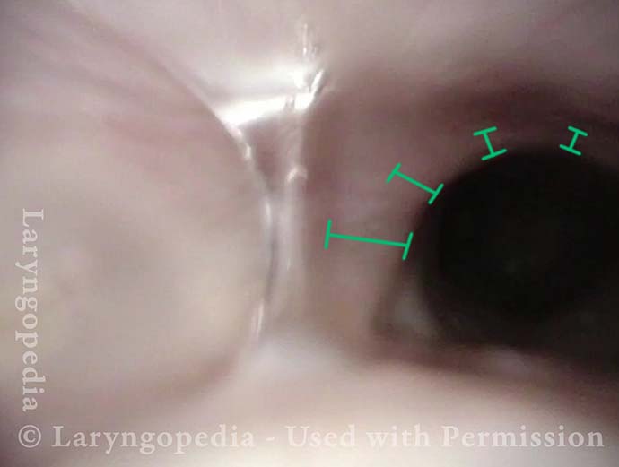
Distended Esophagus (5 of 5)

Now just beyond (below) the cricopharyngeus muscle, and in the upper esophagus. Since the individual is unable to belch, trapped air keeps the esophagus “always open.” The aortic “shelf” is in the distance (at A). “T” marks the tracheal mound. Though not shown here, the esophagus remains open all the way down to the gastroesophageal junction.

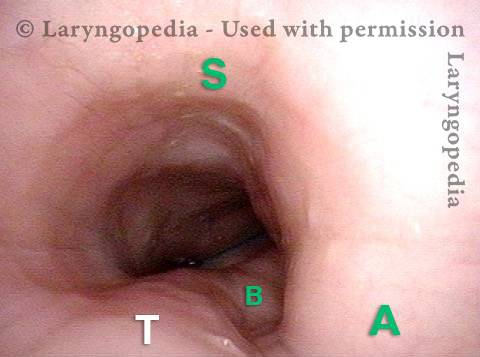
Esophageal Stretching by Unburpable Air in R-CPD

This young man has had the classic syndrome of R-CPD lifelong. His esophageal findings at the end of a videoendoscopic swallow study are classic. The esophagus is mostly a collapsed muscular tube in young people, yet his esophagus is widely open on a continuous basis due swallowed air that he cannot burp up.

Visual Portfolio, Posts & Image Gallery for WordPress

Typical view of an esophagus (1 of 4)

Typical view of the esophagus using an ENT scope without insufflated air. Esophageal walls are collapsed and therefore too close to the lens of the scope to allow visualization of the esophageal wall.

Lower esophagus (2 of 4)

Using ENT (not GI) scope in this young man with R-CPD to view the lower esophagus, which is continuously “open” due to un-burped air. H = heart; Ao = aorta. Blue water previously administered, moves up and down between stomach and lower esophagus due to lower esophageal sphincter incompetence from years of bloating.

Stretched mid-esophagus (3 of 4)

A view in the mid esophagus: Ao = aorta; S = spine; T = trachea. The esophagus remains open continuously due to unburped air. Insufflated air is routinely not necessary in persons with R-CPD.

Esophagus stretches laterally (4 of 4)

At a moment of upward surge of air that cannot escape the upper esophageal sphincter as a burp, the esophagus stretches laterally, almost trying to get around (arrows) the non-compressible trachea (T), accentuating the “tracheal mound.” Note as well the widened lumen as compared with photo 3.

More Interesting Esophageal Findings of R-CPD (Inability to Burp)

Visual Portfolio, Posts & Image Gallery for WordPress

Stretched Esophagus (1 of 4)

Using a 3.7mm ENT scope with no insufflated air, note the marked dilation of the esophagus by swallowed air the patient is unable to belch. T = trachea; A = aortic shelf; S = spine

Tracheal Wall (2 of 4)

The posterior wall of the trachea (T) is better seen here from a little higher in the esophagus. A = aorta

Over-dilation (3 of 4)

The photo is rotated clockwise at a moment when air from below is pushed upward so as to transiently over-dilate the esophagus. Note that the esophagus is almost stretching around the left side of the trachea in the direction of the arrow.

Bronchus is visible (4 of 4)

Now deeper in the esophagus (with it inflated throughout the entire examination by the patient’s own air), it even appears that the left mainstem bronchus (B) is made visible by esophageal dilation stretching around it.

The Esophagus Doesn’t Like Being Stretched for Years Due to Untreated R-CPD

Visual Portfolio, Posts & Image Gallery for WordPress

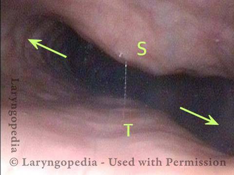
Lateral dilation from R-CPD (1 of 3)

In this middle-aged patient with R-CPD (inability to burp), now fully resolved (burping well for more than a year) after botox therapy. This view is pre-treatment, at mid-esophagus using an ENT scope. No air was insufflated to get this photo; the patient “has her own.” The aortic shelf is prominent, but observe the dramatic lateral dilation (arrows). S = spine; T = trachea.

Lateral dilation in the upper esophagus (2 of 3)

Now in the upper esophagus, arrows again depict the remarkable lateral dilation.

Medial-lateral stretch (3 of 3)

Opening of the esophagus is constant, due to the patient’s retained air, but as air goes downward transiently, the lumen size is reduced, almost accentuating the medial-lateral “stretch” of the esophagus. * denotes the same place in photos 2 and 3, for reference.

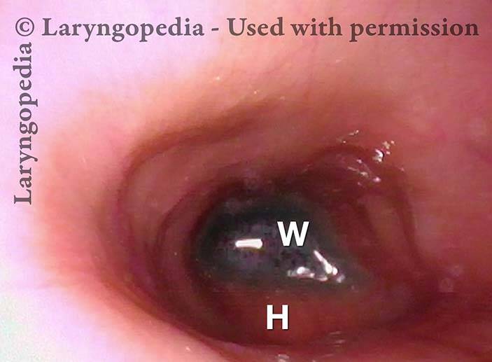
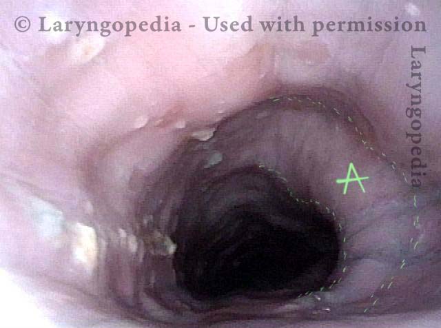
Emerging Esophageal Findings

Visual Portfolio, Posts & Image Gallery for WordPress

Aortic shelf (1 of 3)

A view of the mid-esophagus in a young person (early 30’s). The esophagus is kept open by the patient’s un-burped air. Note the “aortic shelf” at A, delineated by dotted lines.

Bony spur emerges due to stretched esophagus (2 of 3)

A moment later, additional air is pushed upwards from the stomach to dilate the mid-esophagus even more. A bony “spur” in the spine is thrown into high relief by the stretched esophagus.

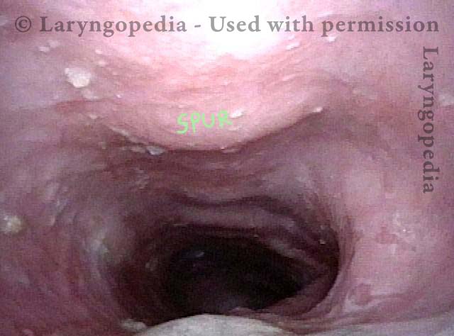
Stretched esophagus due to unburpable air (3 of 3)
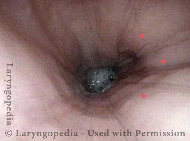
A view of the upper esophagus (from just below the cricopharyngeus muscle sphincter) shows what appears to be remarkable lateral dilation (arrows) caused over time by the patient’s unburpable air. Dilation can only occur laterally due to confinement of the esophagus by trachea (anteriorly) and spine (posteriorly), as marked.

Example 2

Visual Portfolio, Posts & Image Gallery for WordPress

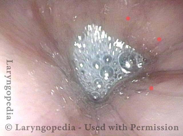
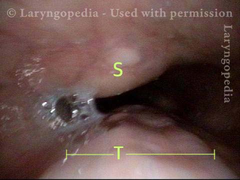
Saliva bubbles (1 of 5)

Saliva bubbles in the lower esophagus. Note that no air has been insufflated to obtain this view.

Symptoms point to GERD (2 of 5)

Approximately 0.5 second later, saliva wells up from below while the esophagus itself remains unchanged (Red dots in photos one and two mark identical reference points). Either abdominal wall or stomach wall compression lifts a column of stomach contents superiorly (retrograde) in the esophagus, explaining significant rates of GERD symptoms in this group, especially after treatment, which allows air to evacuate and might potentiate a higher rise of the material in the stomach/ lower esophagus.

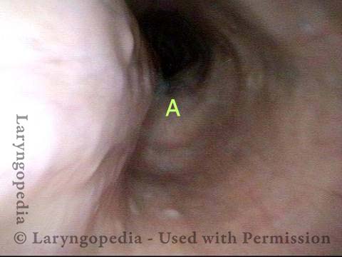
Pocket in the aorta (3 of 5)

Seen here is what almost appears like a pocket (arrow) at the upper surface of the aorta (A). Compare with the next photo.

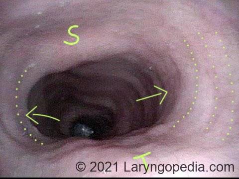
Trapped air expands the esophagus (4 of 5)

The patient’s unburped air further expands the esophagus and makes more of a “shelf” of the upper surface of the aorta (A).

Exaggerated laterally-stretched esophagus (5 of 5)

Now visualizing the upper esophagus, just below the cricopharyngeus muscle (UES): It appears that there is exaggerated lateral “stretch” of the esophagus. It cannot stretch posteriorly due to immovable spine (S) nor can it expand anteriorly due to the non-collapsible trachea (T). Consequently, it can only dilate laterally (arrows).

Dramatic Lateral Dilation of the Upper Esophagus

Visual Portfolio, Posts & Image Gallery for WordPress

Remarkable lateral dilation (1 of 3)

This photo is at the level of (estimated) C6 of the spine (at S). This person has known cervical arthritis, accounting for the prominence. Opposite the spine is the trachea (T). Note the remarkable lateral dilation (arrows) in this picture obtained with with no insufflated air using a 3.6mm ENF-VQ scope. It is the patient’s own air keeping the esophagus open for viewing.

Dilated upper esophagus (2 of 3)

At a moment when air from below further dilates the upper esophagus, the tracheal outline is particularly well-seen (T) opposite the spine (S). The “width” of the trachea indicated further emphasizes the degree of lateral dilation, which is necessary because spine and trachea resist anteroposterior dilation.

Aortic shelf (3 of 3)

Just for interest, at mid-esophagus, the familiar aortic “shelf” is seen. Again, this esophagus is being viewed with a 3.6 mm scope with only the patient own (un-burped) air allowing this view.

Dramatic Dilation of the Esophagus in a Person with R-CPD due to Buildup of Swallowed Air that He Cannot Belch.

Visual Portfolio, Posts & Image Gallery for WordPress

View of the mid-esophagus (1 of 2)

This view in the mid-esophagus was obtained with a 3.6mm scope without an air channel. The dilation is from the patient’s own unbelchable air. Note quite major lateral dilation of the esophagus, indicated by concentric dotted lines and arrows. Dilation is not possible in the direction of unyielding spine (S) and trachea (T).

View of the mid-esophagus (2 of 2)

A view that shows more clearly the indentation of trachea (T). Persons with this much dilation of esophagus often complain as much of chest pressure as they do abdominal bloating. This man has experienced “large” reduction of R-CPD symptoms after botulinum toxin injection into his upper esophageal sphincter (cricopharyngeus muscle).
R-CPD in X-ray Pictures: Misery vs. Crisis from Inability to Burp
Play Video about R-CPD in X-ray Pictures: Misery vs. Crisis from Inability to Burp

R-CPD in X-ray Images

Why do persons with R-CPD experience such daily misery? These X-ray images provide the explanation, as well as the rare “abdominal crisis” in this group is also explained.

In a new video format, Dr. Bastian will discuss various photo essays found across Laryngopedia, and provide in-depth descriptions on their origins and what is going on behind the scenes during the time of capture.

Tags

· ·

Table of Contents

Subscribe
Notify of

1 Comment
Newest
Oldest Most Voted
Inline Feedbacks
View all comments
1
0
Click to see all comments.x