An encyclopedia about voice, swallowing, airway, coughing, & other head + neck disorders.

Radiation Mucositis

Inflammation of mucosa caused by cancer-treating radiation. Mucositis is to mucosa as dermatitis is to skin. This inflammation appears reddish with patches of greyish superficial necrosis or ulceration. Typically, radiation mucositis fully resolves four to six weeks after the last radiation treatment.

Photo Essays

Visual Portfolio, Posts & Image Gallery for WordPress

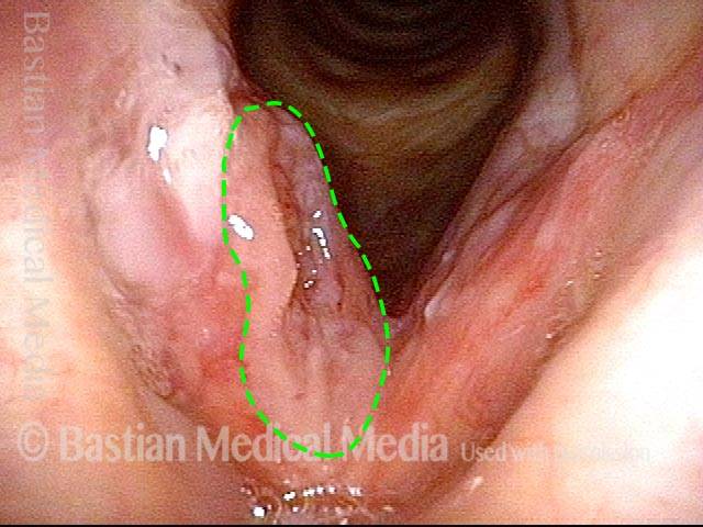
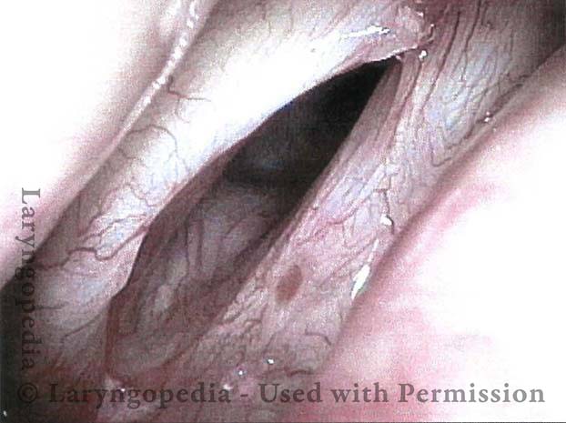
Prior to start of radiation (1 of 4)

Patient with vocal cord carcinoma, primarily of the right true cord (left of picture). This is before radiation therapy began, so there is not yet any radiation mucositis.

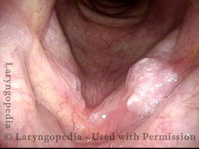
Radiation mucositis, 1 week after radiation (2 of 4)
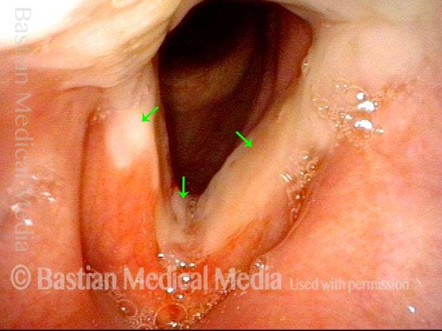
One week after the end of radiation therapy. The tumor has disappeared. Radiation mucositis is evident from the patches of grey (arrows), which are superficial ulceration.

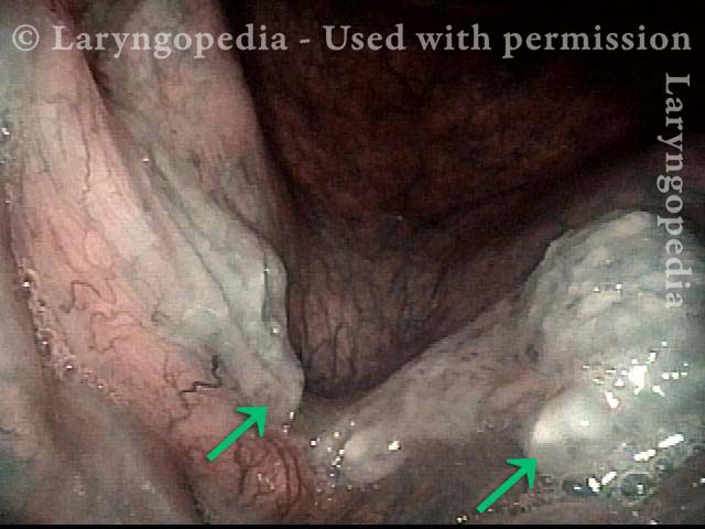
Radiation mucositis, 4 weeks after radiation (3 of 4)
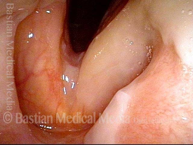
Almost four weeks after the end of radiation therapy. Note that the mucositis has begun resolving, especially on the right cord (left of picture).

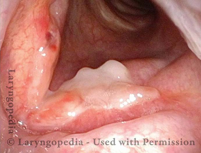
Disappearing radiation mucositis, 10 weeks after radiation (4 of 4)
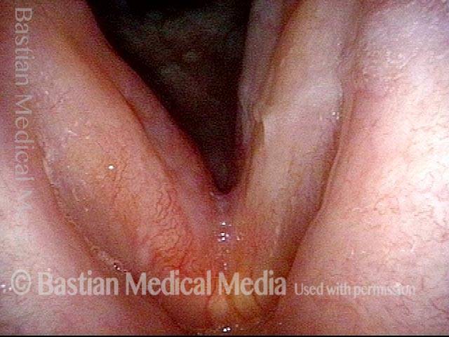
Almost ten weeks after the end of radiation therapy. The mucositis is virtually gone.

Radiation Induced Web

Visual Portfolio, Posts & Image Gallery for WordPress

Post radiation therapy (1 of 4)

Marked laryngeal swelling and inflammation in a woman undergoing radiation therapy to the neck for recurrent thyroid cancer. The larynx is unavoidably in the radiation field. Note swelling of the left arytenoid (right of photo) as compared with right (left of photo). The X’s indicate the location of the arytenoids. In the distance, surrounded by the dotted line, is the web between the vocal cords.

Web formation (2 of 4)

At closer range, the superficial ulceration is indicated by the dotted line. Dashed line indicates where the cords should remain separated but are not, due to formation of a web.

Closer view of ulceration and web (3 of 4)

Closer view. The dotted line is yet again seen around the ulceration and web. Dashed line shows where the cords should remain separate.

Flexible scope used to separate vocal cords​ (4 of 4)

Using only topical anesthesia with patient in chair and not in the O.R., the flexible scope has been used several times to break the cords apart. Voice is instantly restored. The dotted line helps to see the separation of the forming scar ban.

Vocal Cord Cancer, before, during, and after Radiation

Visual Portfolio, Posts & Image Gallery for WordPress

Vocal cord cancer (1 of 8)

Superficial cancer involving both vocal cords. This is stage 1 disease (T1B). The greatest bulk is on the right posterior cord (left of image), but the majority of both cords is involved with at least superficial disease. A faint dotted rectangle indicates the zoomed-in area seen in photo 2.

Vocal cord cancer (2 of 8)

Close-up view of only the anterior half of the cords. Notice the irregular surface, and areas of leukoplakia within this squamous cell carcinoma.

Vocal cord cancer, during radiation (3 of 8)

Just over midway through radiation treatment. One can see that the tumor is melting away.

Vocal cord cancer, during radiation (4 of 8)

Postcricoid / hypopharyngeal mucositis. In this view, the patient is performing a so-called trumpet maneuver to splay open the lower throat. The radiation delivered to the vocal cords (which inhabit the airway but are hidden here due to the momentary constriction of the laryngeal vestibule, at arrows) also causes superficial ulceration of the swallowing passage (upper half of the photo), directly behind the vocal cords. On occasion, if tissue reaction and mucositis are much more severe than seen here, a stricture can form, requiring dilation.

Vocal cord cancer, 2 months after radiation (5 of 8)

Two months after radiation is complete, showing that the tumor is gone, and the mucositis has resolved. There is a small anterior commissure web (at arrow) just below the free margin of the cords. The patient’s voice can nevertheless pass for normal.

Vocal cord cancer, 4 months after radiation (6 of 8)

Now four months after the end of radiation. Close-up view of the postcricoid / hypopharynx regions (compare with photo 4 in this series). Mucositis here is resolved as well, and there is no stricture.

Vocal cord cancer, 6 months after radiation (7 of 8)

Now six months after the end of radiation. Strobe illumination, open phase of vibration. Note that the contours of the vocal cords are not perfectly normal, even though voice is very good.

Vocal cord cancer, 6 months after radiation (8 of 8)

Strobe illumination, nearly closed phase of vibration. Oscillatory flexibility is preserved, but the vocal cord margins are not perfectly straight.

Laser Removal of Vocal Cord Cancer with Bilateral Disease

For treatment of early vocal cord cancer, both laser excision and radiotherapy are in competition as good treatment modalities. See also Early Vocal Cord Cancer: Remove with a Laser, or Radiate? Often, radiation is used when disease is bilateral, in the interest of preserving voice. This is an example of the ability to do fairly extensive laser surgery bilaterally, yet preserving good voice. This man had a friend who had severe difficulty with radiation, and he was therefore opposed to that option.

Visual Portfolio, Posts & Image Gallery for WordPress

Vocal cord cancer (1 of 10)

This 70-something man is a longterm smoker. Here you see an obvious cancer of his left vocal cord (biopsy-proven), but disease on the right side as well that is more superficial.

Stippling (2 of 10)

At higher magnification and using narrow band light, some of the vascular abnormality (stippling) is better seen (arrows).

1 week after excision (3 of 10)

A week after definitive excision of his cancer. Typical early wound appearance, with a suggestion of granulation on the left side (right of photo).

Reparative Granuloma emerges (4 of 10)
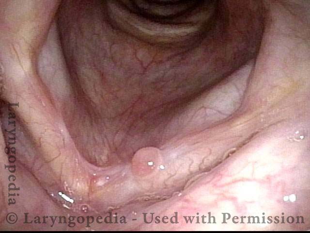
Six weeks later, healing is nearly complete other than a typical reparative granuloma on the left (right of photo).

Granuloma interferes with voicing (5 of 10)
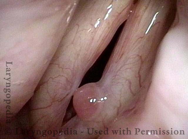
During voicing, the granuloma interferes with closure, explaining in part his ongoing severe hoarseness. Note also the typical medial-to-lateral capillary reorientation.

Granuloma fades away (6 of 10)

Now 3 months postop, the granuloma is smaller. Classic capillary reorientation is again seen.

Closer view (7 of 10)

Under strobe light, closed phase of vibration. Voice is highly functional, since the granuloma no longer interferes with closure.

Granuloma cleft (8 of 10)

Open phase of vibration under strobe light shows the bilobed, clefted nature of the granuloma, where the right vocal fold “fits into” the granuloma (arrows at cleft).

Blood tattoo (9 of 10)

At nearly 5 months postop, the granulation tissue has auto-detached, leaving only a small “blood tattoo.” Here, under strobe light and closed phase of vibration.

Voice is improved (10 of 10)

Voice is somewhat hoarse but highly serviceable, and “better than it has been in years,” according to the patient.

Resources for Further Research

D’Aguillo C, Husain Q, Patel C, Soly Baredes, Eloy JA. Mucositis as a Complication of Radiation Therapy in Nasopharyngeal Carcinoma: A Systematic Review. Otolaryngology. 2013;149(S2). doi:10.1177/0194599813496044a115

Share this article

Support Laryngopedia
Are you a patient or doctor who loves Laryngopedia? If we've helped you, help us help others too!

Table of Contents