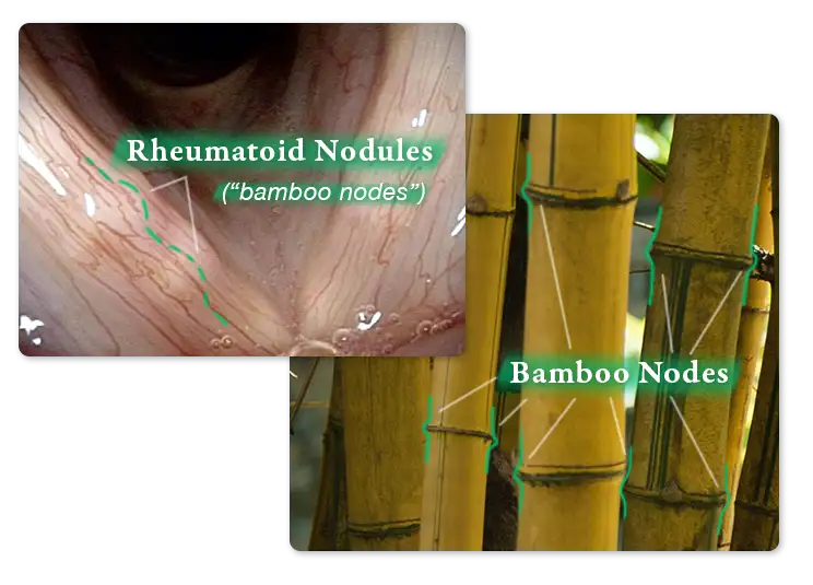
 Rheumatoid nodules are white, fibrous submucosal nodules that can be located on the vocal cords. In that location, they are sometimes described as “bamboo nodes,” because they can be multiple, as seen in the figure to the right.
Rheumatoid nodules in other parts of the body tend to occur at pressure points such as at the elbow, knuckles, or flexor surfaces of the forearms. They are seen in some location in an estimated 25% of persons diagnosed with rheumatoid arthritis (RA). Pathological specimens show fibrosis and palisading granuloma formation. Use of methotrexate for RA is known to increase the occurrence of rheumatoid nodules.
Characteristics of Rheumatoid Nodules of the Vocal Cords
Features that distinguish rheumatoid nodules from the only other potential diagnosis (epidermoid cyst) are the medial-to-lateral oval or bamboo joint-like orientation of the submucosal lesion. An epidermoid cyst is either spherical—or if slowly leaking contents, oval—in an anterior-posterior orientation. Other distinguishing features of rheumatoid nodules are not only their bilaterality, but the presence of multiple lesions on each fold.
Diagnosis of Rheumatoid Nodules
Vocal cord rheumatoid nodules can be the sole initial harbinger of subsequent autoimmune disease before the patient has any other symptoms besides hoarseness. In one case at Bastian Voice Institute, rheumatoid arthritis was diagnosed a few years later; in another, systemic lupus erythematosis with severe renal involvement was diagnosed only 5 or more years after the initial voice complaint and findings of rheumatoid nodules.
Treatment of Rheumatoid Nodules
In other body sites, steroid injection, surgical removal, and more recently rituximab have been utilized for symptomatic rheumatoid nodules, such as at the elbows. In the vocal cords, intralesional injection of triamcinolone has anecdotally provided significant benefit to voice, at least in the short term of weeks to a couple of months.
Dissection of a very large rheumatoid nodules in one case gave surprisingly good voice results but in other cases, neutral to poor results. Of course, in order for the patient to experience surgery as having been beneficial, the stiffening effects of incision and dissection must be less than the stiffening effect of nodules themselves.
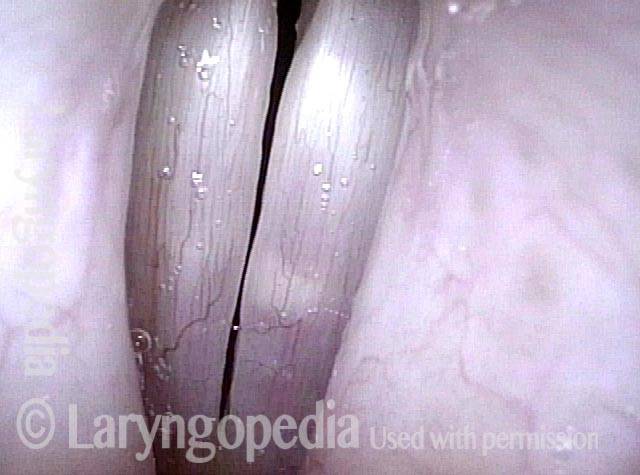
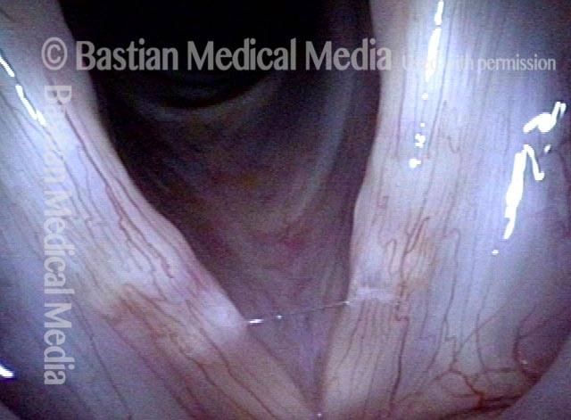
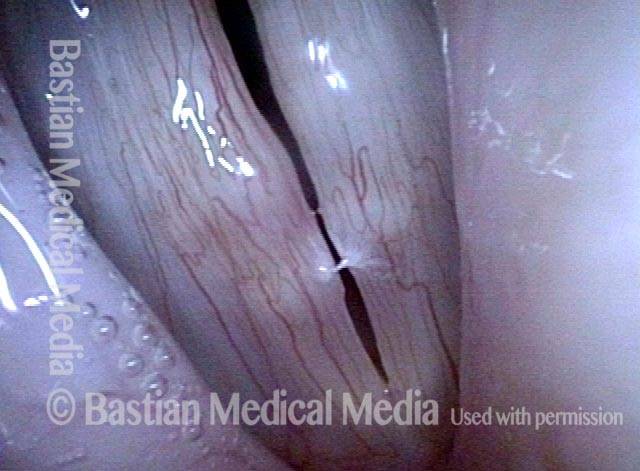
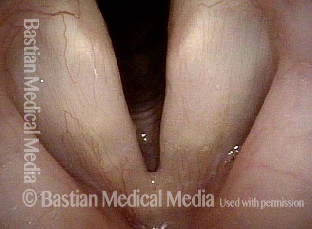
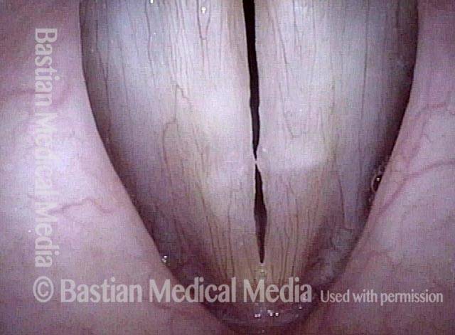
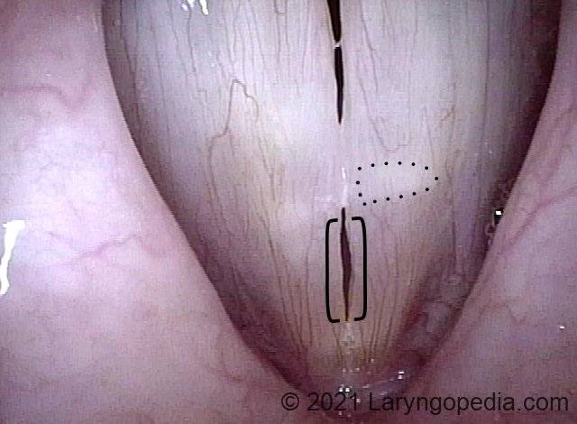
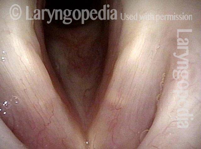
Multiple Rheumatoid Nodules Under 3 Kinds of Light
Standard light, submucosal lesions seen (1 of 4)
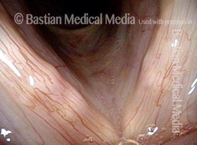
Narrow band light, accentuated capillaries (2 of 4)
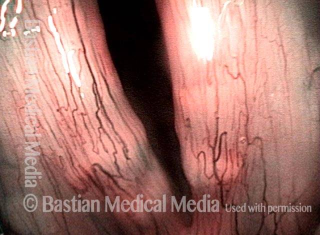
Strobe light, nodules (3 of 4)
Strobe light during phonation (4 of 4)
Rheumatoid Nodules and Crohn’s Disease
Woman with hoarseness (1 of 4)
White submucosal markings (2 of 4)
Submucosal lesions (3 of 4)

Remission from Crohn's (4 of 4)
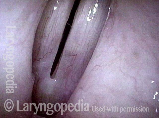
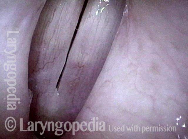
This Subtle, Submucosal Mass is made more Evident with High Pitch. It is likely a Rheumatoid Nodule
Submucosal mass (1 of 4)
Open phase (2 of 4)
Closed phase (3 of 4)
Much higher pitch (4 of 4)