An encyclopedia about voice, swallowing, airway, coughing, & other head + neck disorders.

Tonsils

Tonsils are accumulations of lymphoid tissue found in the upper aerodigestive tract. Lymph nodes are also part of the lymphatic system, but are encapsulated and found buried in tissue of the neck, chest, abdomen, etc.

When one refers to “tonsils” the usual reference is to the palatine tonsils, comprised of semi-encapsulated lymphoid tissue located on the lateral walls of the pharynx. These are often easily seen by opening the mouth widely and shining a light towards the posterior wall of the pharynx while saying “ah.”

Adenoid tissue is very similar but found in the extreme back of the nose, above the palate. They can be referred to as the “nasopharyngeal tonsils.”

A third main location for similar lymphoid tissue is the base of the tongue, where they are called “lingual tonsils.”

We sometimes refer to “micro-tonsils” which are the small salmon-colored bumps seen on the wall of the throat, especially during/after pharyngitis.

Massive Tonsillar Hypertrophy in a Singer

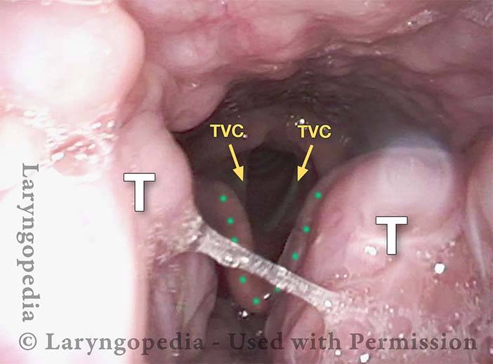
This approximately 30-year-old man is a serious classical singer experiencing symptoms of sleep apnea. While he has not yet been officially diagnosed, he has an upcoming sleep study. His more immediate concern is vocal strain following extensive singing during an illness. At the time of evaluation, his voice and vocal cords appear relatively normal, validating his sense that he has recovered.  However, his tonsils are significantly hypertrophied, making a tonsillectomy likely in his future.

There is no indication of a short palate issue (palatal insufficiency), so a tonsillectomy that preserves the muscles of the palate and pharynx should not negatively impact his voice. In fact, some singers report that their voice “grows” after a period of healing and adjustment while he adapts to the changes in his throat post-surgery.

Visual Portfolio, Posts & Image Gallery for WordPress

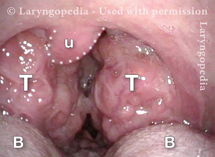
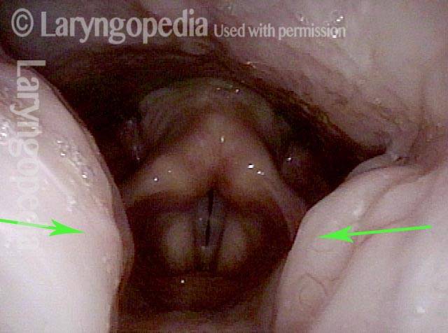
Palatine tonsils (1 of 6)

Looking straight back into the throat through the mouth, with line of sight parallel to the surface of the tongue. For orientation, note the uvula (U), Base of tongue (B and B). Then note the enormous tonsils (T).

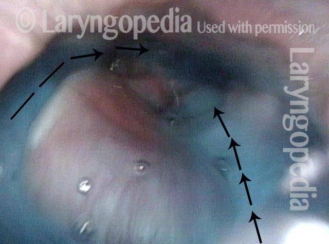
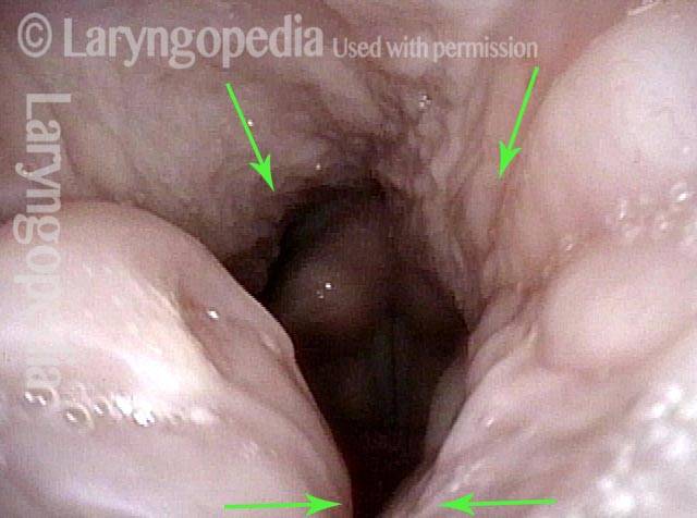
View into the Pharynx (2 of 6)

Beginning to look downward, the epiglottis is seen in the distance (arrow and dotted line).

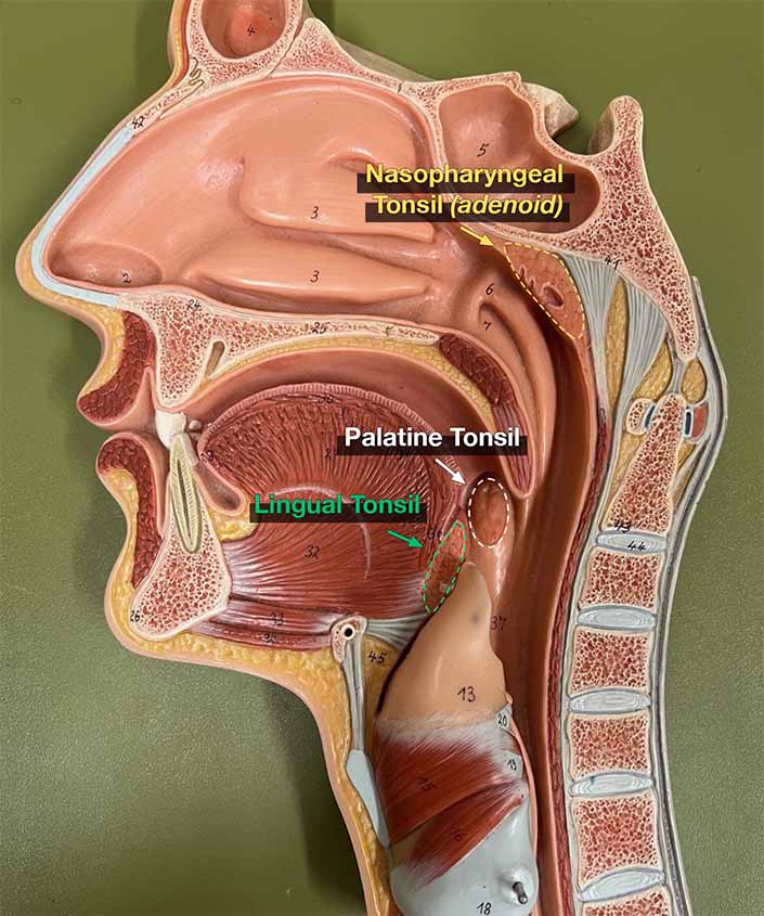
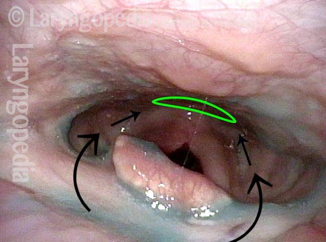
Nasopharyngeal tonsil (3 of 6)

Now looking via the nasal passage directly at the posterior wall of the nasopharynx. The adenoid (nasopharyngeal tonsil) is also markedly enlarged, especially for this far into adulthood. Palate elevation and closure is complete.

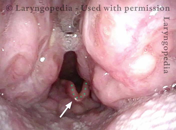
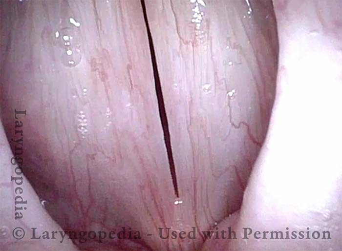
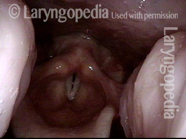
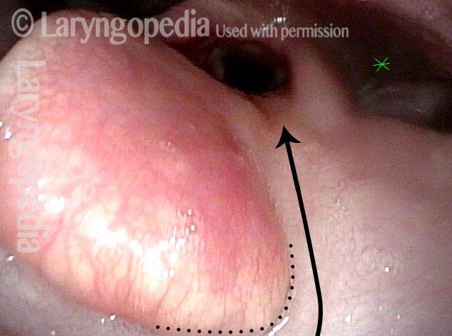
Tonsils meet (4 of 6)

Now looking downward from the nasopharynx, the tonsils nearly meet in the midline during quiet breathing. The vocal cords (two arrows) are open for breathing. Epiglottis is indicated by the dotted line. T = tonsils.

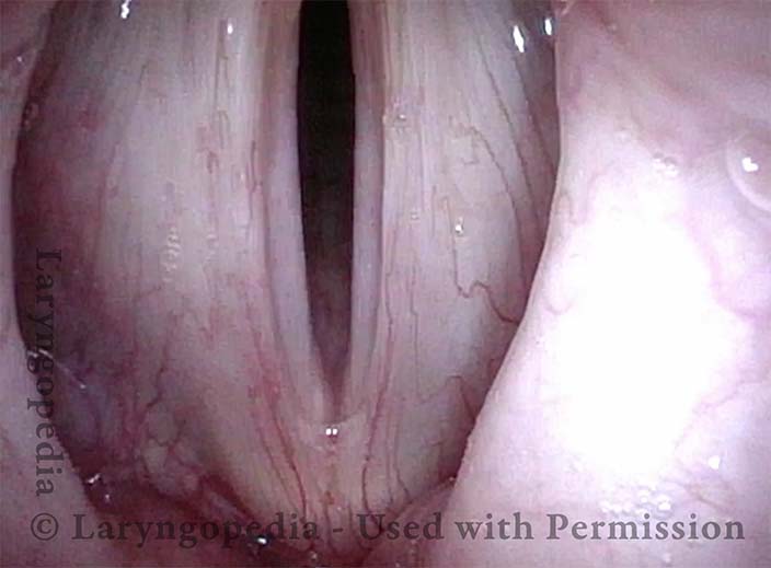
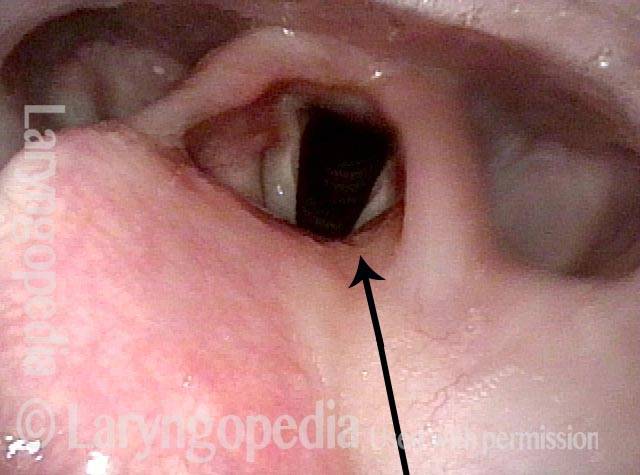
No vocal cord injury (5 of 6)

During phonation, under strobe light, near-closed phase of vibration. The vocal cords are not injured; slight capillary prominence may indicate acid reflux.

Voice is not affected (6 of 6)

Open phase of vibration showing equal vibratory waves and again, no injury.

“Kissing” Tonsils

Visual Portfolio, Posts & Image Gallery for WordPress

Tonsils enlarged (1 of 3)

A singer with very large tonsils seen on either side of the photo as she sings A3 (220 Hz). The line of sight is looking straight down from the nasopharynx.

Higher pitch (2 of 3)

At an octave above, A4 (440 Hz), a slight pharynx contraction brings the tonsils closer together.

Tonsils in contact (3 of 3)

At nearly an octave higher again, G5 (784 Hz), the pharynx has contracted more (upper arrows), causing the tonsils to come into contact just out of the view (lower arrows)–hence the term “ kissing tonsils.” This phenomenon can often be seen by looking at the tonsils through the mouth on an “ah” vowel.

Scarring Diverts Swallowed Materials Directly into the Larynx

Visual Portfolio, Posts & Image Gallery for WordPress

Post tonsillectomy (1 of 4)

A young woman struggles to swallow after extensive cauterization of severe bleeding after tonsillectomy elsewhere. The arrows here show the path food and liquid should follow to get into the esophagus (opening indicated by flat oval).

Closer view (2 of 4)

Closer view shows that the epiglottis is tethered to base of tongue at the dotted line. Furthermore, the “ski jump” scar appears to be ready to divert swallowed material directly into the larynx ( arrow) rather than into the pyriform sinus at *.

The “chute” (3 of 4)

A closer view shows even better the “chute” into the larynx.

Abnormal diversion (4 of 4)

While swallowing blue-colored water, arrows indicate the normal path on the left (right of photo) and the abnormal diversion into the larynx on the right (left of photo). The patient manages, but must swallow carefully, especially since the epiglottis cannot invert since it is scarred to the base of tongue as shown in photo 2.

Share this article

Subscribe
Notify of

1 Comment
Newest
Oldest Most Voted
Inline Feedbacks
View all comments
1
0
Click to see all comments.x