Some polyps are covered by mucosa that is opaque. Some are filled with blood (hemorrhagic polyp). On the other hand, some have a thin and delicate mucosa, and a watery content that is not transparent, yet transmits some light. Unlike a blister, which they could be construed as resembling, and which typically resolves itself, most translucent polyps end up requiring surgery for their resolution.
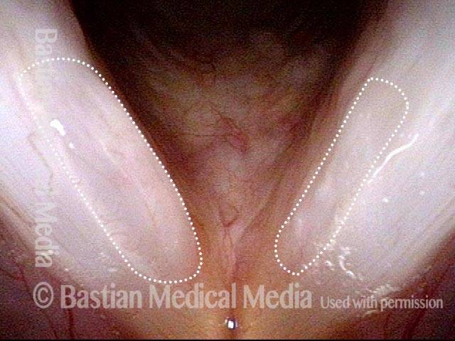
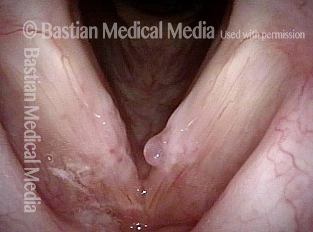
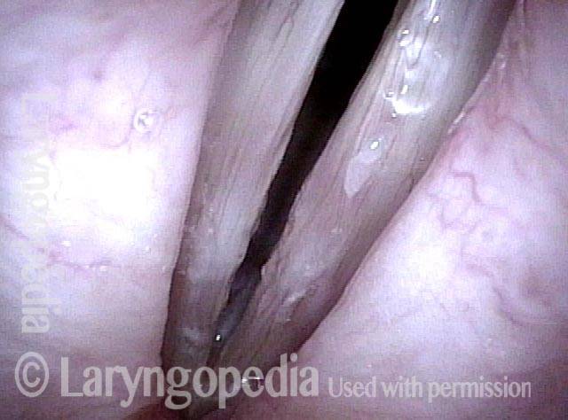
Translucent polyp (1 of 4)
Close-range view with vocal cords in abducted position. This is not the best view to see translucence but faintly “grey” tone of polyps (circled by dotted lines) is indicator of translucence.
Translucent polyp (1 of 4)
Close-range view with vocal cords in abducted position. This is not the best view to see translucence but faintly “grey” tone of polyps (circled by dotted lines) is indicator of translucence.
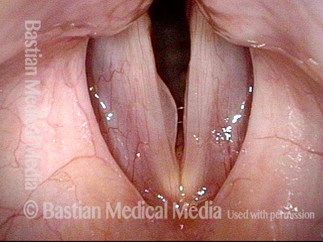
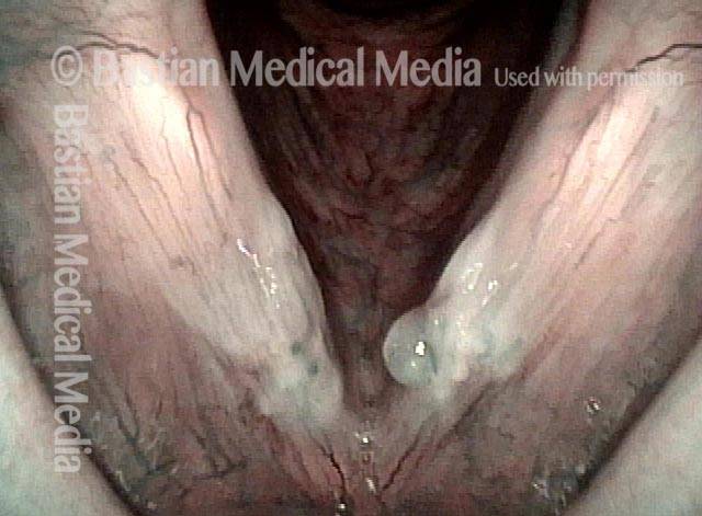
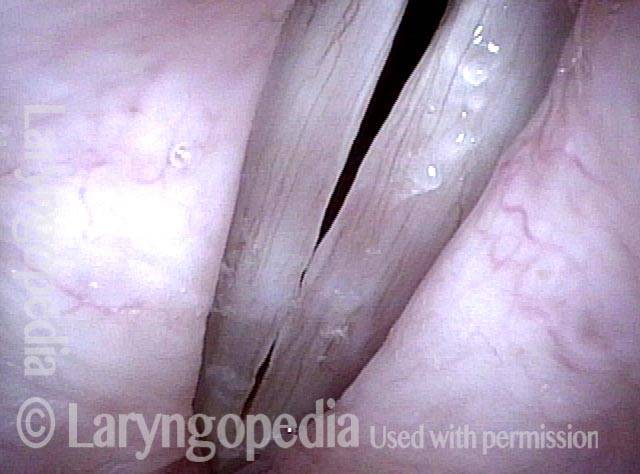
Translucent polyp (2 of 4)
As vocal cords are coming towards adduction, grey indicator of translucence.
Translucent polyp (2 of 4)
As vocal cords are coming towards adduction, grey indicator of translucence.
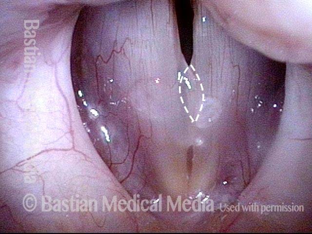
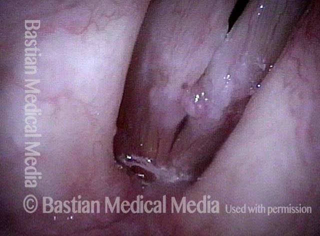
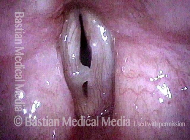
Translucent polyp (3 of 4)
Similar view, with elicitation of rapid inspiration to reveal polyps better, especially on left (right of image).
Translucent polyp (3 of 4)
Similar view, with elicitation of rapid inspiration to reveal polyps better, especially on left (right of image).
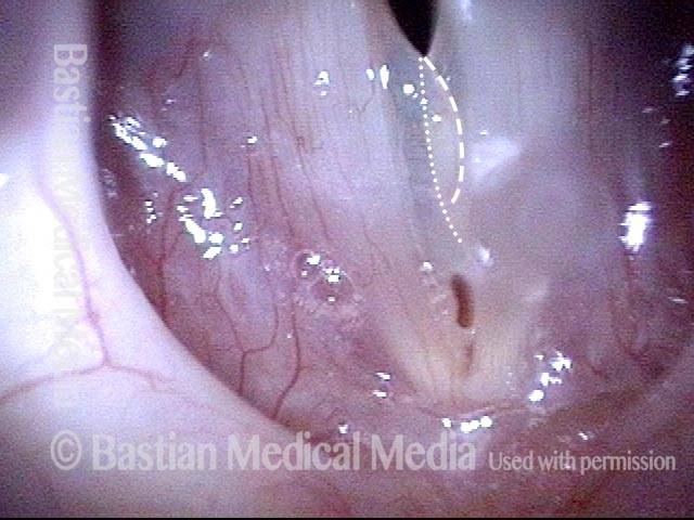
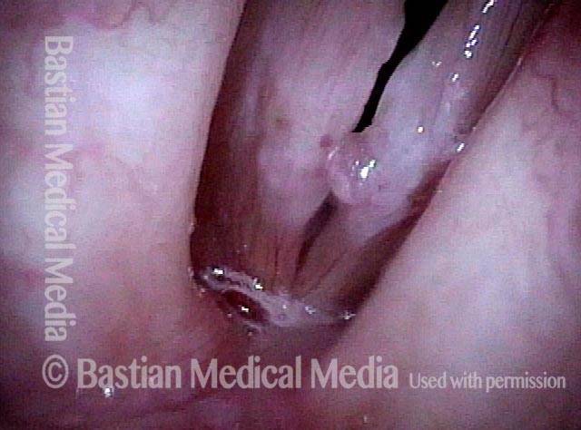
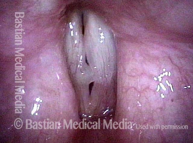
Translucent polyp (4 of 4)
During strobe illumination, translucence especially of the right vocal cord (left of image), is seen best. Note that the larger polyp rides on the margin of the left vocal cord (right of image).
Translucent polyp (4 of 4)
During strobe illumination, translucence especially of the right vocal cord (left of image), is seen best. Note that the larger polyp rides on the margin of the left vocal cord (right of image).
An Extreme Example of Protective Fibrosis Deposits
Extroverted elementary teacher (1 of 6)
Elementary teacher and major extrovert is grossly hoarse. Here you can see the fibrotic-appearing injuries bilaterally and an extra translucent polypoid component on the left cord (right of photo).
Extroverted elementary teacher (1 of 6)
Elementary teacher and major extrovert is grossly hoarse. Here you can see the fibrotic-appearing injuries bilaterally and an extra translucent polypoid component on the left cord (right of photo).
Submucosal fibrosis (2 of 6)
Under narrow band light, the white area is not hazy leukoplakia, but instead submucosal fibrosis, deposited as a protection against mucosal vibratory collision/ shearing injury.
Submucosal fibrosis (2 of 6)
Under narrow band light, the white area is not hazy leukoplakia, but instead submucosal fibrosis, deposited as a protection against mucosal vibratory collision/ shearing injury.
Phonatory view (3 of 6)
Under strobe light, closure is imperfect due to the mid-cord elevations.
Phonatory view (3 of 6)
Under strobe light, closure is imperfect due to the mid-cord elevations.
Open phase (4 of 6)
Open phase of vibration with small amplitude and absent “mucosal wave” due to stiffness of the mucosa.
Open phase (4 of 6)
Open phase of vibration with small amplitude and absent “mucosal wave” due to stiffness of the mucosa.
Post microsurgery, open phase (5 of 6)
A week after vocal cord microsurgery, voice is markedly improved. No attempt was made to remove all of the fibrosis, but only to straighten the vocal cord margins. Open phase of vibration at F5.
Post microsurgery, open phase (5 of 6)
A week after vocal cord microsurgery, voice is markedly improved. No attempt was made to remove all of the fibrosis, but only to straighten the vocal cord margins. Open phase of vibration at F5.
Post microsurgery, closed phase (6 of 6)
Closed phase of vibration at same pitch shows that some margin swelling remains. The patient also has MTD; posterior cords are widely separated.
Post microsurgery, closed phase (6 of 6)
Closed phase of vibration at same pitch shows that some margin swelling remains. The patient also has MTD; posterior cords are widely separated.
The Mucosa’s Expression of Injury Varies
Vocal cord injuries (1 of 4)
Vocal cord injuries of overuse are often bilaterally similar, but here we have two quite different expressions of injury: fibrosis and capillary ectasia on left (right of photo); translucent polypoid injury (not a cyst) on the right (left of photo).
Vocal cord injuries (1 of 4)
Vocal cord injuries of overuse are often bilaterally similar, but here we have two quite different expressions of injury: fibrosis and capillary ectasia on left (right of photo); translucent polypoid injury (not a cyst) on the right (left of photo).
Narrow band lighting (2 of 4)
Now under narrow band light, the left cord (right of picture) has a flatter, fibrotic expression with tiny ectatic capillaries.
Narrow band lighting (2 of 4)
Now under narrow band light, the left cord (right of picture) has a flatter, fibrotic expression with tiny ectatic capillaries.
Strobe lighting (3 of 4)
Under strobe light, the translucent, polypoid nodule of the right cord (left of photo) distorts vibratory closure.
Strobe lighting (3 of 4)
Under strobe light, the translucent, polypoid nodule of the right cord (left of photo) distorts vibratory closure.
Phonation (4 of 4)
This is the best closure this grossly hoarse person can achieve.
Phonation (4 of 4)
This is the best closure this grossly hoarse person can achieve.
Translucence of a Polyp to the Point of Near-Transparency
Vocal cord polyp (1 of 4)
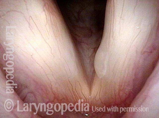
This singer has had marked vocal impairment for many months. In this standard light abducted position at medium distance, a polyp is seen on the left vocal cord (right of photo).
Vocal cord polyp (1 of 4)
This singer has had marked vocal impairment for many months. In this standard light abducted position at medium distance, a polyp is seen on the left vocal cord (right of photo).
Distant view (2 of 4)
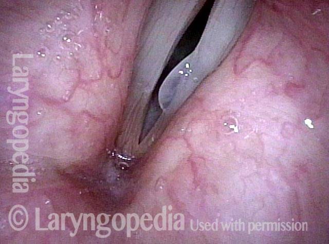
In a more distant view under strobe light, translucence is noted as the darker, more grey center of the polyp ‘looking through’ to the darkness below the polyp.
Distant view (2 of 4)
In a more distant view under strobe light, translucence is noted as the darker, more grey center of the polyp ‘looking through’ to the darkness below the polyp.
Translucent polyp with mucus (3 of 4)
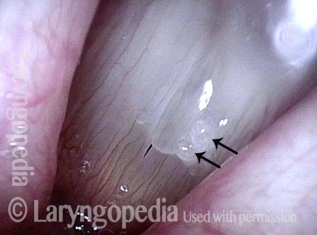
At higher magnification still under strobe light, notice that the linear antero-posterior blood vessels in the floor of the polyp can, remarkably, be seen through the overlying polyp. The white ‘blobs’ (arrows) are mucus.
Translucent polyp with mucus (3 of 4)
At higher magnification still under strobe light, notice that the linear antero-posterior blood vessels in the floor of the polyp can, remarkably, be seen through the overlying polyp. The white ‘blobs’ (arrows) are mucus.
Different phase of vibration (4 of 4)
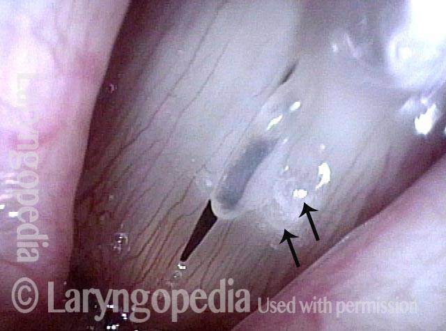
At a different phase of vibration also at C#4 (277 Hz), the dark center of the polyp is the result of 'seeing through' this nearly transparent polyp to the dark glottal chink below it. The arrows again indicate mucus.
Different phase of vibration (4 of 4)
At a different phase of vibration also at C#4 (277 Hz), the dark center of the polyp is the result of 'seeing through' this nearly transparent polyp to the dark glottal chink below it. The arrows again indicate mucus.
Tags
[cool_tag_cloud on_single_display=”local” style=”green” largest=”16″ font_family=”Helvetica” font_weight=”bold” number=”0″ show_count=”yes”]
Tagged Photos
