An encyclopedia about voice, swallowing, airway, coughing, & other head + neck disorders.

A peculiar and uncommon type of laryngitis that causes the mucosa to ulcerate. With ulcerative laryngitis, there is usually a white area of apparent mucosal necrosis with a surrounding rim of intense erythema. Recovery of voice is slow, often taking several weeks, in contrast to more typical forms of laryngitis. The causative agent is not known to us at our practice, though we have some theories.

Ulcerative Laryngitis in a Professional Singer

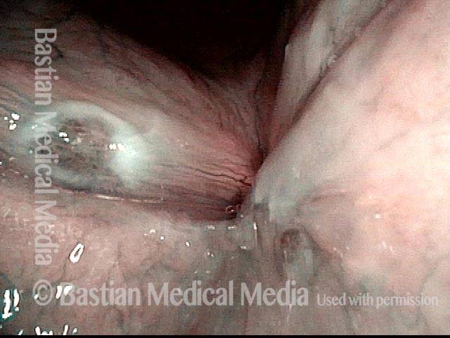
This professional singer had struggled with severe hoarseness for several weeks before being examined.  Identification of superficial ulcerative laryngitis (rather than garden-variety viral or bacterial types) helped counsel him that it would be several more weeks before his voice would be restored. This kind of laryngitis does not seem to respond to antibiotics, antifungals, or steroids, and must just take its course to healing.  Unfortunately, a common time required for healing is 6 and even 8 weeks.  Surprisingly, with healing, the voice typically returns fully to its baseline capabilities.

Visual Portfolio, Posts & Image Gallery for WordPress

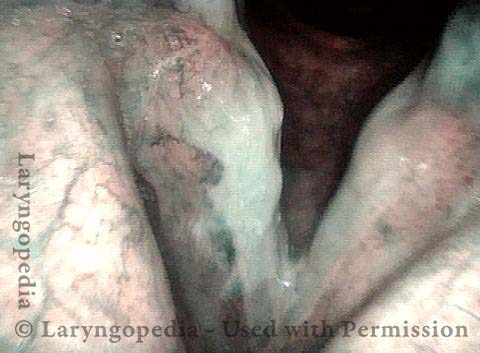
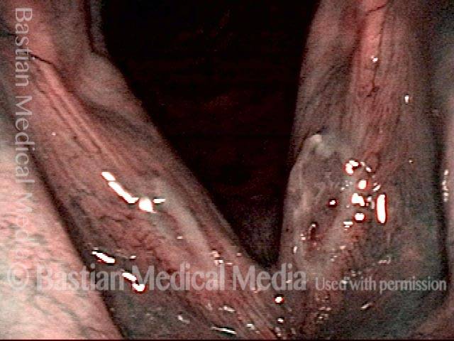
Ulcerative Laryngitis (1 of 4)

Notice that the right vocal cord (left of photo) has a hazy white area that is strongly demarcated from a very red “surround.” Overall, there is diffuse redness and inflammatory appearance throughout the larynx, too.

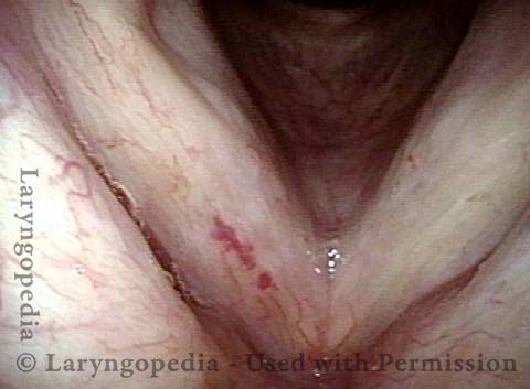
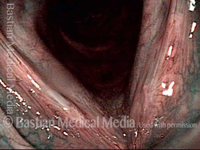
Ulceration of the right cord (2 of 4)

Under narrow band light, the superficial ulceration of the right cord is even more evident. Rapid inspiration in this view is making the margin convex.

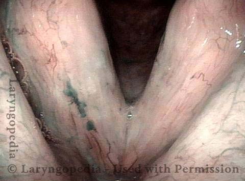
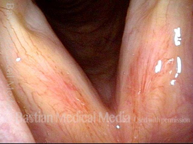
Re-mucosalization well under way (3 of 4)

A month later, healing is not yet complete, but re-mucosalization is well under way. The general color of the larynx as a whole is paler, signifying subsidence of the inflammatory process. Compare with photo 1.

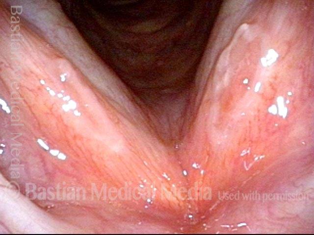
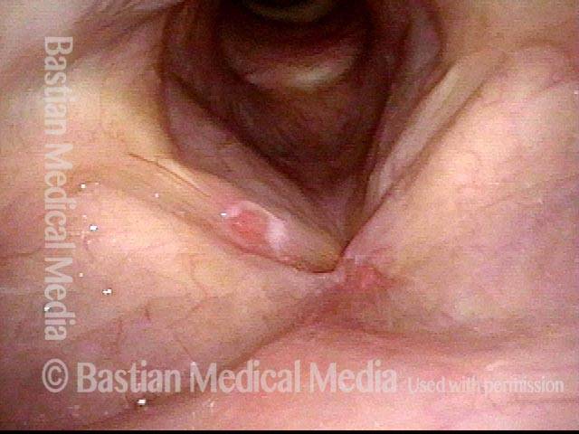
Near complete healing (4 of 4)

Under narrow band light, the formerly demarcated, ulcerated area is much fainter. Compare with photo 2.

Gradual Healing of Ulcerative Laryngitis

Visual Portfolio, Posts & Image Gallery for WordPress

Ulcerative laryngitis (1 of 4)

Ulcerated area, free margin of both vocal cords. When diagnosed at this early stage, the patient is notified that recovery will likely require as much as six weeks.

Ulcerative laryngitis (2 of 4)

Same view, but using narrow band illumination to accentuate the ulcers.

1 month later: ulcerative laryngitis healing (3 of 4)

One month later, under narrow band illumination. Ulceration dramatically diminished.

3 months later: ulcerative laryngitis virtually all healed (4 of 4)

At three months, standard illumination. Virtually complete healing, with excellent return of voice. The prominent vascularity at area of ulcer will eventually fade.

Vocal Cord Ulcer

Visual Portfolio, Posts & Image Gallery for WordPress

Vocal cord ulcer (1 of 2)

In a patient who had had hoarseness lasting several months, this inflammatory lesion of the right vocal cord (left of image) was identified. There is hazy leukoplakia surrounding a central intensely erythematous ulcer. It most resembles an aphthous ulcer, though these are not previously reported on the vocal cord, and this lesion's duration is longer than the typical aphthous ulcer.

Vocal cord ulcer (2 of 2)

Closer view, under narrow-band light. The vessel pattern looks inflammatory and not neoplastic. There is heaped-up leukoplakia surrounding the lesion. After this lesion persisted for more than four months, it was removed. Tissue examination showed inflammatory response and keratosis without atypia.

Ulcerative Laryngitis and Resulting Synechia – Fixed!

Visual Portfolio, Posts & Image Gallery for WordPress

Synechia (1 of 3)

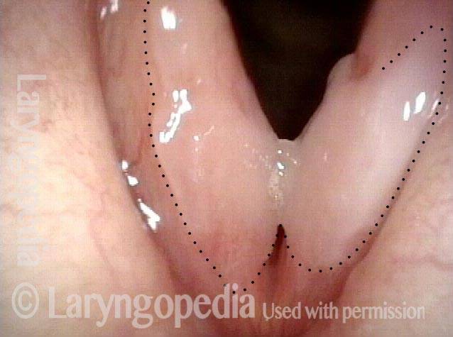
This woman developed a sore throat and lost her voice a week after a chemotherapy treatment for her metastatic breast cancer. Here, 6 weeks later, note the hazy area representing resolving “ulcerative” laryngitis (surrounded by tiny dotted line). There is a synechia attaching the cords together.

Attempted to detach (2 of 3)

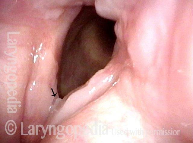
The flexible scope has been used once to “twang upwards” from below in order to detach the cords from each other. At the arrow, slight separation can be seen.

Successfully detached (3 of 3)

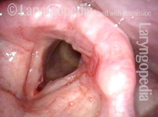
Just after the second attempt. That is, for the second time, the scope was passed below the cords, angulated sharply underneath the synechia, and then pulled upwards. The adhesion has been released. Voice is instantly and dramatically restored (though still hoarse, of course).

Healed Mucosa After Ulcerative Laryngitis

Visual Portfolio, Posts & Image Gallery for WordPress

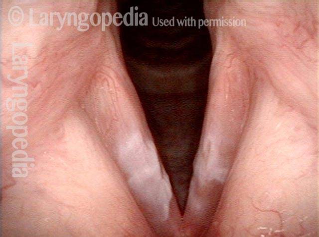
Ulcerative laryngitis (1 of 4)

Severe ulcerative laryngitis. Mucosa has sloughed away in the area of the white patches, not yet re-mucosalized.

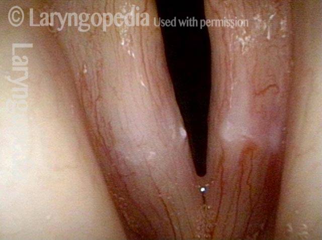
Six weeks later (2 of 4)

As is typical for ulcerative laryngitis, the mucosa takes 6 weeks or more to regenerate. This is six weeks after photo 1. Soon after this examination, voice returned to nearly-normal.

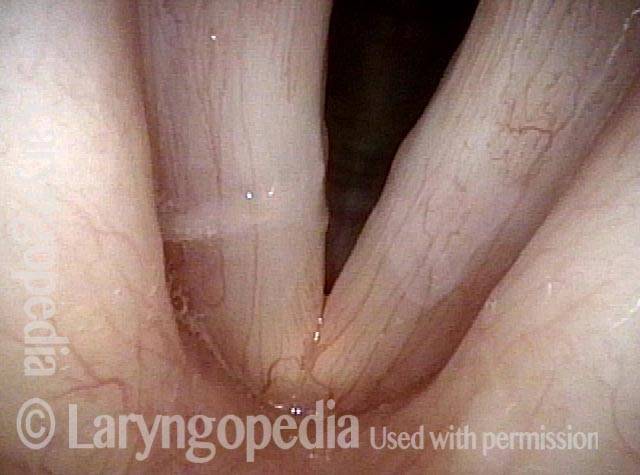
7 years later (3 of 4)

About 7 years later, the voice remains essentially normal. Still, in this standard light view, one can see a faint white area in the area of the original ulceration. This is better appreciated in the next photo.

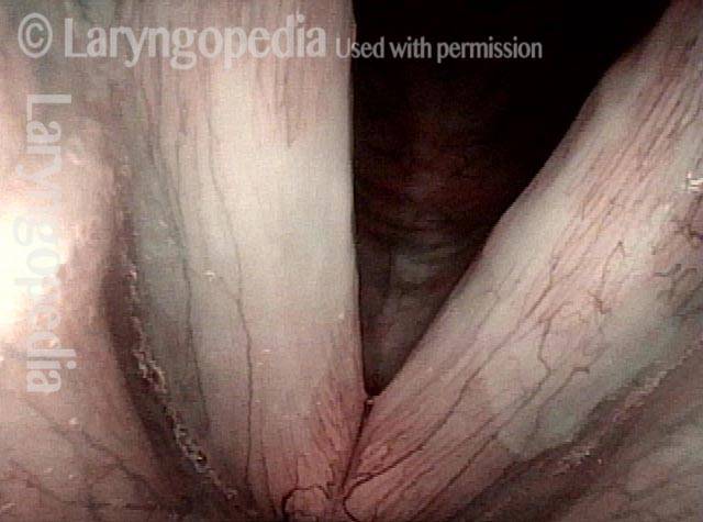
Narrow band lighting (4 of 4)

Under narrow band light, the (originally) more deeply ulcerated right vocal cord (left of photo) remains more avascular than the left (right of photo), where the capillary pattern has returned, but still with a demarcated area of reduced micro-capillaries causing a whiter appearance.