A projection of the anterior arytenoid cartilage, to which is attached the membranous vocal cord.
Arytenoid’s Vocal Process
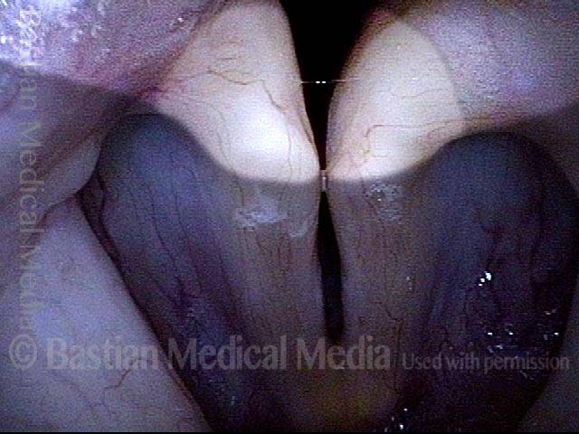
Vocal process of the arytenoid, artificially highlighted
Strobe light, as the vocal cords are just coming into contact for phonation. The vocal process of each arytenoid is brightly highlighted; the extension of each vocal process back into the arytenoid is moderately highlighted.
Vocal process of the arytenoid, artificially highlighted
Strobe light, as the vocal cords are just coming into contact for phonation. The vocal process of each arytenoid is brightly highlighted; the extension of each vocal process back into the arytenoid is moderately highlighted.
Vocal Processes of the Arytenoid Cartilages, Accentuated by Vocal Cord Atrophy
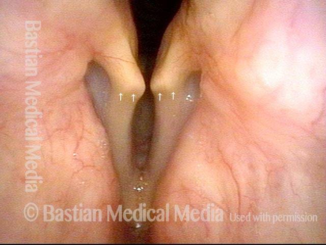
Vocal processes, accentuated by vocal cord atrophy (1 of 3)
Panoramic view of the vocal cords just before voicing. Notice the obvious outline of both vocal processes. (For orienting, the processes are bounded at one end by small arrows.) The processes shine through particularly clearly due to the marked atrophy of the vocal cords as a whole.
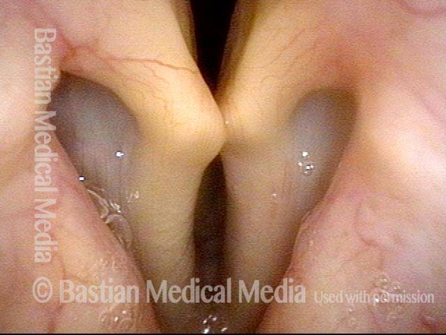
Vocal processes, accentuated by vocal cord atrophy (2 of 3)
Closer view.
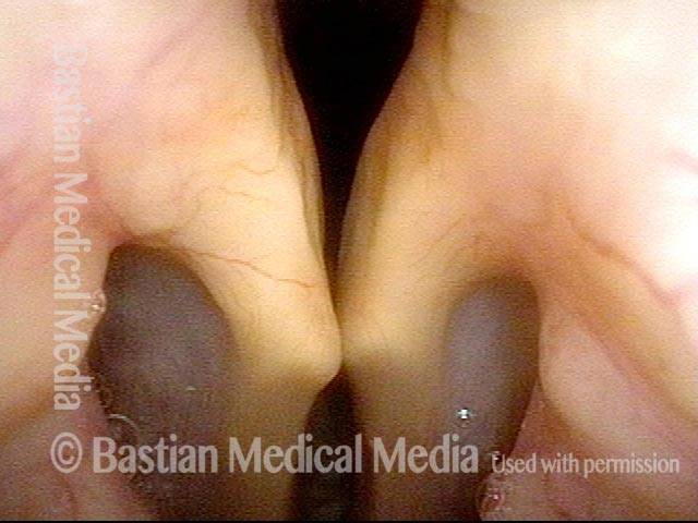
Vocal processes, accentuated by vocal cord atrophy (3 of 3)
Notice that the overlying mucosa is very thin and tightly adherent along the entire medial surface of the arytenoid cartilages. This shows very graphically why the posterior third of the cords do not participate in vibration.
Vocal processes, accentuated by vocal cord atrophy (3 of 3)
Notice that the overlying mucosa is very thin and tightly adherent along the entire medial surface of the arytenoid cartilages. This shows very graphically why the posterior third of the cords do not participate in vibration.
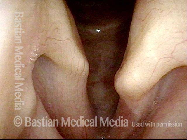
Vocal processes, accentuated by vocal cord atrophy (1 of 4)
The vocal processes in this patient are extremely visible because the rest of the vocal cord on each side is atrophic and bowed.
Vocal processes, accentuated by vocal cord atrophy (1 of 4)
The vocal processes in this patient are extremely visible because the rest of the vocal cord on each side is atrophic and bowed.
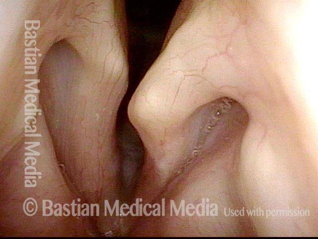
Vocal processes, accentuated by vocal cord atrophy (2 of 4)
The vocal cords approach each other for voicing. Note the evident asymmetry between the vocal processes. The left vocal process (right of image) projects further anteriorly than does the opposite process. It is also at a higher (more cephalad) level.
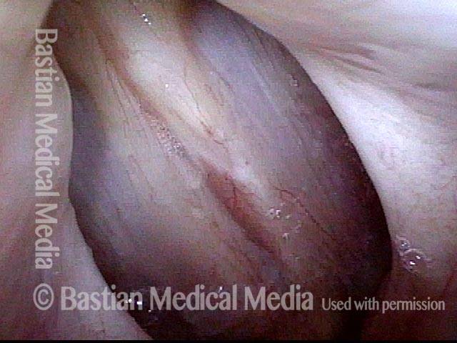
Vocal processes, accentuated by vocal cord atrophy (3 of 4)
Phonation, closed phase of vibration, under strobe lighting. Note the overlap (scissoring) of the left vocal process (right of image) on top of the other process.
Vocal processes, accentuated by vocal cord atrophy (3 of 4)
Phonation, closed phase of vibration, under strobe lighting. Note the overlap (scissoring) of the left vocal process (right of image) on top of the other process.
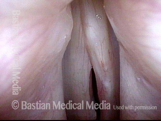
Vocal processes, accentuated by vocal cord atrophy (4 of 4)
Phonation, at a higher pitch, at which the scissoring of the left vocal process (right of image) on top of the other becomes even more evident.
Vocal processes, accentuated by vocal cord atrophy (4 of 4)
Phonation, at a higher pitch, at which the scissoring of the left vocal process (right of image) on top of the other becomes even more evident.
Tagged Anatomy & Physiology, Education, Photos
