Candida Laryngitis and Pharyngitis
Infection with candida albicans, an ubiquitous commensal organism in the upper aerodigestive tract, usually on the vocal cord mucosa. While this organism normally causes no problem, under certain circumstances it can overgrow. These circumstances include:
- When other (competing) normal flora are killed through administration of antibiotics.
- When surface immunity of the mucosa is decreased via inhalation of steroid medication (e.g. asthma).
- When the individual is immunosuppressed by disease (e.g. diabetes) or other drugs.
Symptoms and Treatment
Typical symptoms of candida laryngitis and pharyngitis include slight sore throat and hoarseness. Treatment may consist of reducing or withdrawing listed potentiators, or using an antifungal agent such as fluconazole.
Candida Can Resolve Quickly
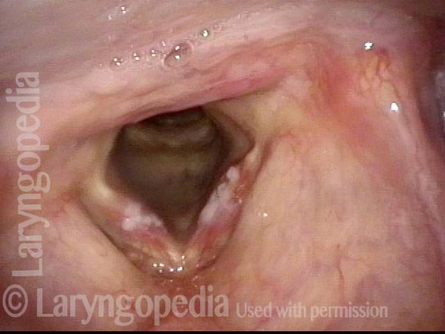
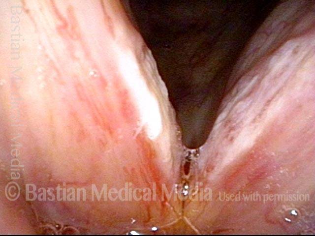
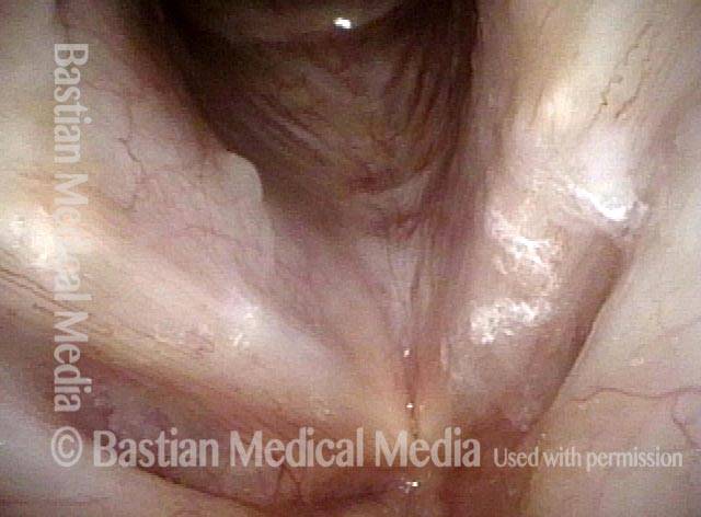
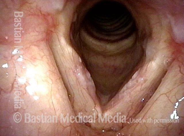
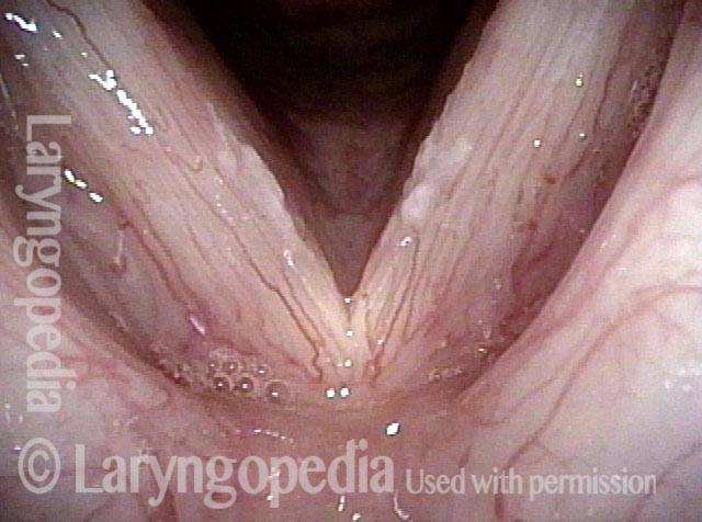
Steroid-induced candida growth (1 of 4)
The additive effects of high-dose systemic steroids, inhaled steroids for asthma, and multiple courses of antibiotics have precipitated this severe case of vocal cord candida growth.
Steroid-induced candida growth (1 of 4)
The additive effects of high-dose systemic steroids, inhaled steroids for asthma, and multiple courses of antibiotics have precipitated this severe case of vocal cord candida growth.
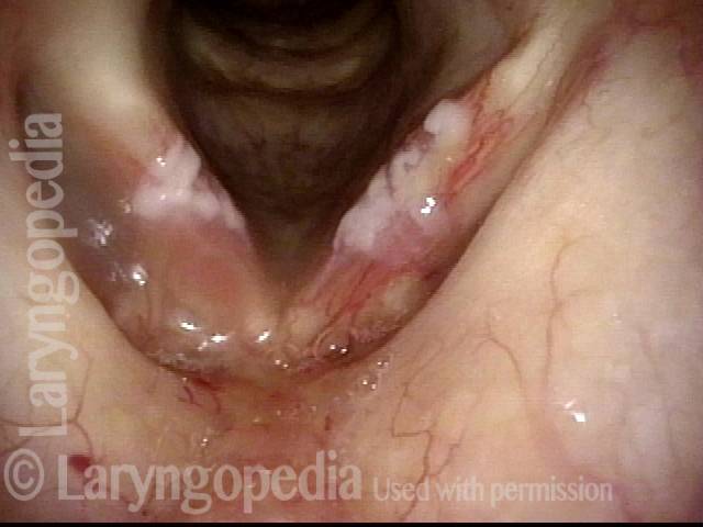
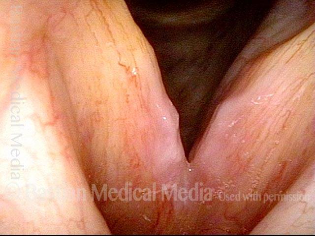
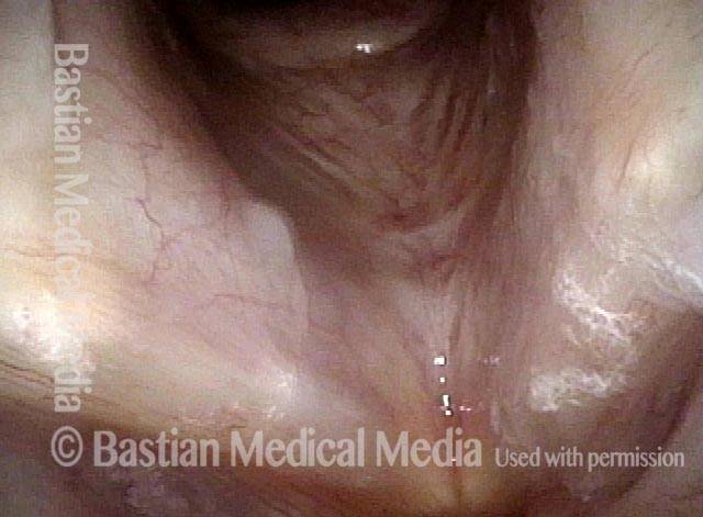
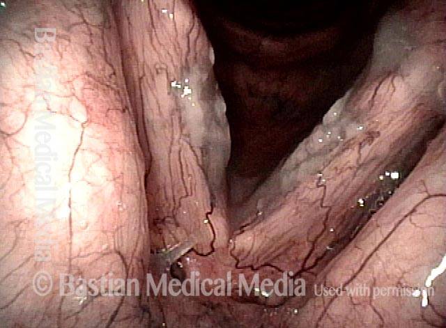
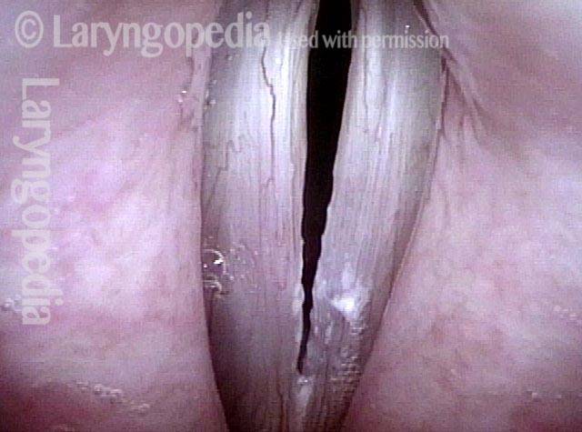
Candida (2 of 4)
In this closer view, the “inflammatory surround” typical of candida (but not garden-variety leukoplakia) is better seen.
Candida (2 of 4)
In this closer view, the “inflammatory surround” typical of candida (but not garden-variety leukoplakia) is better seen.
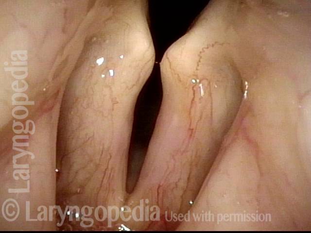
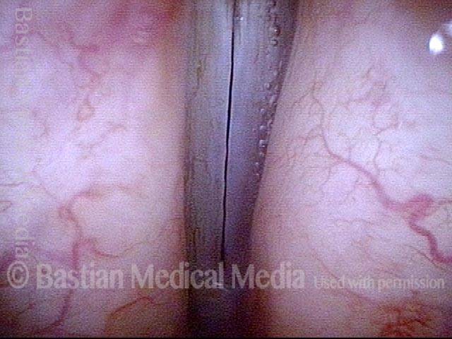
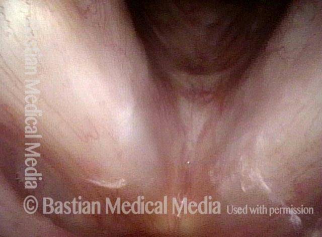
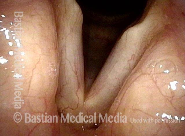
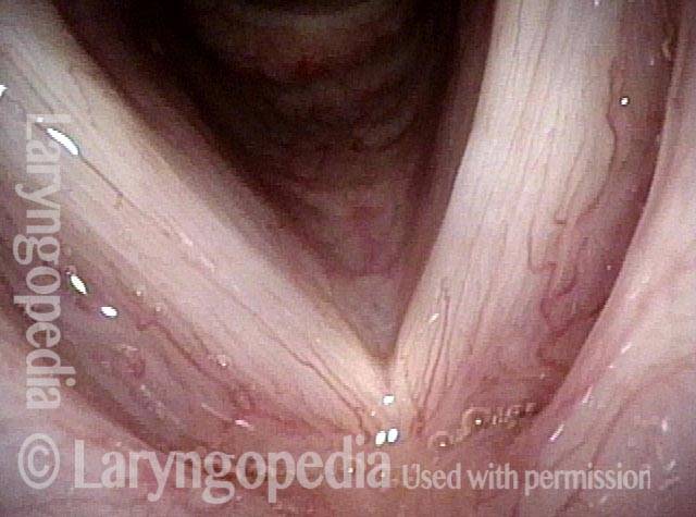
2 weeks after fluconazole treatment (3 of 4)
After just 2 weeks of fluconazole, 100mg per day, the candida colonies and surrounding inflammation are nearly resolved. Only faint haziness remains, along with subtle pinkness.
2 weeks after fluconazole treatment (3 of 4)
After just 2 weeks of fluconazole, 100mg per day, the candida colonies and surrounding inflammation are nearly resolved. Only faint haziness remains, along with subtle pinkness.
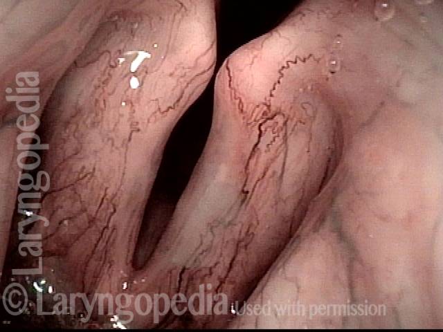
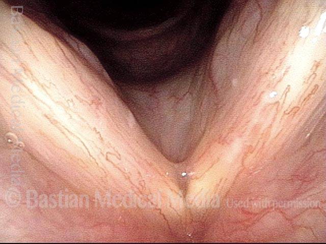
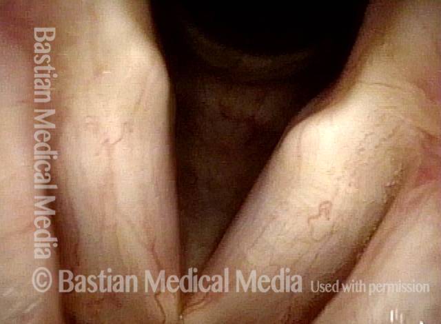
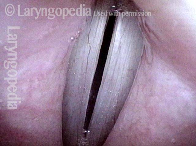
Fluconazole (4 of 4)
Narrow band light shows the residual hazy white especially on the left vocal cord. The patient’s heavy steroid / antibiotic treatment had to continue for several more weeks, and during this time, she remained on fluconazole 100 mg twice a week, which was sufficient to keep candida from regrowing.
Candida Laryngitis, Before & After Treatment
Candida laryngitis (1 of 4)
Severe laryngeal candidiasis, in a person using inhaled steroids at high dose. Standard light.
Candida laryngitis (1 of 4)
Severe laryngeal candidiasis, in a person using inhaled steroids at high dose. Standard light.
Candida laryngitis (2 of 4)
Closer view shows more clearly not only the white areas, but also surrounding inflammation. Standard light.
Candida laryngitis (2 of 4)
Closer view shows more clearly not only the white areas, but also surrounding inflammation. Standard light.
Candida laryngitis, 15 days after starting treatment (3 of 4)
After 15 days of oral fluconazole. Obvious improvement, but incomplete resolution of tissue changes.
Candida laryngitis, 15 days after starting treatment (3 of 4)
After 15 days of oral fluconazole. Obvious improvement, but incomplete resolution of tissue changes.
Candida laryngitis, several months later (4 of 4)
After longer-term fluconazole, along with reduction of inhaled steroid dose, complete resolution. Strobe light, closed phase of vibration at high vocal pitch.
Candida laryngitis, several months later (4 of 4)
After longer-term fluconazole, along with reduction of inhaled steroid dose, complete resolution. Strobe light, closed phase of vibration at high vocal pitch.
Example 2
Candida laryngitis (1 of 4)
Candidiasis in patient using inhaled steroids for asthma. Under standard light, the lesions are vague, hazy, and best seen anteriorly on the right cord (left of image).
Candida laryngitis (1 of 4)
Candidiasis in patient using inhaled steroids for asthma. Under standard light, the lesions are vague, hazy, and best seen anteriorly on the right cord (left of image).
Candida laryngitis (2 of 4)
Same patient, narrow-band illumination. This not only emphasizes vascularity, but brings out the candida colonies.
Candida laryngitis (2 of 4)
Same patient, narrow-band illumination. This not only emphasizes vascularity, but brings out the candida colonies.
Candida laryngitis, after treatment (3 of 4)
After treatment with fluconazole, the colonies have virtually disappeared.
Candida laryngitis, after treatment (3 of 4)
After treatment with fluconazole, the colonies have virtually disappeared.
Candida laryngitis, after treatment (4 of 4)
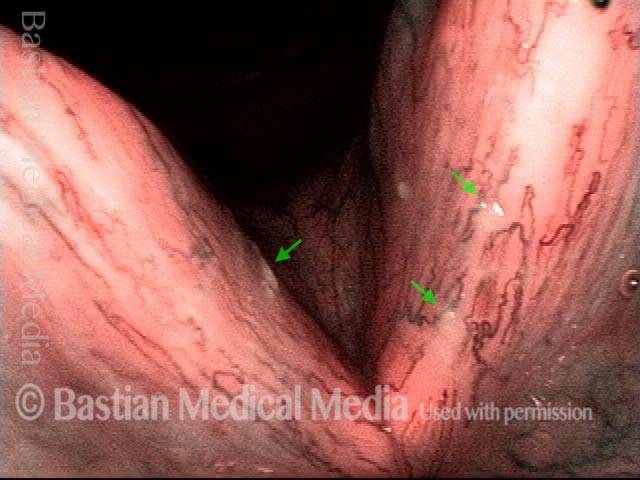
Same post-treatment examination, under narrow-band illumination. Note that there are normal specks of mucus (such as at the arrows) in the view.
Candida laryngitis, after treatment (4 of 4)
Same post-treatment examination, under narrow-band illumination. Note that there are normal specks of mucus (such as at the arrows) in the view.
Example 3
Candida laryngitis (1 of 3)
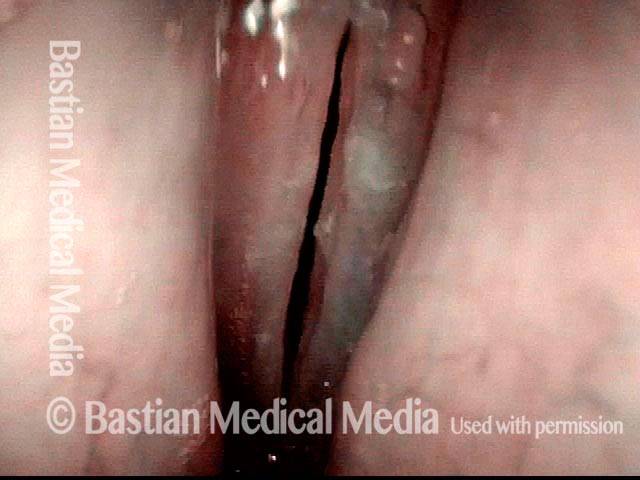
Elderly woman with a history of laryngeal amyloidosis requiring laser sculpting several years earlier. Now using high-dose inhaled steroids, antibiotics, and oral steroids for unrelated pulmonary problem. Marked increase of hoarseness, and whitish discoloration, especially of the left vocal cord (right of image).
Candida laryngitis (2 of 3)
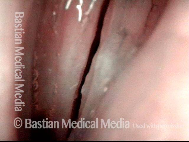
Closer view of hazy white areas and irregular right cord margin (left of image), presumed to be candida overgrowth. Empiric treatment with fluconazole is justified, given history and findings.
Candida laryngitis (2 of 3)
Closer view of hazy white areas and irregular right cord margin (left of image), presumed to be candida overgrowth. Empiric treatment with fluconazole is justified, given history and findings.
Candida laryngitis, after starting treatment (3 of 3)
Two weeks after starting fluconazole; the white areas are completely resolved. The patient’s voice had improved markedly within three or four days of starting the treatment.
Candida laryngitis, after starting treatment (3 of 3)
Two weeks after starting fluconazole; the white areas are completely resolved. The patient’s voice had improved markedly within three or four days of starting the treatment.
Candida Pharyngitis
Candida pharyngitis (1 of 2)
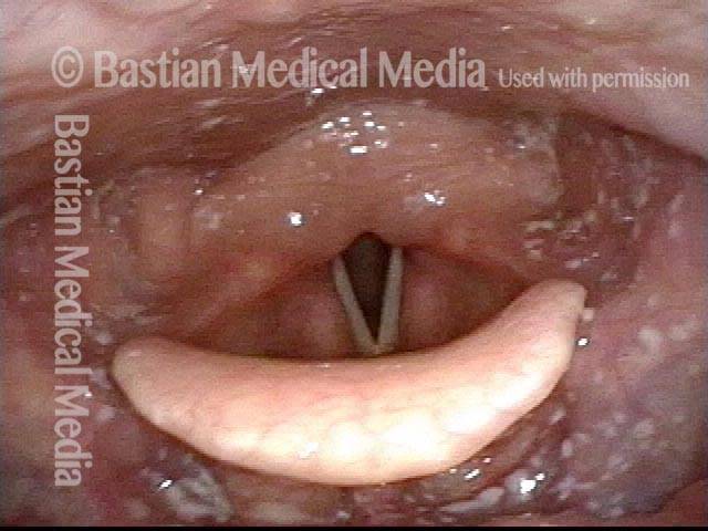
Countless candida colonies in the hypopharynx (lower throat) of a patient who, for treatment of an auto-immune disorder, is not only inhaling steroids but also taking high-dose steroids orally. Each tiny white dot represents a colony of the fungus.
Candida pharyngitis (1 of 2)
Countless candida colonies in the hypopharynx (lower throat) of a patient who, for treatment of an auto-immune disorder, is not only inhaling steroids but also taking high-dose steroids orally. Each tiny white dot represents a colony of the fungus.
Candida pharyngitis (2 of 2)
An even more dramatic case of candidiasis, in a different patient. Here, the colonies are more obvious and nearly confluent.
Candida pharyngitis (2 of 2)
An even more dramatic case of candidiasis, in a different patient. Here, the colonies are more obvious and nearly confluent.
Candida Masquerader!
White lesions (1 of 3)
This man uses a steroid inhaler for his asthma. During esophagoscopy, white lesions were seen in the larynx and his GI Doctor sent him for evaluation of probable candida overgrowth. In all routine views, such as this one, white lesions are seen.
White lesions (1 of 3)
This man uses a steroid inhaler for his asthma. During esophagoscopy, white lesions were seen in the larynx and his GI Doctor sent him for evaluation of probable candida overgrowth. In all routine views, such as this one, white lesions are seen.
Candida colonies (2 of 3)
Candida colonies are routinely surrounded by a zone of erythema (see other photo series). No redness is seen here.
Candida colonies (2 of 3)
Candida colonies are routinely surrounded by a zone of erythema (see other photo series). No redness is seen here.
After throat clearing (3 of 3)
After aggressive throat clearing, the pattern of white lesions has changed, and this is of course another indication that we are not dealing with candida colonies here, but simple adherent mucus.
After throat clearing (3 of 3)
After aggressive throat clearing, the pattern of white lesions has changed, and this is of course another indication that we are not dealing with candida colonies here, but simple adherent mucus.
Candida Before and After Fluconazole
Patient with chronic hoarseness (1 of 4)
Chronic hoarseness across many months in the context of long term steroid inhaler use for asthma. Note the irregular margins and “leukoplakia” which is actually candida overgrowth.
Patient with chronic hoarseness (1 of 4)
Chronic hoarseness across many months in the context of long term steroid inhaler use for asthma. Note the irregular margins and “leukoplakia” which is actually candida overgrowth.
Candida colonies (2 of 4)
Narrow band light at closer range accentuates the abnormalities. Candida colonies on the vocal cords do not usually have as discrete boundaries as they do elsewhere, because of the “smearing” effect of vibration.
Candida colonies (2 of 4)
Narrow band light at closer range accentuates the abnormalities. Candida colonies on the vocal cords do not usually have as discrete boundaries as they do elsewhere, because of the “smearing” effect of vibration.
Fluconazole treatment (3 of 4)
The voice was noticeably improved by about day 4 of fluconazole treatment. Here we see the result after a month of treatment of this longterm, deep-seated infection.
Fluconazole treatment (3 of 4)
The voice was noticeably improved by about day 4 of fluconazole treatment. Here we see the result after a month of treatment of this longterm, deep-seated infection.
Microvascularity (4 of 4)
At closer range, the microvascularity is easily visible. No residual candida remains.
Microvascularity (4 of 4)
At closer range, the microvascularity is easily visible. No residual candida remains.
Laryngeal Candida in A Singer
Candida colonies (1 of 4)
A voice performance major just through a siege of bronchitis with wheezing requiring use of antibiotics, inhaled steroid, and a brief burst of systemic steroids. This is the classic triad that can produce the (presumed) candida colonies shown here.
Candida colonies (1 of 4)
A voice performance major just through a siege of bronchitis with wheezing requiring use of antibiotics, inhaled steroid, and a brief burst of systemic steroids. This is the classic triad that can produce the (presumed) candida colonies shown here.
Phonation, open phase (2 of 4)
Producing voice under strobe light, the irregular margins and white lesions are seen.
Phonation, open phase (2 of 4)
Producing voice under strobe light, the irregular margins and white lesions are seen.
Recovered voice (3 of 4)
After a course of fluconazole, voice has recovered fully. The white lesions are gone. Compare with photo 1.
Recovered voice (3 of 4)
After a course of fluconazole, voice has recovered fully. The white lesions are gone. Compare with photo 1.
Straight margins (4 of 4)
Again producing voice under strobe light, the margins are straight and no lesions are seen. Compare with photo 2.
Straight margins (4 of 4)
Again producing voice under strobe light, the margins are straight and no lesions are seen. Compare with photo 2.
Candida Infection with Cheesy Residue Before and After Fluconazole
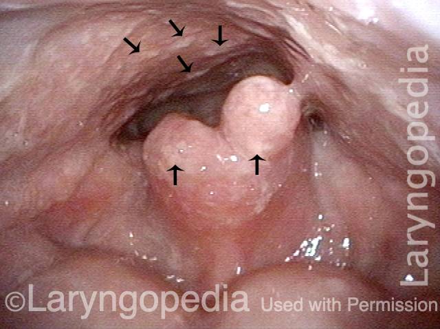
white lesion (1 of 4)
This patient was treated years earlier with radiotherapy for larynx cancer. The esophageal defect is chronic. The main finding here is white lesions on residual epiglottis and posterior pharyngeal wall. Arrows point to some examples of the countless candida colonies.
white lesion (1 of 4)
This patient was treated years earlier with radiotherapy for larynx cancer. The esophageal defect is chronic. The main finding here is white lesions on residual epiglottis and posterior pharyngeal wall. Arrows point to some examples of the countless candida colonies.
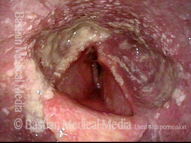
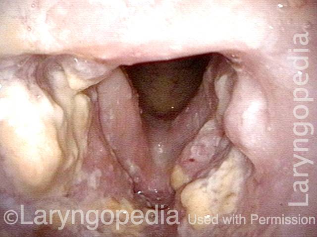
Inside the laryngeal vestibule (2 of 4)
Now viewing inside the laryngeal vestibule, notice the cheesy residue clinging in large areas, representing large amounts of candida overgrowth.
Inside the laryngeal vestibule (2 of 4)
Now viewing inside the laryngeal vestibule, notice the cheesy residue clinging in large areas, representing large amounts of candida overgrowth.
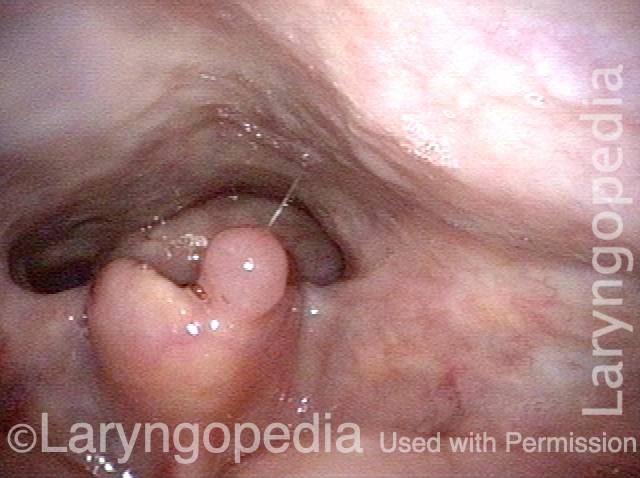
White Residue Is Gone (3 of 4)
After 15 days of fluconazole, notice the white residue on esophageal remnant and posterior pharyngeal wall is gone. Compare with photo 1.
White Residue Is Gone (3 of 4)
After 15 days of fluconazole, notice the white residue on esophageal remnant and posterior pharyngeal wall is gone. Compare with photo 1.
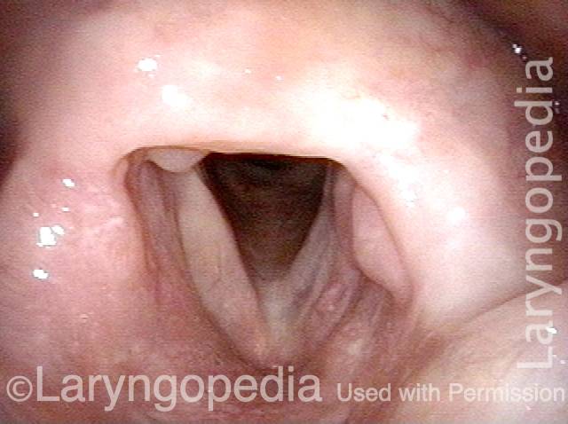
After treatment with fluconazole (4 of 4)
Again viewing inside the laryngeal vestibule, after treatment with fluconazole, the copious cheesy residue is also gone. Compare with photo 2.
After treatment with fluconazole (4 of 4)
Again viewing inside the laryngeal vestibule, after treatment with fluconazole, the copious cheesy residue is also gone. Compare with photo 2.
Share this article
