An encyclopedia about voice, swallowing, airway, coughing, & other head + neck disorders.

Carcinoma

Carcinoma (especially squamous cell carcinoma [SCCA] in the head and neck) is a malignancy originating in the cells that comprise the skin, or that line the respiratory and digestive tracts. SCCA may also be called “epidermoid carcinoma.”

Types of SCCA

SCCA may have subvariants such as spindle cell, verrucous, and papillary forms. The “angriness is described as a degree of differentiation: mildly-, moderately-, or poorly-differentiated.

See also: cancer, carcinoma in situ, and verrucous carcinoma.

Papillary Squamous Cell Cancer

As in this case, there is sometimes a debate in the mind of the pathologist peering through the microscope, between papillary and verrucous squamous cell carcinoma. The latter (verrucous) form tends to have a pushing margin at the interface with normal tissue, the mitotic rate is low, and there is a cauliflower-like, keratotic surface with clear demarcation between the tumor and normal tissues.

The prognosis for treatment is very good as this is a relatively “lazy” variant of SCCA. Papillary SCCA has features more like garden variety SCCA: the tumor is more infiltrating at the interface with normal tissue and a there is usually a higher mitotic rate.  The reader will note strong visual similarity between verrucous SCCA and papillary SCCA, however.

Visual Portfolio, Posts & Image Gallery for WordPress

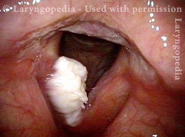
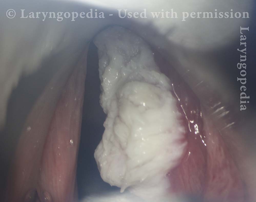
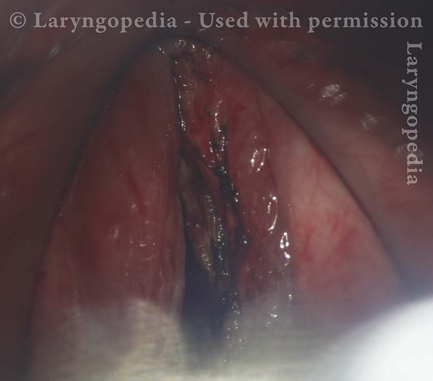
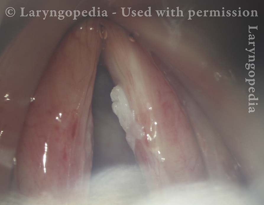
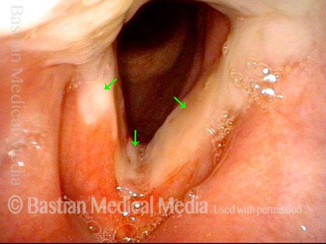
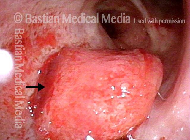
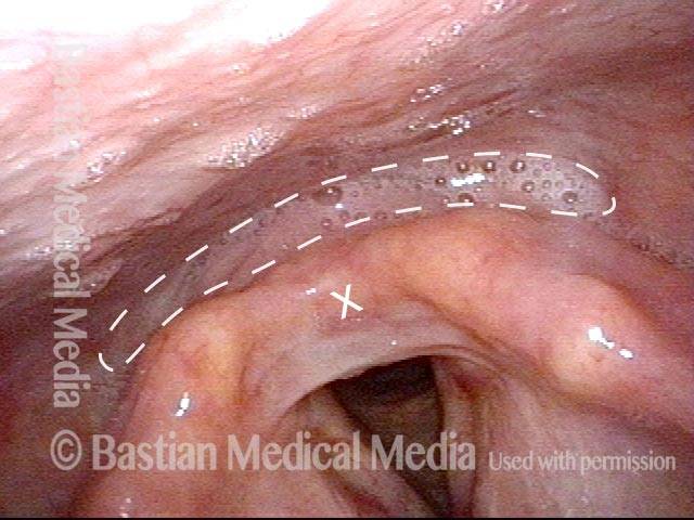
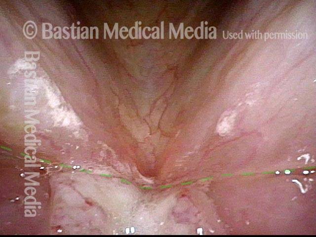
Bulky White Lesion (1 of 17)

This man has a grossly hoarse voice due to this bulky white lesion of the right vocal cord (left of photo). Open breathing position. Note the rough surface of the left cord (right of photo) is not so much contact reaction, but instead the result of his vocally exuberant and taxing lifestyle and work.

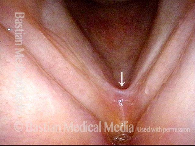
Normal Vocal Cord Movement (2 of 17)

Even though the tumor is so bulky, there is no impairment of vocal cord movement. Both come to the midline and close completely.

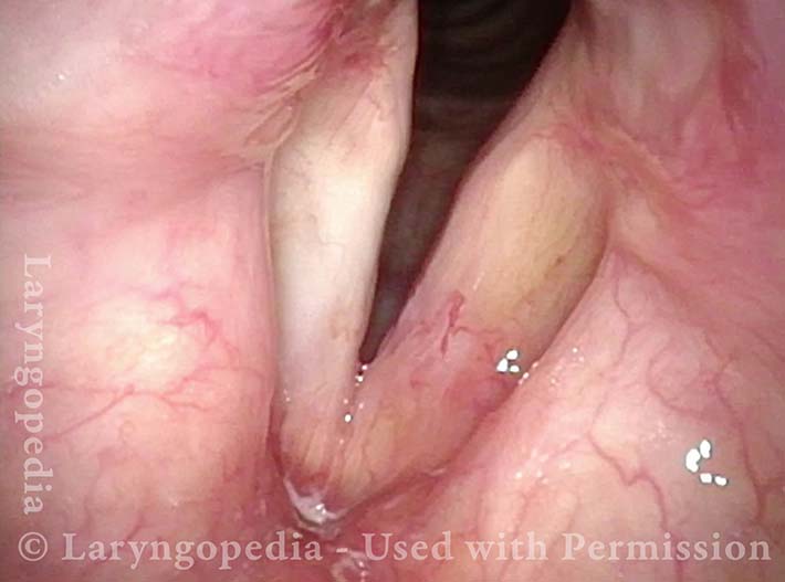
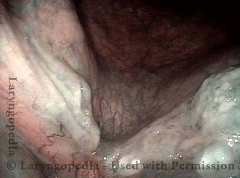
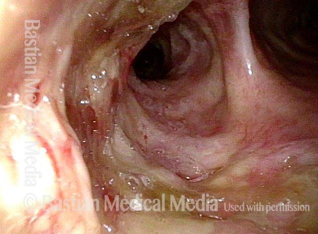
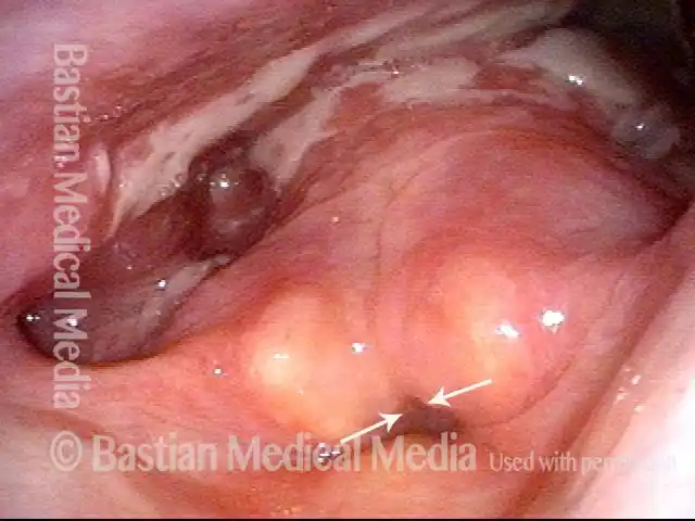
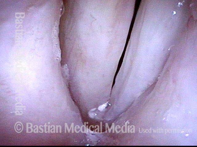
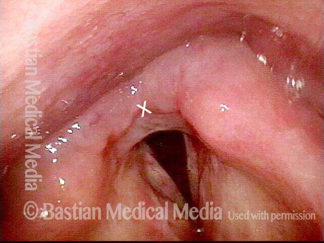
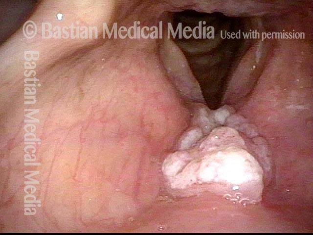
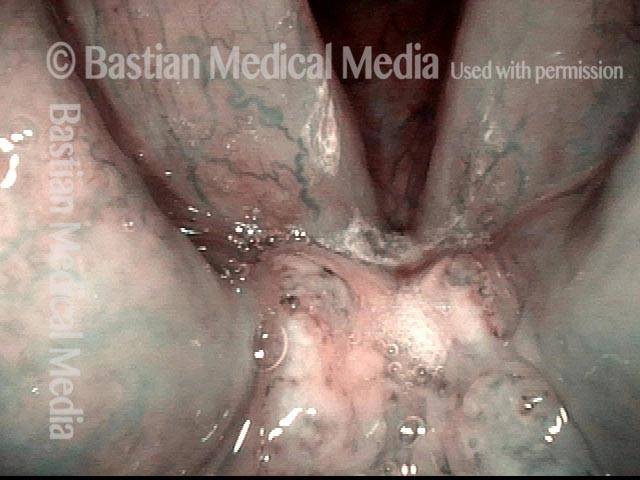
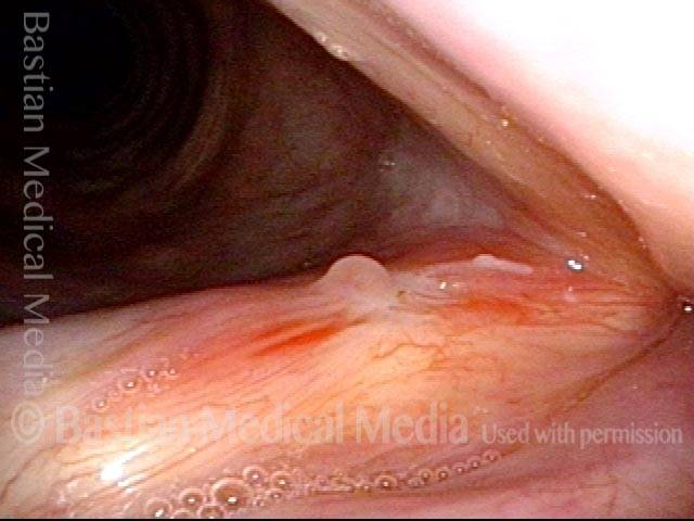
Initial View During Microlaryngoscopy (3 of 17)

The bulky nature and rough, keratotic surface is again noted.

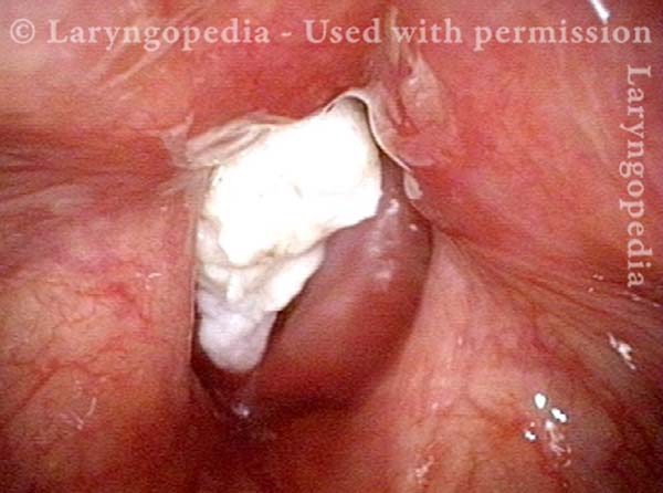
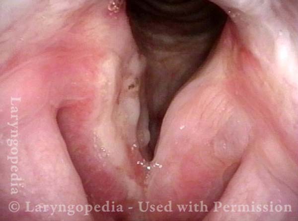
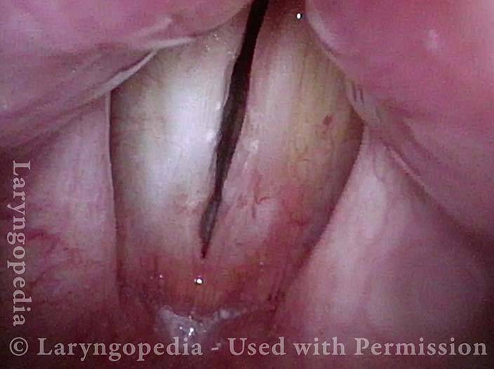
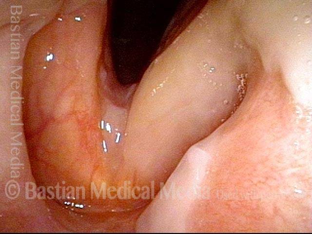
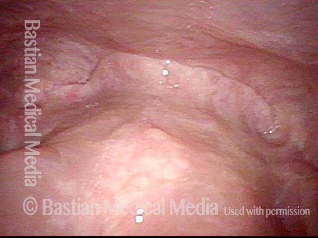
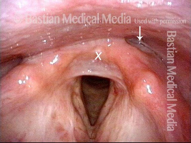
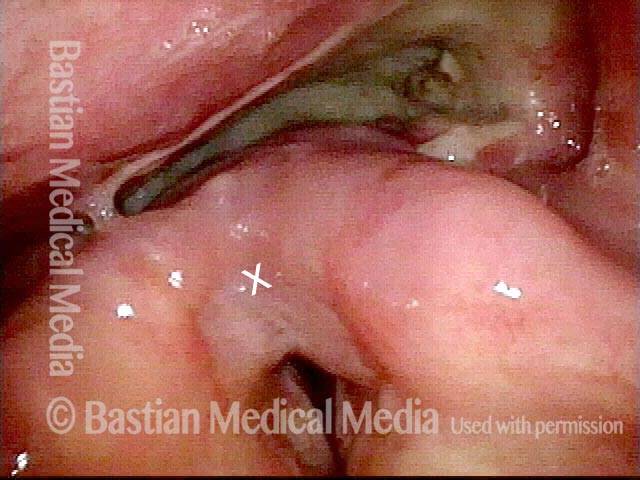
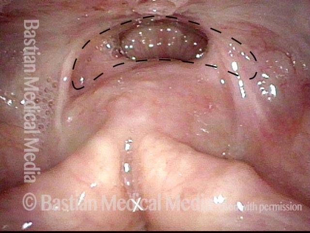
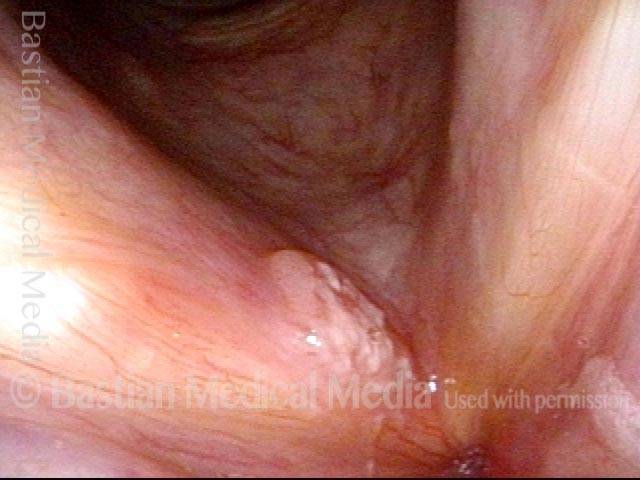
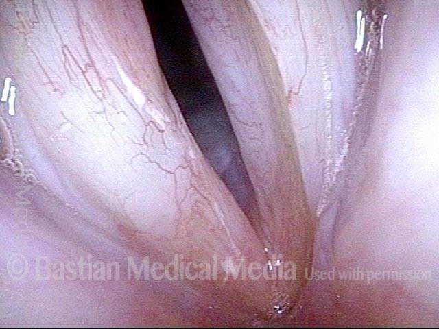
Clear Demarcation from Normal (4 of 17)

The suction cannula retracts the tumor to show that the boundary between normal and abnormal is clear.

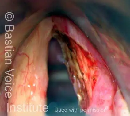
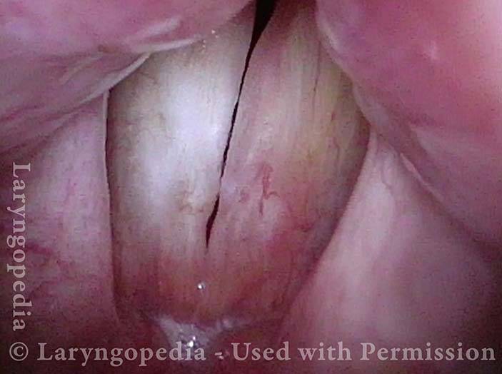
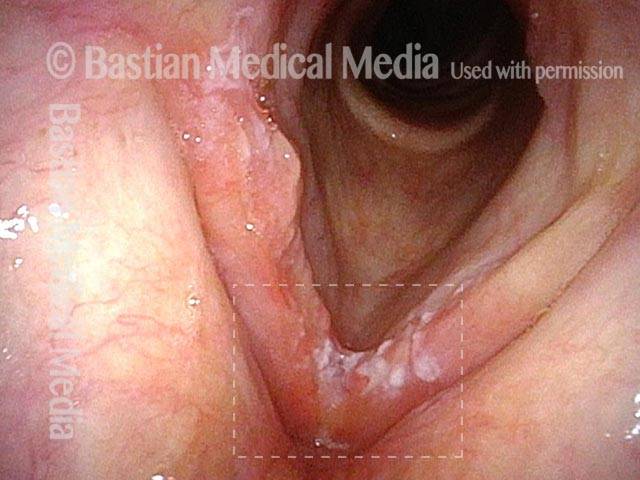
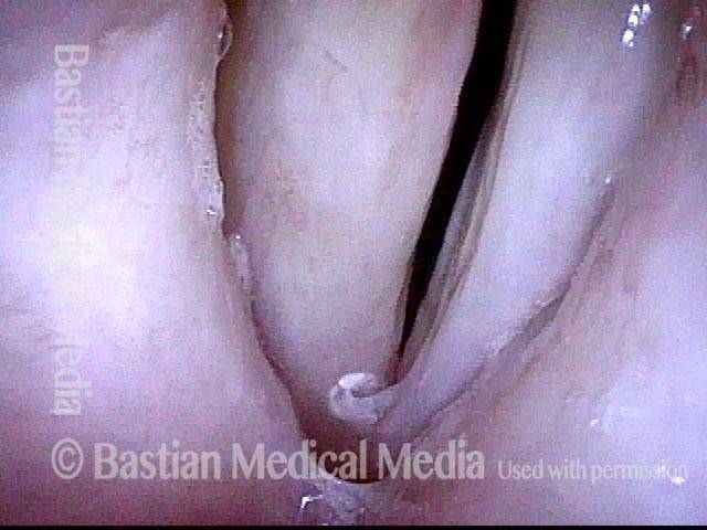
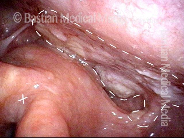
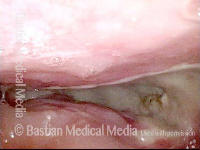
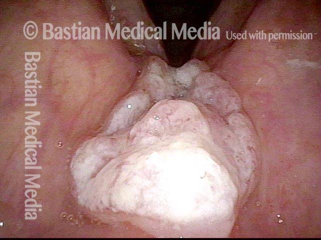
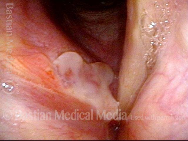
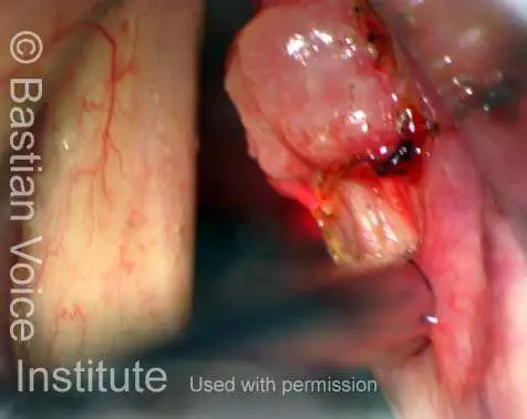
Narrow Margin (5 of 17)

We call the vocal cord “valuable real estate” and therefore use a narrow margin of resection (arrows point to narrow strip of mucosa taken with the tumor). This is to preserve voice as much as possible. Exophytic and well demarcated tumors permit this, and careful follow up keeps the patient safe, in case of a small recurrence.

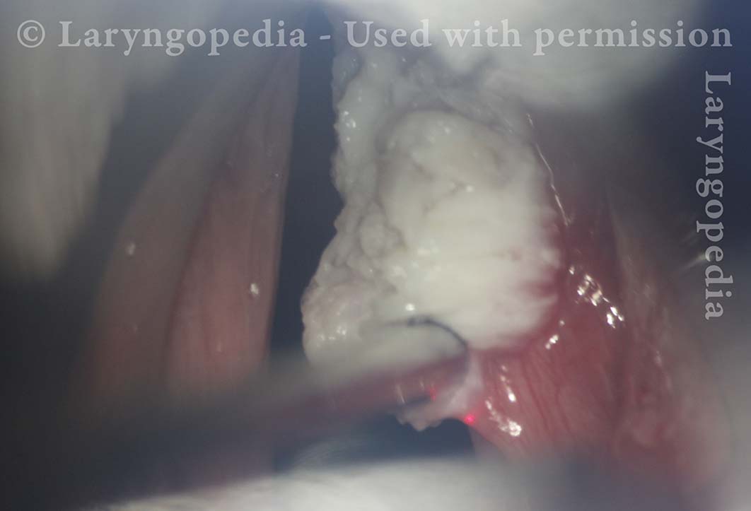
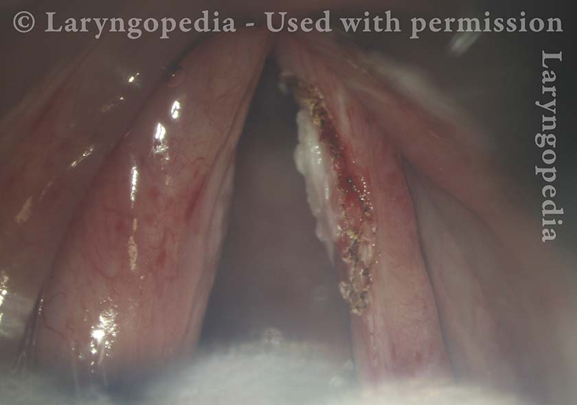
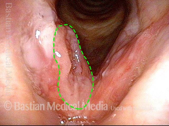
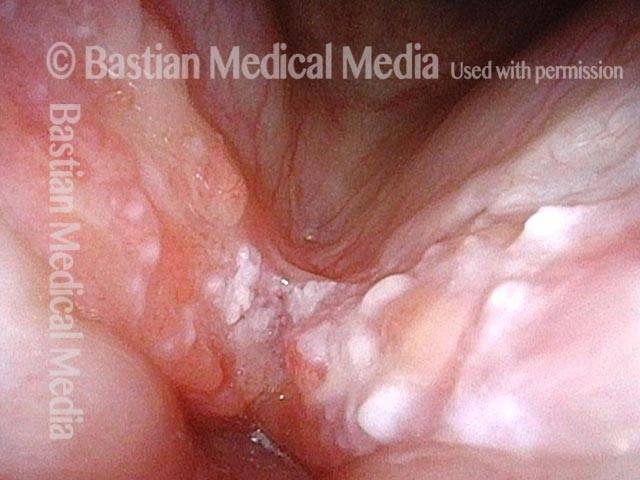
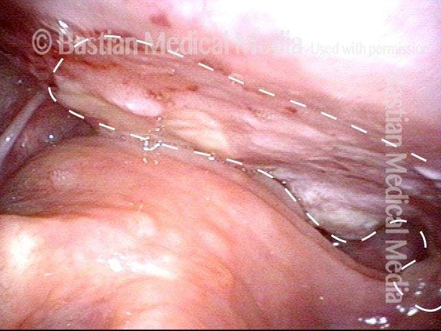
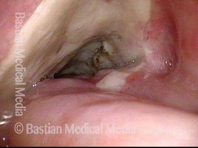
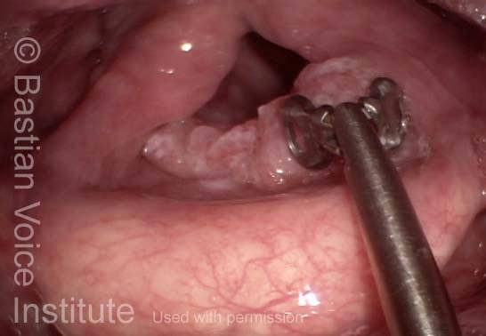
Added Excision (6 of 17)

The focus of apparent tumor circumscribed by the arrows is to be additionally removed with the specimen.

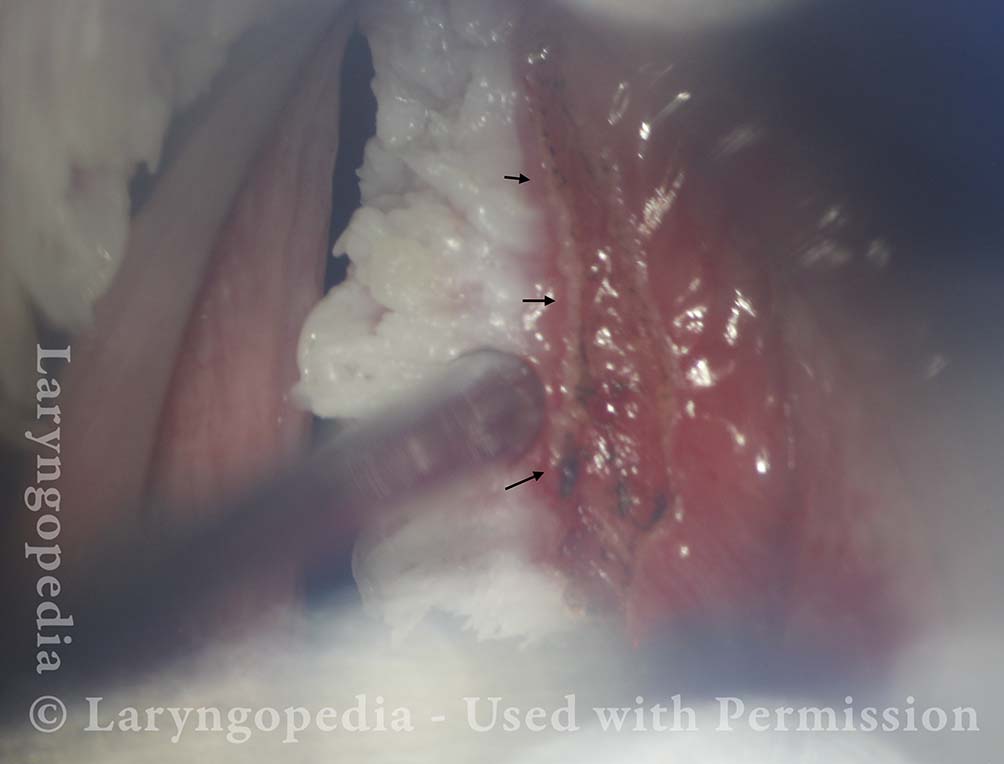
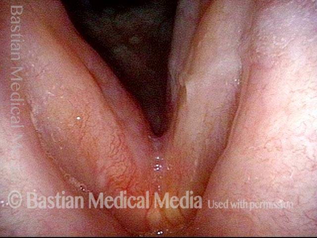
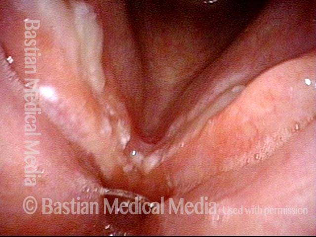
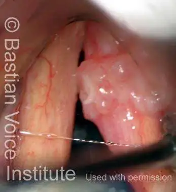
Excision Complete (7 of 17)

The tumor has been removed with only a millimeter of normal tissue, again to preserve voice.

At One Week, Voice Better (8 of 17)

The resulting wound will close in across time, but the right cord will likely not vibrate but serve as a partner to meet the left vocal cord. Note margin swelling due to overuse of the left, unoperated, cord (right of photo).

Voice Already Better (9 of 17)

The match of the cords is much better with tumor gone. Vibratory blur of the left cord explains his highly functional voice.

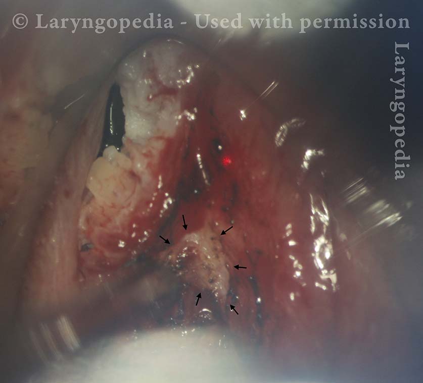
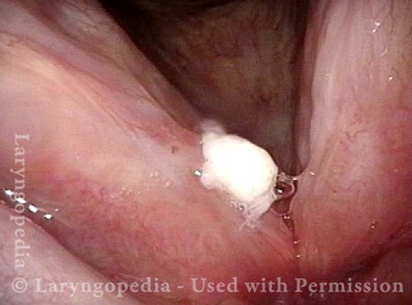
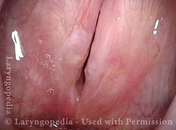
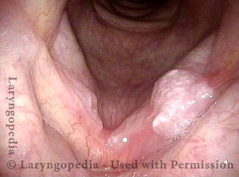
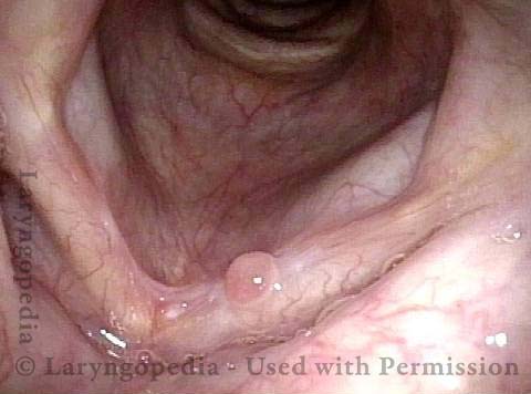
Small recurrence (10 of 17)

At six weeks postop, a small recurrence, which is not entirely surprising given the bulk of the original lesion and intentional narrow margin excision. The discussion was “radiation vs. second removal.” The latter was chosen.

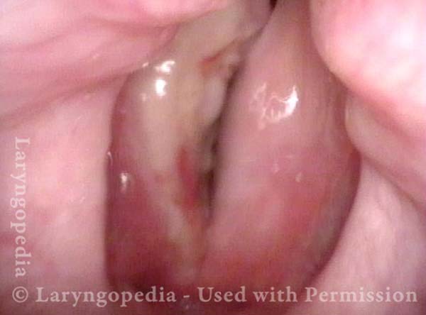
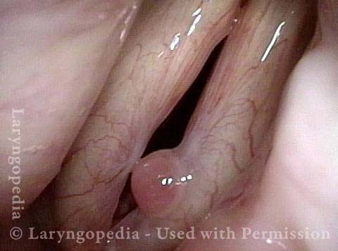
Vocal Cord Microsurgery (11 of 17)

View during second surgery

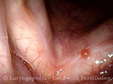
Excision in progress (12 of 17)

Outline only of the second surgical removal (completion excision photo was not taken).

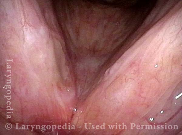
Post-surgical divot (13 of 17)

A week after excision #2, the right cord “divot” due to surgical excision is seen (arrow). The left cord (right of photo) shows margin swelling due to vocal overuse.

Voice is still good (14 of 17)

Match of the two cord margins is already reasonably good.

No recurrence 7 years later (15 of 17)

Seven years after surgical excision #2, there has been no recurrence. Voice is extremely functional but a little rough, partly due to occupational and personality-based overuse. The right cord (left of photo) is white due to scarring from the two removals.

Strobe light view (16 of 17)

Open phase of vibration under strobe light. Even the right cord oscillates laterally. The margin swellings are from vibratory trauma of vocal overuse.

Good match (17 of 17)

Closed phase of vibration shows quite good match, consistent with the patient’s preserved vocal capability, and good if imperfect voice quality.

Radiation Mucositis

Visual Portfolio, Posts & Image Gallery for WordPress

Prior to start of radiation (1 of 4)

Patient with vocal cord carcinoma, primarily of the right true cord (left of picture). This is before radiation therapy began, so there is not yet any radiation mucositis.

Radiation mucositis, 1 week after radiation (2 of 4)

One week after the end of radiation therapy. The tumor has disappeared. Radiation mucositis is evident from the patches of grey (arrows), which are superficial ulceration.

Radiation mucositis, 4 weeks after radiation (3 of 4)

Almost four weeks after the end of radiation therapy. Note that the mucositis has begun resolving, especially on the right cord (left of picture).

Disappearing radiation mucositis, 10 weeks after radiation (4 of 4)

Almost ten weeks after the end of radiation therapy. The mucositis is virtually gone.

Laser Surgery for Bilateral Vocal Cord Cancer

Visual Portfolio, Posts & Image Gallery for WordPress

Squamous cell carcinoma (1 of 6)

This man developed hoarseness across a few months. Biopsy elsewhere revealed squamous cell carcinoma, seen here on both vocal cords. Based upon a friend’s experience, he rejected radiotherapy, opting for laser resection, knowing it could be very hard on voice.

Tumor on the vocal cords (2 of 6)

At closer range and under narrow band (blue-green) light to accentuate the vascular abnormalities associated with this tumor.

Granuloma delays voice recovery (3 of 6)

Six weeks after superficial laser cordectomy, the larynx is almost healed with the exception of a small granuloma, left vocal cord (right of photo). When healing includes granulation, voice recovery is delayed as the granuloma resolves.

Closer view of granuloma (4 of 6)

He has hoarse but functional voice, but under strobe light, the granuloma prevents vibratory closure. Note the medial-to-lateral capillary reorientation so typical after laser cordectomy.

Granuloma is smaller (5 of 6)

Now 3 ½ months from surgery, voice has improved further and he considers it “75%” of original…One can see that the granuloma is smaller.

Granuloma doesn’t impede voice (6 of 6)

Note that the granuloma no longer prevents vibratory closure and this explains further improvement of voice. Compare with photo 4.

Tumor in Trachea

Visual Portfolio, Posts & Image Gallery for WordPress

Biopsy (1 of 4)

Tumor growing through wall of trachea, from a paratracheal lymph node. Biopsy forceps are about to close to take a tissue fragment for study.

After biopsy (2 of 4)

After biopsy was taken at arrow. The result: squamous cell carcinoma thought to be an unusual metastasis from unusually aggressive larynx cancer.

Tumor gone (3 of 4)

Soon after radiation therapy, the tumor has melted away, leaving a depression in the tracheal wall.

Slow return (4 of 4)

Eighteen months later, the patient has experienced a fairly durable response, with very slow return of tumor.

Vocal Cord Cancer, before, during, and after Radiation

Visual Portfolio, Posts & Image Gallery for WordPress

Vocal cord cancer (1 of 8)

Superficial cancer involving both vocal cords. This is stage 1 disease (T1B). The greatest bulk is on the right posterior cord (left of image), but the majority of both cords is involved with at least superficial disease. A faint dotted rectangle indicates the zoomed-in area seen in photo 2.

Vocal cord cancer (2 of 8)

Close-up view of only the anterior half of the cords. Notice the irregular surface, and areas of leukoplakia within this squamous cell carcinoma.

Vocal cord cancer, during radiation (3 of 8)

Just over midway through radiation treatment. One can see that the tumor is melting away.

Vocal cord cancer, during radiation (4 of 8)

Postcricoid / hypopharyngeal mucositis. In this view, the patient is performing a so-called trumpet maneuver to splay open the lower throat. The radiation delivered to the vocal cords (which inhabit the airway but are hidden here due to the momentary constriction of the laryngeal vestibule, at arrows) also causes superficial ulceration of the swallowing passage (upper half of the photo), directly behind the vocal cords. On occasion, if tissue reaction and mucositis are much more severe than seen here, a stricture can form, requiring dilation.

Vocal cord cancer, 2 months after radiation (5 of 8)

Two months after radiation is complete, showing that the tumor is gone, and the mucositis has resolved. There is a small anterior commissure web (at arrow) just below the free margin of the cords. The patient’s voice can nevertheless pass for normal.

Vocal cord cancer, 4 months after radiation (6 of 8)

Now four months after the end of radiation. Close-up view of the postcricoid / hypopharynx regions (compare with photo 4 in this series). Mucositis here is resolved as well, and there is no stricture.

Vocal cord cancer, 6 months after radiation (7 of 8)

Now six months after the end of radiation. Strobe illumination, open phase of vibration. Note that the contours of the vocal cords are not perfectly normal, even though voice is very good.

Vocal cord cancer, 6 months after radiation (8 of 8)

Strobe illumination, nearly closed phase of vibration. Oscillatory flexibility is preserved, but the vocal cord margins are not perfectly straight.

Hypopharyngeal Cancer, before and after Surgery

Visual Portfolio, Posts & Image Gallery for WordPress

Hypopharyngeal cancer (1 of 10)

Years after successful radiotherapy for glottic cancer, during a routine, yearly follow-up examination, a new hypopharyngeal cancer (invasive squamous cell carcinoma) is barely seen, at arrow. (For reference, X marks the same location in the larynx throughout this series.)

Hypopharyngeal cancer (2 of 10)

Trumpet maneuver begins, to splay open the post-arytenoid part of the posterior pharyngeal wall (arrows and dotted lines), as well as the pyriform sinuses on each side. The tumor is now seen clearly. (Note the change of perspective from photo 1, using X as a reference point.)

Hypopharyngeal cancer (3 of 10)

Similar view to photo 2 (still with the trumpet maneuver), but now with the tumor outlined.

Hypopharyngeal cancer (4 of 10)

Closer view than photos 2 and 3 (again, still with the trumpet maneuver), with the tumor still outlined. Radiation is not an option for this tumor because of prior larynx irradiation; an attempt at laser surgery was selected.

Hypopharyngeal cancer: 1 week after surgery (5 of 10)

One week after laser excision of the tumor. Note the widespread redness, and the swollen arytenoid mounds, especially on the right side of the image. Compare with photo 1.

Hypopharyngeal cancer: 1 week after surgery (6 of 10)

Panoramic view, with the patient beginning the trumpet maneuver. The surgical wound is coming into view. Compare with photo 2 (again, using X as a reference point).

Hypopharyngeal cancer: 1 week after surgery (7 of 10)

Maximum trumpet maneuver. Closer view of the surgical wound. Compare with photo 4.

Hypopharyngeal cancer: 1 week after surgery (8 of 10)

View into the left pyriform sinus, where the tumor was most bulky.

Hypopharyngeal cancer: several months after surgery (9 of 10)

Months after laser resection, panoramic view, showing the swallowing “crescent” (within dotted lines), at the upper limit of where the laser resection occurred. There is surgical stenosis at the entrance to the esophagus (shown in the next photo). This stenosis affects swallowing of solid food, but the patient says this is no problem for him, if he eats a little more slowly and chews well.

Hypopharyngeal cancer: several months after surgery (10 of 10)

With the trumpet maneuver, again splaying open the hypopharynx, as in photo 2. Notice the hypopharyngeal/ postcricoid stenosis; the dotted lines represent what would be a normal-sized opening.

Supraglottic Cancer

Visual Portfolio, Posts & Image Gallery for WordPress

Supraglottic cancer (1 of 4)

Middle-aged woman with sore throat, discovered to have a circumscribed exophytic squamous cell carcinoma at the petiole and anterior false cords.

Supraglottic cancer (2 of 4)

Closer view, showing the aberrant tumor vessels especially in the inferior half of the tumor, with less “white-out” from light overexposure.

Supraglottic cancer (3 of 4)

As is often the case, this tumor “respects” the ventricle, meaning it does not cross the plane of the ventricle (entrance at dotted line) to invade the true cords. An instrument could lift the inferior border of the tumor to show that the true cords aren’t invaded; the tumor is simply overlapping them.

Supraglottic cancer (4 of 4)

Under narrow-band lighting, the tumor’s aberrant vessels and its relationship to both the true and false cords are clearer. This is a supraglottic (not transglottic) tumor, likely amenable to outpatient endoscopic laser resection, and management of lymphatic compartments of the neck, depending on individual circumstances, via observation, radiotherapy, or selective neck dissection.

Vocal Cord Cancer, before and after Surgery

Visual Portfolio, Posts & Image Gallery for WordPress

Vocal cord cancer (1 of 4)

Squamous cell carcinoma, right vocal cord (left of image), standard light.

Vocal cord cancer, 1 week after surgery (2 of 4)

One week after laser excision. See irregular granulation especially at lower margin of excision.

Vocal cord cancer, 1 month after surgery (3 of 4)

Approximately one month after excision, healing progressing.

Vocal cord cancer, after complete healing (4 of 4)
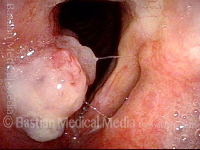
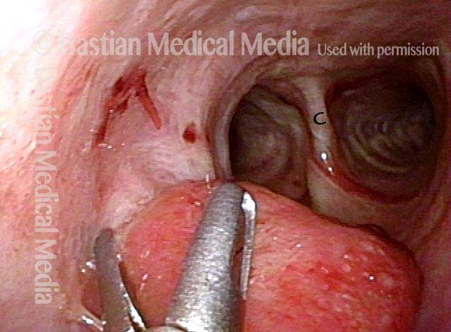
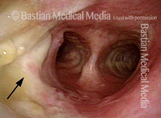
After complete healing, patient has a voice that passes for normal. Under strobe light, right cord oscillates well except at very high vocal pitch. Note, however, the mild pseudo-bowing of the right cord due to tissue loss, and that there is a mucosal wave on the left, but not on the right.

Biopsy of Carcinoma

Visual Portfolio, Posts & Image Gallery for WordPress

Biopsy, epiglottis (1 of 1)

Biopsy of lesion involving the petiole (low laryngeal surface of epiglottis). The pathology report revealed squamous cell carcinoma, usually caused by smoking.

Glottic Cancer, Laser Removal

Visual Portfolio, Posts & Image Gallery for WordPress

Glottic cancer, laser removal (1 of 3)

Early right vocal fold carcinoma, operative view (OR).

Glottic cancer, laser removal (2 of 3)

Same lesion, at the start of laser removal (OR).

Glottic cancer, laser removal (3 of 3)

Same larynx, after removal is complete. With healing over the next several months, the deficit “fills in” and voice result is often surprisingly good (OR).

Share this article

large lobule projecting upwards
Carcinoma right vocal cord with a large lobule projecting upwards and medial to the false cord, too.

Table of Contents